Introduction
Murine intraperitoneal cysticercosis is caused by the taeniid T. crassiceps and it has been useful to explore the physiological host factors associated with porcine cysticercosis and, to some degree, with human neurocysticercosis (NCC) (Smith et al., Reference Smith, Esch and Kuhn1972; Larralde et al., Reference Larralde1990; Huerta et al., Reference Huerta1992), because this model produces controlled and reproducible experimentation. In murine cysticersosis, female mice of any strain are more susceptible than male mice, which suggests a relationship to the sex-steroid microenvironment (Larralde et al., Reference Larralde1995). 17β-estradiol (E2) induces an increase in the parasite load by directly promoting parasite growth and reproduction (Escobedo et al., Reference Escobedo2005). Previously, we reported that E2 increases the in vitro reproduction of T. crassiceps, whereas androgens inhibit it (Morales-Montor et al., Reference Morales-Montor2002a; Escobedo et al., Reference Escobedo2004, Reference Morales-Montor2010a; Vargas-Villavicencio et al., Reference Vargas-Villavicencio2005, Reference Vargas-Villavicencio, Larralde and Morales-Montor2006). Steroid hormone effects are not only restricted to cestode parasites but also extend to nematodes such as Ancylostoma caninum (Bhai and Pandey, Reference Bhai and Pandey1982) and Trichinella spiralis. In taeniids, such as T. solium, in vitro treatment with progesterone increases the scolex evagination and worm growth (Escobedo et al., Reference Escobedo2011). These effects on parasites occur in a concentration- and time-independent fashion (Hernández-Bello et al., Reference Hernández-Bello2011). Direct exposure of T. crassiceps cysticerci to E2 and progesterone (P4) induces a differential proteomic map pattern in which actin, tubulin and myosin are highly expressed, with an apparent effect on the assembly and function of their flame cells, key elements of the excretion system of the parasite (Ambrosio et al., Reference Ambrosio2014). In vivo assays have also demonstrated the effects of testosterone (T4) or dihydrotestosterone (DHT) in T. crassiceps cysticerci, as parasite loads decreased by 80% in castrated and reconstituted (with T4 or DHT) male mice, or substituted (also with T4 or DHT) female mice, meaning that the androgen treatment affects the establishment, growth and reproduction of the parasite (Morales-Montor et al., Reference Morales-Montor2002a). In vitro experiments with these androgens have shown reductions in T. crassiceps viability after 10 days in culture (Ambrosio et al., Reference Ambrosio2015).
Cytoskeletal proteins are the main proteins expressed in tapeworms such as T. solium, Hymenolepis diminuta and Echinococcus granulosus (Almeida et al., Reference Almeida2009; Tsai et al., Reference Tsai2013), which could be expressed in several isoforms, as found for five actin isoforms in T. solium (Ambrosio et al., Reference Ambrosio2003). Several isoforms could be deduced for other single proteins from the genomes of cestodes (Tsai et al., Reference Tsai2013) and these protein isoforms could be affected by the hormone treatment. However, despite the fact that androgens have been shown to have important anti-parasitic activities, to date, knowledge of the exact effects of androgens on the proteomic pattern of the parasite, and their role in the parasite physiology, is null, and there is no clear evidence of the molecular mechanisms by which androgens have cysticidal effects. Nevertheless, an orthologue of the progesterone receptor membrane component (PGRMC) has been identified in the Taenia solium genome (TSM_00613800), which could serve as a promiscuous steroid receptor on the taeniid.
Thus, the aim of this study was to explore the in vitro effects of T4 and DHT on the proteomic map pattern of the metacestode stage of T. crassiceps and to establish if the effects could be related to changes in the expression of several proteins involved in the cellular biology of the parasites related to their cytoskeleton, vesicular traffic and intracellular signalling.
Materials and methods
Animals
Female BALB/c AnN (H2-d) syngeneic mice obtained from Harlan (Mexico City) were used in all of the experiments. Animals were housed in the animal care facilities at the IIB-UNAM, under controlled conditions of temperature (22°C) and 12 h dark–light cycles, with lights on between 07.00 and 19.00. They were fed Purina Diet 5015 (Purina, St. Louis, MO, USA) and purified water ad libitum. The fast-growing ORF strain of T. crassiceps was used for infection. Ten non-budding T. crassiceps cysticerci (c. 2 mm in diameter) were suspended in 0.3 ml sterile phosphate-buffered saline (PBS: 0.15 m NaCl, 0.01 m sodium phosphate buffer, pH 7.2) and carefully injected intraperitoneally into 8-week-old female mice using a 21G gauge needle. Infected mice were housed in separate cages (5 per cage) in the same room of the animal facility. At 16 weeks of infection, mice were euthanized by cervical dislocation after anaesthesia with sodium pentobarbital (Pfizer, Mexico), always at 08.00 am.
Parasites
Cysticerci were obtained from intraperitoneally infected mice and placed in tubes containing sterile PBS (1×) supplemented with 100 U/ml of antibiotics (Fungizone, Gibco, Grand Island, NY, USA). The tubes were centrifuged for 10 minutes at 1700 g and 4°C, and the supernatants were discarded. Cysticerci were washed in this way a total of three times. Packed cysticerci were incubated in DMEM serum-free medium (Gibco 12491).
In vitro treatment effects of T4 and DHT
Culture grade testosterone (T4) and dihydrotestosterone (DHT) were obtained from Sigma and were dissolved in pure ethanol (JT Baker) to the desired stock concentration, and sterilized by passage through a 0.2 μm pore filter. Afterwards, hormones were added to DMEM medium (Gibco 12491) to a final concentration of 8 nm for T4 and 4 nm for DHT and ethanol was left to evaporate. Pure ethanol to a final concentration of 0.05% was also added to media for a vehicle control. Ten viable (complete, translucent and motile cystic structures) non-budding cysticerci of c. 2 mm in diameter were then selected and dispensed into each well of 12-well culture plates (Falcon, Becton Dickinson Labware, Franklin Lakes, New Jersey) in 2 ml of culture medium supplemented with the corresponding treatment and incubated at 37°C and 5% CO2. Cultures were checked daily and their medium was completely replaced every other day.
Cytoskeletal proteins of parasites
Proteins of parasites were prepared and analysed by SDS-PAGE, isoelectrofocusing and 2D electrophoresis, as described by Reynoso-Ducoing et al. (Reference Reynoso-Ducoing2014). Also, the computational analysis of parasite protein expression was assessed in proteomic maps, followed completely as indicated in the same publication: all the considered proteins were resolved in proteomic maps in apparent molecular weight (MW) of 10–250 kDa and isoelectric point (pI) of pH 3–10.
Bioinformatic analysis
After Coomasie blue staining of the gels of the proteomic maps, they were scanned with ChemidocTM XRS Device (Bio-Rad Laboratories, Segrate, Milan, Italy) at 252 dpi resolution and compared using the PDQuest program. Spots that showed different expression after comparing the T4, DHT hormone treatments and the control without treatment were checked for their MW and pI values, to deduce through a bioinformatic search (using the GeneDB and Wormbase databases) their possible correspondent proteins in the T. solium genome (Tsai et al., Reference Tsai2013). Considering that the effect of the hormones could be mediated by PGRMC stimulation and its downstream signalling events (Hlavaty et al., Reference Hlavaty2016), we searched for the proteins involved in this pathway. Other additional criteria were adopted for the bioinformatic searching of those possible proteins, mainly associated with the cytoskeletal restructuration, as described by Papadopoulou et al. (Reference Papadopoulou2009) and Stournaras et al. (Reference Stournaras2014). For the T4 effect, proteins were searched in geneDB for T. solium, assuming they presented an apparent MW over 100 kDa and pIs under 7.
Statistical analysis
Optical density data from three independently performed sets of gels are reported as mean ± standard deviation and analysed with Prism 6® software (GraphPad Software Inc.). One-way ANOVA (α = 0.05) was performed, followed by a Tukey post-hoc test. Differences were considered to be significant when P < 0.05.
Results
In vitro motility and survival of androgen-treated cysticerci
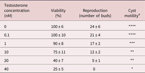
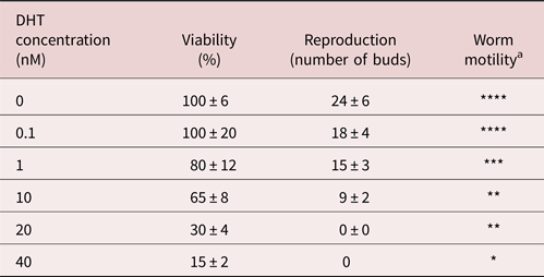
As previously shown by us (Ambrosio et al., Reference Ambrosio2015), we extended and reproduced the cysticidal effects of androgens. There was a clear concentration-response pattern in the presence of androgens. DHT had a greater effect than T4, but both reached 75% (table 1) and 85% (table 2) of mortality at the higher dose used (40 ng) in culture. The effect of androgens was also characterized by a 100% loss of motility and 100% inhibition of reproduction (tables 1 & 2).
Reproduction of cultured Taenia crassiceps cysticerci in response to various concentrations of testosterone (T4). Data represent mean ± SD from three experiments, with 10 cysticerci per well, six wells per dose, during 10 days of culture.

aInjury to worms was recognized microscopically by progressive internal disorganization, by development of whitish opaque areas on the parasite's tegument, and by loss of motility. Dead worms were immobile, opaque and disorganized structures.
* 0% of worms motile and 25% alive.
** 50% of worms motile, and 80% alive.
*** 80% of worms motile, and 90% alive.
**** Very motile, 100% alive and healthy in their appearance.
Reproduction of cultured Taenia crassiceps cysticerci in response to various concentrations of dihydrotestosterone (DHT). Data represent mean ± SD from three experiments, with 10 cysticerci per well, six wells per dose, during 10 days of culture.

aInjury to worms was recognized microscopically by progressive internal disorganization, by development of whitish opaque areas on the parasite's tegument, and by loss of motility. Dead worms were immobile, opaque and disorganized structures.
* 0% of worms motile and 25% alive.
** 50% of worms motile, and 80% alive.
*** 80% of worms motile, and 90% alive.
**** Very motile, 100% alive and healthy in their appearance.
Protein expression patterns from control, T4- and DHT-treated cysticerci by 2-DE
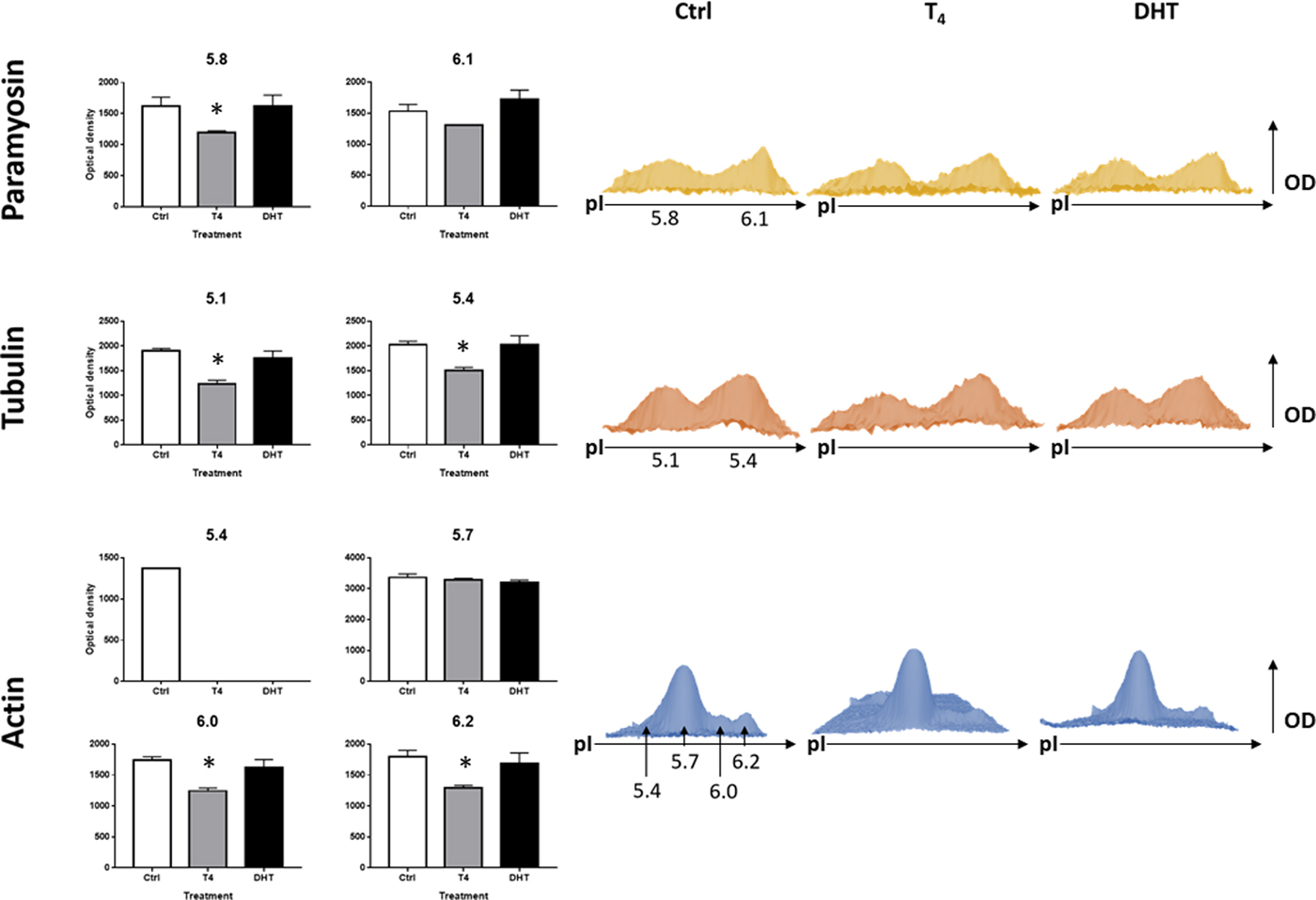
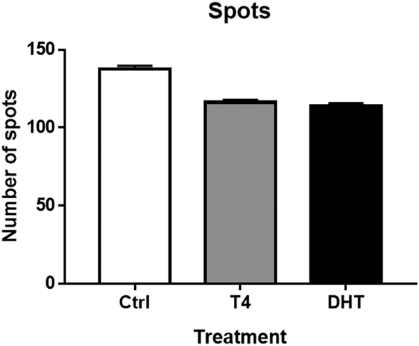
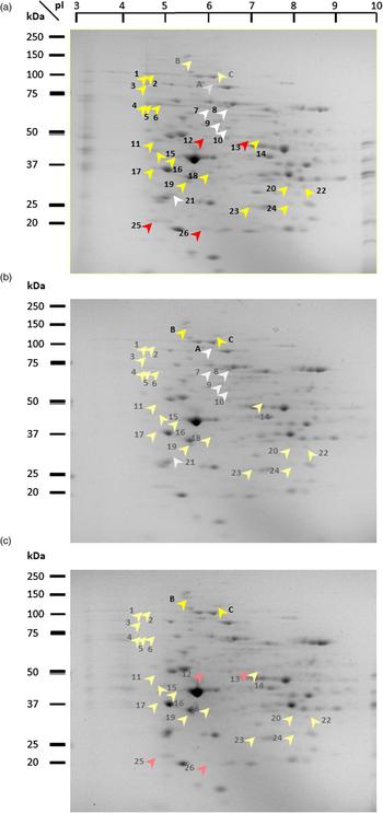
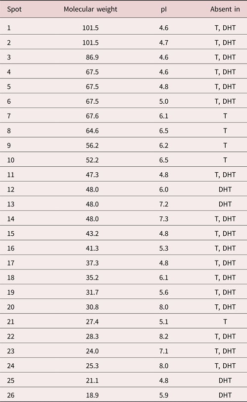
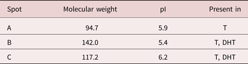
We previously reported that androgens exert a cysticidal effect that seemed to be mediated by an alteration in the cytoskeletal proteins associated with flame cell assembly. In order to dissect particular differences in cytosleketal protein expression, a 2DE proteomic analysis was performed, with emphasis on the identification of actin, tubulin and paramyosin isoforms. As depicted in fig. 1, a general decrease was found for cytoskeleton proteins revealed in the T4-treated group. These results are consistent with our previous findings. However, the main finding was that androgen treatment apparently blocks the expression of a specific actin isoform sited at 5.4 of pI and details of those changes were clearly registered after 3D image analysis, as shown in fig. 1. Regarding the rest of the protein profile, the initial analysis showed that the total number of spots was decreased in the androgen-treated samples as deduced by the densitometric analysis of 2D gels (fig. 2), although not at a significant level, which suggested that only a few proteins were absent or greatly downregulated. Upon analysis, it was found that some spots were not detectable on the androgen-treated gels, detailed in table 3. Representative gel images of three experimental treatments are shown in fig. 3, in which it was found that 17 spots were consistently absent (or greatly downregulated) in both T4- and DHT-treated samples, indicated with yellow arrowheads in the control gel (fig. 3a). The places where the spots should appear are indicated by pale yellow arrowheads on the testosterone and DHT- treated gels (fig. 3b, c). Besides, five spots specifically depleted by T4 treatment, as well as four spots absent only in the DHT-treated samples, were detected (table 3), which are indicated in the control gel using white and red arrowheads, respectively (fig. 3a). Light grey and pink arrowheads are used to indicate the places where these spots should be on androgen-treated gels (fig. 3b, c). Evaluating the consistency of the expression of some spots, it was noted that some of them were detected only in the androgen-treated samples but were absent in the control samples. One of them was specifically induced by testosterone treatment and was named spot A, shown as a white arrowhead on fig. 3b, and a light grey arrowhead on the control gel (fig. 3a), while spots B and C were found in T4 and DHT samples (yellow arrowheads on fig. 3b, c and light yellow arrowheads on fig. 3a). The electrophoretical parameters of those specific spots are detailed in tables 3 and 4.
Quantitative analysis of cytoskeleton protein isoforms. Left panel: optical density values for each protein isoform. Right panel: representative 3D model of spots corresponding to analysed isoforms. Optical density values and 3D models were obtained from PDQuest software. Data from triplicate gels are expressed as mean ± SD. *, P < 0.05.

Total number of spots detected. Spot number yielded after automated spot detection with same sensitivity threshold. Data from triplicate gels are expressed as mean ± SD.

Representative gels showing differentially expressed spots. (a) Control sample; yellow arrowheads: spots missing in both androgen-treated samples; pale yellow arrowheads: places where spots B and C would appear; white arrowheads: spots missing only in T4-treated samples; grey arrowheads: place where spot A would appear; red arrowheads: spots missing only in DHT-treated samples. (b) T4-treated sample and (c) DHT-treated sample; yellow arrowheads: spots arising in both androgen-treated samples; pale yellow arrowheads: spots missing in both androgen-treated samples; white arrowhead: spot arising only in T4-treated sample; grey arrowheads: spots missing only in T4-treated sample; pink arrowheads: spots missing only in DHT-treated samples. Numbers and letters correspond to details in tables 3 and 4.

Spots whose expression was lost or significantly downregulated by androgen treatments (see gels depicted in fig. 3). T, testosterone; DHT, dihydrotestosterone.

Spots whose expression emerge by androgen treatment. T, testosterone; DHT, dihydrotestosterone (see gels depicted in fig. 3).

Considering that the recent identification of a PGRMC orthologue (TSM_000613800 from T. solium genome) in T. crassiceps could be involved in the binding of multiple steroids, with different affinities, and that it could serve as a promiscuous steroid receptor, it was investigated if the protein could be expressed in the samples evaluated in the present study. One spot (spot 25), with electrophoretic properties of MW = 21.13 kDa, pI = 4.75, was found to be expressed in the controls but its expression was downregulated after DHT treatment.
Bioinformatic analysis
The bioinformatic approach was focused on the search for proteins involved in the cytoskeleton function and the signalling pathways associated with PGRMC. As there is not currently a genomic database for T. crassiceps, and assuming a high homology among the genus Taenia, meaning the variations in molecular weight and isoelectrical point would be minimal, we based the search on T. solium orthologues.
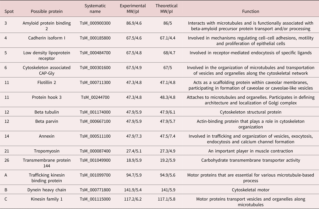
Regarding cytoskeletal physiology, we found that some spots could correspond to structural cytoskeleton proteins such as β-tubulin (MW and pI) and tropomyosin (MW and pI), as well as cytoskeleton-associated proteins such as β-parvin (MW and pI) and trafficking kinesin binding protein (MW and pI).
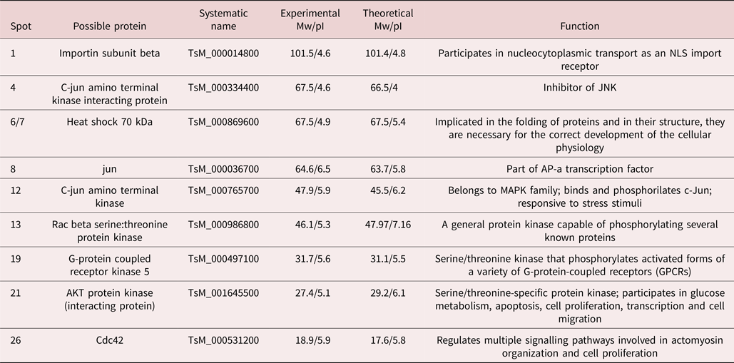
As for the proteins associated with PGRMC signalling, we found that two spots could correspond to Rac (MW and pI) and Cdc42 (MW and pI) orthologues, which play a role in actin reorganization upon PGRMC signalling. This receptor is also involved in apoptosis control via MAPKs and we were surprised to find that the kinase JNK (MW and pI) and the transcription factor c-jun (MW and pI) orthologues corresponded to two of the spots regulated by androgen treatment. Interestingly, we also found that two other spots downregulated by androgen treatment could correspond to AKT (MW and pI) and HSP70 (MW and pI) orthologues, both of them negative regulators of apoptosis. All the results are detailed in tables 5 and 6.
Prospective identification of androgen-regulated proteins by bioinformatic analysis related to cytoskeleton and vesicular traffic.

Prospective identification of androgen-regulated proteins by bioinformatic analysis related to signalling.

Discussion
Previously, we found that gender and circulating T4 and DHT levels in host mice crucially affected the dynamics of parasite loads in mice infected with cysticerci of T. crassiceps (Larralde et al., Reference Larralde1995; Morales-Montor et al., Reference Morales-Montor2002b). The present work confirms and extends the notion that androgens act directly upon T. crassiceps cysticerci without need for the host's participation: T4 and DHT significantly lead to their destruction. This finding offers an alternative explanation of why T. crassiceps cysticerci grow better in a low androgen environment, as in the female, and do not grow well in a high androgen environment, as in the male, emphasizing the molecular cross-talk between host and parasite, which is in turn differentially influenced by the hormonal microenvironment of each gender (Larralde et al., Reference Larralde1995; Morales-Montor et al., Reference Morales-Montor2002b).
However, so far, there is no description of an androgen receptor in T. crassiceps that can explain its effects. Perhaps passive diffusion through the tegument is the explanation for how androgens easily reach the inner parasite tissues. It has been established that passive diffusion is the main entry of some drugs, with similar structure to androgens, through the cuticle of nematodes and the tegument of cestodes and trematodes (Alvarez et al., Reference Alvarez, Mottier and Lanusse2007). On the other hand, steroids have proven to exert their effects on cells lacking canonical steroid receptors, acting through non-classical receptors in the membrane.
Following this argument, we have recently identified a protein analogue to a progesterone receptor membrane component (PGRMC) in T. solium (Aguilar-Díaz et al., Reference Aguilar-Díaz2018), a phylogenetic relative of T. crassiceps. Furthermore, we found that a spot of the analysed tissues is identical to that protein reported in T. solium.
One of the signalling pathways associated with PGRMC is the MAP kinase/ERK pathway (Migliaccio et al., Reference Migliaccio1998; Lange, Reference Lange2004). Many of the proteins of this pathway are well known to have orthologues in helminths, and ERK-like proteins have been found to play similar roles in the taeniid physiology (Escobedo et al., Reference Escobedo2010b). It is interesting that c-jun transcription factor was downregulated upon T4 treatment (Johnson and Lapadat, Reference Johnson and Lapadat2002), as it plays a key role in the JNK pathway (MAPK subfamily), regulating cellular differentiation and apoptosis (Escobedo et al., Reference Escobedo2004).
The bioinformatic analysis performed in this study found that proteins of the JNK pathway are possibly affected by hormonal treatment. No effect on c-fos was found, but it is known that both proteins are complementary to various cell functions mentioned above (Glover and Harrison, Reference Glover and Harrison1995). There also appear to be changes in several protein kinases related to the JNK signalling pathway, and to hormone synthesis. The membrane component of the progesterone receptor (PGRMC), a molecule capable of binding steroids to the plasma membranes of cells, has been reported also in other in helminth parasites (Hu et al., Reference Hu2003; Aguilar-Díaz et al., Reference Aguilar-Díaz2018). One of the bioinformatically identified proteins that seem to show changes in response to androgens in T. crassiceps is precisely this receptor, which could influence the synthesis and degradation mechanisms of T4 and DHT.
During the analysis, we also found several proteins fulfilling functions related to the cytoskeleton, such as tropomyosin and β-parvin, which bind to actin (Yamaji et al., Reference Yamaji2001), and also proteins related to vesicular trafficking, such as annexin and importin (Corbett and Silver, Reference Corbett and Silver1997; Gerke and Moss, Reference Gerke and Moss2002). This would imply an important process of steroid hormones affecting vital processes of the parasite, which seems to corroborate the effects that have been seen previously in T. crassiceps, where changes in the expression of proteins are located, including specific sites such as flame cells (Ambrosio et al., Reference Ambrosio2014).
Based on the deductions presented in this paper, we propose that the high reproducibility of the experimental strategy relied on 2D proteomic map analysis and, associated with the bioinformatic analysis, allows a precise approximation to the proposal initially formulated by us (Escobedo et al., Reference Escobedo2010b). This proposal is an approach as to how signal transduction via MAPK could have an effect on cytoskeletal restructuring, as has been previously demonstrated in other cell types (Gu et al., Reference Gu2009; Papadopoulou et al., Reference Papadopoulou2009).
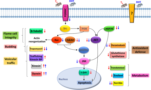
Gathering the experimental data and the potential identity of the proteins that are regulated upon T4 and DHT treatments, we propose a preliminary interactome, in order to show a better and integral understanding of the molecular basis for the deleterious effects of androgens upon T. crassiceps cysticerci. As illustrated in fig. 4, T4 and DHT may bind to PGRMC or other unknown membrane receptors. Upon binding to PGRMC, androgens can trigger a signalling pathway via SRC and PI3K that further activates Rac, which leads to actin reorganization. As Rac is one of the proteins affected by DHT treatment, the actin reorganization could be perturbed from this point on. Rac may also interact with Cdc42 to activate MAPKs, which further activate JNK and c-jun, leading to apoptosis. This pathway is negatively regulated by pro-survival AKT and HSP-70, which are both downregulated by androgen treatment. Regardless of the way of entrance of androgens, several cytoskeleton proteins, such as β-tubulin, tropomyosin and certain actin isoforms, are downregulated, which affects flame cell assembly and interferes with the budding process necessary for the reproduction of the cysticerci. The expression of cytoskeleton-associated proteins was increased, which could reflect the vesicular traffic distress caused by cytoskeletal alterations.
Proposed interactome for the physiological effects of androgens on T. crassiceps cysticerci. Upon androgen treatment, the expression of some cytoskeleton proteins, such as β-tubulin, tropomyosin and certain actin isoforms, is downregulated, which affects flame cell assembly and interferes with the budding process necessary for the reproduction of this parasite. The expression of cytoskeleton-associated proteins is increased, probably reflecting the vesicular traffic distress caused by cytoskeletal alterations. Binding of androgens to PGRMC can trigger a signalling pathway via Src (presumed) and PI3K (presumed) that may lead to erratic actin reorganization, as Rac is affected. This same pathway can regulate apoptosis, which can be promoted, as two important negative regulators, namely HSP70 and AKT, are downregulated by androgen treatment. Furthermore, androgen treatment may weaken the parasite by decreasing the expression of important proteins involved in antioxidant defense and metabolism, such as glutathione synthetase and ferritin, respectively. Red arrows: up- or downregulation upon T4 treatment; blue arrows: up- or downregulation upon DHT treatment.

Androgen treatment may weaken the parasite by decreasing the expression of peroxiredoxin and glutathione synthetase, both important for antioxidant defense, and further damage may be caused or reflected by the downregulation metabolism-involved proteins such as transketolase, enolase and ferritin.
The knowledge of the proteins whose expression is modulated by androgens may contribute to a better understanding of the biology of helminth parasites, considering that hormones have been shown to be a crucial factor in the infection process. Furthermore, the identification of molecular targets, as well as the mechanisms involved, can lead us to the development of novel therapeutic strategies.
Financial support
Grants PE-21115, IN-216213 and IN-224316 to Javier Ambrosio, and Grant IN-208715 to Jorge Morales-Montor, were obtained from Programa de Apoyo a Proyectos de Investigación e Innovación Tecnológica (PAPIIT) from Dirección General de Asuntos del Personal Académico (DGAPA), Universidad Nacional Autónoma de México (U.N.A.M.), and Grant 151747 to Pedro Ostoa-Saloma is from Programa de Fondos Sectoriales CB-SEP, Consejo Nacional de Ciencia y Tecnología (CONACyT). Diana G. Ríos-Valencia is a PhD student at Programa de Doctorado en Ciencias Quimico-Biológicas IPN, and has a scholarship from CONACyT.
Ethical standards
Animal care and experimentation practices at the Instituto de Investigaciones Biomédicas of the Universidad Nacional Autónoma de Mexico (IIB-UNAM) are constantly evaluated by the Institute's Animal Care and Use Committee, adhering to the official Mexican regulations (NOM-062-ZOO-1999). Mexican regulations are in strict accordance with the recommendations in the Guide for the Care and Use of Laboratory Animals of the National Institute of Health (NIH) of the USA to ensure compliance with established international regulations and guidelines.