Tetralogy of Fallot was first described anatomically by Etienne-Louis Fallot in 1888. This defect is due to antero-cephalad deviation of the outlet septum during development consists of the following components: an overriding aorta, right ventricular outflow tract obstruction, a ventricular septal defect, and usually consequent right ventricular hypertrophy.Reference Huehnergarth, Gurvitz and Stout1
Tetralogy of Fallot is the most common form of cyanotic congenital heart disease, accounting for 10% of congenital heart disease patients, and occurring in one in 3600 births.Reference Apitz, Webb and Redington2 It carries a recurrence risk of 3% in siblings, with genetic microdeletion in 22q11 in 15–25% of Tetralogy of Fallot patients. It is associated with DiGeorge and velocardiofacial syndromes.Reference Apitz, webb and Reding3
In all, 85% of children survive into adulthood after repair of Tetralogy of Fallot.4 In our adult congenital heart disease clinic, these patients account for 22% of our total number of patients followed.Reference Dearani, Connolly, Martinez, Fontanet and Webb5 Most patients function at New York Heart Association Class I and II. A number of patients are now approaching their 50s and 60s and experiencing comorbidities of ageing.
Surgical palliation was achieved with classic Blalock–Taussig shunts – direct subclavian artery to pulmonary artery connections – in 1944. Other forms of palliation, such as other types of systemic-to-pulmonary artery shunts, followed. All have some effect on the growth and development of the pulmonary and subclavian arteries. Over time, the classic Blalock–Taussig shunt has given way to the modified Blalock–Taussig shunt – gortex interposition graft. The Potts and Waterson shunts are no longer utilised.
These shunts all have in common the aim of increasing the pulmonary blood flow either by anastomosing a systemic artery or vena cava to the pulmonary artery. The resultant increased pulmonary flow reduces the level of cyanosis (Fig 1).6
Blalock–Taussig shunt, classic and modified, central shunt, Potts and Waterston Shunts. CBTS = Classic Blalock-Taussig Shunt; MBTS = Modified Blalock-Taussig Shunt.

Surgical repair began in the 1950s with the advent of cardiopulmonary bypass. Tetralogy of Fallot surgical repair usually involves patch closure of the ventricular septal defect with either transannular patching of the right ventricular outflow tract or creation of an right ventricular to pulmonary artery valved conduit; occasionally, the annulus can be spared (Fig 2).7
Pre- and post-repair illustrations by Steven P. Goldberg, MD. VSD = ventricular septal defect.
In this article, we will attempt to outline what to expect from this patient population as they age, and outline a rational way to manage these patients.
European recommendations
Patients should be followed up at a centre for adults with congenital cardiac disease at least annually with serial testing as outlined by the European Society of Cardiology.Reference Baumgartner, Bonhoeffer, De Groot, de Haan, Deanfield, Galie, Gatzoulis, Gohlke-Baerwolf, Kaemmerer, Kilner, Meijboom, Mulder, Oechslin, Oliver, Serraf, Szatmari, Thaulow, Vouhe and Walma8
On the electrocardiogram, right bundle branch block is usually present with the QRS duration reflecting the extent of right ventricular dilation. A QRS duration >180 ms correlates to a risk of ventricular arrhythmias or sudden death;Reference Gatzoulis, Till, Somerville and Redington9 in addition, a rate of change of QRS duration >3 ms/year is associated with adverse outcomes.Reference Gatzoulis, Till, Somerville and Redington9
Echocardiography enables assessment of residual haemodynamic issues, such as right ventricular outflow tract obstruction, pulmonary regurgitation, or residual ventricular septal defect, in addition to left heart issues of left ventricular function, aortic regurgitation, and aortic root dilation.
Cardiac magnetic resonance imaging is excellent for the assessment of right ventricular volumes and function, which are useful for decision making regarding pulmonary valve replacement when the right ventricular end-diastolic volume is >170 cc/m2. Gadolinium enhancement shows fibrosis, which may help in risk stratification for sudden death.
Computed axial tomography scanning can provide information about coronary arteries and similar information to magnetic resonance imaging in patients with pacemakers.
Metabolic stress testing provides an objective assessment of functional and exercise capacity. Post-operative patients may demonstrate low oxygen consumption when not yet symptomatic.
Assessment of arrhythmias with Holter monitoring, loop recording, and electrophysiological studies is performed as indicated. Arrhythmias are common in patients with Tetralogy of Fallot, with estimated rates of atrial flutter/fibrillation in 10% of patients, ventricular arrhythmias in 12%, and a rate of sudden death of 8% at mean 21.1 years of follow-up.Reference Gatzoulis, Balaji and Webber10 These arrhythmias have been linked to areas of fibrosis from previous surgery, although fibrosis has also been seen in native tissue.
Significant pulmonary regurgitation leads to replacement of the pulmonary valve, via surgery usually with bioprosthetic valves, homograft or animal tissue, and occasionally with mechanical valves or man-made synthetic valves, or percutaneously with transcatheter replacement of the pulmonary valve.Reference Apits, Webb and Redington11, Reference Bonhoeffer, Boudjemline and Qureshi12
Appropriate timing may be difficult. Percutaneous pulmonary valve implants may help to reduce the total number of operations in the patient with Tetralogy of Fallot.
Pregnancy in post-operative Tetralogy of Fallot patients with good haemodynamics is low risk, but should be managed within a multi-disciplinary team with joint obstetrical and cardiology input. Vaginal delivery with epidural anaesthesia is generally recommended; however, one must consider caesarean section for obstetrical indications.Reference Baumgartner, Bonhoeffer, De Groot, de Haan, Deanfield, Galie, Gatzoulis, Gohlke-Baerwolf, Kaemmerer, Kilner, Meijboom, Mulder, Oechslin, Oliver, Serraf, Szatmari, Thaulow, Vouhe and Walma8 The unoperated patient with Tetralogy of Fallot represents a high-risk pregnancy due to cyanosis, arrhythmias, thromboembolic risks, and endocarditis.
Antibiotic prophylaxis against endocarditis in patients with Tetralogy of Fallot is recommended.
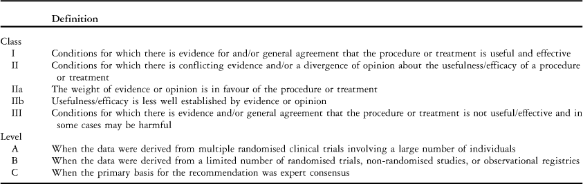
Canadian recommendations (Table 1)
Tetralogy of Fallot
Class I
• In patients with sustained ventricular tachyarrhythmia and/or those resuscitated from sudden cardiac death with no clear identified reversible cause, implantable cardioverter defibrillators are indicated for secondary prevention. (Level B)
• Patients who require surgery for Tetralogy of Fallot should be operated on by congenital heart surgeons. (Level C)
Table 1Definitions for grading of evidence.Reference Silversides, Marelli and Beauchesen13

Class IIa
• Following palliative surgery, complete intracardiac repair should be considered in all patients, in the absence of severe irreversible pulmonary hypertension or extremely unfavourable anatomy, such as severely hypoplastic inadequate pulmonary arteries. In palliated patients, the following situations particularly warrant complete repair:
○ Worsening symptoms. (Level C)
○ Cyanosis with erythrocytosis. (Level C)
○ Reduction or absence of the continuous shunt murmur – suspected shunt stenosis or occlusion. (Level C)
○ Aneurysm formation in the shunt. (Level C)
○ Left ventricular dilation due to aortic regurgitation or a residual shunt. (Level C)
Class IIa: re-interventions for Tetralogy of Fallot
The following situations may warrant intervention after repair:
• Free pulmonary regurgitation associated with progressive or moderate to severe right ventricular enlargement (right ventricular end-diastolic volume >170 ml/m2), moderate to severe right ventricular dysfunction, important tricuspid regurgitation, atrial or ventricular arrhythmias, or symptoms such as deteriorating exercise performance. (Level C)
• Residual ventricular septal defect with a shunt greater than 1.5:1. (Level C)
• Residual pulmonary stenosis with right ventricular pressure at least two-thirds the systemic pressure – either the native right ventricular outflow or valved conduit if one is present. (Level C)
• Significant aortic regurgitation associated with symptoms and/or progressive left ventricular systolic dysfunction. (Level C)
• Aortic root enlargement of at lease 55 mm in diameter. (Level C)
• Sustained clinical arrhythmias, most commonly either atrial flutter or fibrillation or sustained monomorphic ventricular tachycardia. When any of these arrhythmias occur, the patient should also be evaluated for a treatable haemodynamic cause of the arrhythmia. (Level C)
• The combination of residual ventricular septal defect and/or residual pulmonary stenosis and regurgitation, all mild–moderate but leading to substantial right ventricular enlargement, reduced right ventricular function, or symptoms. (Level C)
• Patients deemed to be at particularly high risk for sudden cardiac death may benefit from implantable cardioverter defibrillators for primary prevention. (Level B)
• Patients who require re-operation for Tetralogy of Fallot should be operated on by congenital heart surgeons. (Level B)
ACC/AHA recommendations (Table 1)
The following are the recommendations for the evaluation and follow-up of the patient after repair:
1. Post-operative patients with Tetralogy of Fallot should be seen annually by a cardiologist who has expertise in adults with congenital heart disease. (Level C)
2. Echocardiograms and magnetic resonance imaging evaluations should be performed by staff with expertise in adults with congenital heart disease. (Level C)
3. Screening for heritable causes of their condition (e.g. 22q11 deletion) should be offered to all patients. (Level C)
4. Before pregnancy, or if a genetic syndrome is identified, consultation with a geneticist should be made for patients with Tetralogy of Fallot. (Level B)
5. Patients with unrepaired or palliated forms of Tetralogy of Fallot should be formally evaluated at a centre for adults with congenital cardiac disease regarding possibility of repair. (Level B)
Key post-operative issues
• Residual pulmonary regurgitation.
• Right ventricular dilation and or dysfunction due to pulmonary regurgitation, possibly with associated tricuspid regurgitation.
• Residual right ventricular outflow tract obstruction.
• Branch pulmonary stenosis or hypoplasia.
• Sustained ventricular tachycardia.
• Sudden cardiac death.
• Atrioventricular block, atrial flutter, and/or atrial fibrillation.
• Progressive aortic regurgitation with aortic root dilation (55 mm).Reference Naguib, Dob and Gatzoulis14
• Syndromic associations.
Recommendations for imaging ACC/AHA
The following are some of the imaging techniques to be performed in adults with congenital heart disease to evaluate anatomy and haemodynamics in patients with repaired Tetralogy of Fallot (Level B):
1. Echocardiography is performed for the following reasons:
a. Right ventricular size and function (right ventricular myocardial performance index, right ventricular area fractional shortening, DP/DT from tricuspid valve regurgitation jet) may be assessed, but Echo (2D/3D) is not reliable for right ventricular volumes.
b. The presence and degree of pulmonary incompetence can be assessed, along with the severity of pulmonary stenosis.
c. The tricuspid valve can be assessed and the tricuspid valve regurgitant jet may be used to assess right ventricular peak systolic pressure.
d. Right atrial size can be measured and followed, residual ventricular septal defect can be assessed, and the size of the aortic root with and without aortic regurgitation can be followed.
2. Magnetic resonance imaging (Fig 3) is currently the best way to assess right ventricular volumes and right ventricular function.16
a. Magnetic resonance imaging is also useful to quantify pulmonary regurgitation and to assess the pulmonary arteries.
Figure 3Images obtained from a 44-year-old patient undergoing echocardiography and cardiac magnetic resonance imaging on the same day. (a) Doppler trace showing significant pulmonary regurgitation and antegrade flow in the pulmonary artery in late diastole (arrowed), indicative of restrictive physiology. (b) Right ventricular outflow tract view showing extensive late enhancement of an akinetic region of the right ventricular outflow tract (short arrows) and enhancement in the right ventricular outflow tract free wall (short arrows) and along the moderator band (long arrow). (c) Left ventricular outflow tract view showing late enhancement in the free wall of the right ventricular outflow tract (short arrows) and along the moderator band (long arrow). (d) Short-axis view showing extensive endomyocardial late gadolinium enhancement (short arrows).15
b. Magnetic resonance imaging is also useful to assess the aorta and left ventricle.
c. Magnetic resonance imaging is utilised by the interventional cardiologist to size the right ventricular outflow tract and main pulmonary artery for percutaneous valve insertion
3. Computerised tomography scanning is useful to assess the coronary arteries (origins and course) (Fig 4).
a. In patients with pacemakers and/or defibrillators, much of the same information obtained from magnetic resonance imaging can also be obtained by computed tomography angiography in adults with Tetralogy of Fallot.17
Figure 4Anomalous coronary artery origin in a patient with Tetralogy of Fallot. Oblique axial image from a whole-heart 3D SSFP acquisition shows anomalous origin of the LAD coronary artery from the RCA. Note that the left anterior descending coronary artery crosses the RVOT. A = aorta; br = branch; LAD = left anterior descending; LCX = left circumflex coronary artery; RCA = right coronary artery; RVOT = right ventricular outflow tract; SSFP = steady-state free precession.
The following are some of the recommendations for diagnostic and interventional catheterisation for adults with Tetralogy of Fallot.
Class I
1. Catheterisation of adults with Tetralogy of Fallot should be performed in regional centres with expertise in adults with congenital heart disease. (Level C)
2. Coronary artery delineation should be performed before any intervention for the right ventricular outflow tract. (Level C)
3. Interventional catheterisation in an adults with congenital cardiac disease is indicated for patients with previously repaired Tetralogy of Fallot with the following indications:
a. To eliminate residual native or palliative systemic-to-pulmonary artery shunts.
b. To manage coronary artery disease. (Level B)
Class IIa
1. Interventional catheterisation in patients with repaired Tetralogy of Fallot to eliminate a residual atrial septal defect or ventricular septal defect with a left to right shunt greater than 1.5:1, if it is in an appropriate anatomic location (Level C).
Class IIb
1. In adults with repaired Tetralogy of Fallot, cathertisation may be considered to better define potentially treatable causes of otherwise unexplained left ventricular or right ventricular dysfunction, fluid retention, chest pain or cyanosis. In these circumstances, transcatheter interventions may include:
a. Elimination of residual shunts or aortopulmonary collateral vessels. (Level C)
b. Dilation – with or without stent implantation – of right ventricular outflow tract obstruction. (Level B)
c. Elimination of additional muscular or patch-margin ventricular septal defect. (Level C)
d. Elimination of residual atrial septal defect. (Level B)
Interventional catheterisation in patients with previously repaired Tetralogy of Fallot should be carefully planned with the medical and surgical team in a centre for adults with congenital cardiac disease. The experience with percutaneous stent-valve implants in the right ventricular outflow for patients with pulmonary regurgitation and significant right ventricular dilation is promising and growing. We will discuss it below.
For the unusual case of a patient with Tetralogy of Fallot who has undergone palliation with a surgical shunt, catheterisation should be performed to assess potential for repair. Heart catheterisation is not routinely used in assessment of patients who have undergone repair, except to assess the pulmonary arteries or the coronary arteries.
Angioplasty of the branch pulmonary arteries is to be considered under the following conditions:
1. If right ventricular pressure is >50% of the systemic level, or at a lower level if right ventricular dysfunction is present.
2. If there is unbalanced pulmonary blood flow greater than 75%/25%, or otherwise unexplained dyspnoea with severe vascular stenosis.
Recommendations for surgery in adults with previous repair for Tetralogy of Fallot
Class I
1. Surgeons with training and expertise in congenital heart disease should perform operations in adults with previous repair of Tetralogy of Fallot. (Level C)
2. Pulmonary valve replacement is indicated for severe pulmonary regurgitation and symptoms of decreased exercise tolerance. (Level B)
3. Coronary artery anatomy, specifically the possibility of an anomalous anterior descending coronary artery across the right ventricular outflow tract, should be ascertained before operative intervention. (Level C)
Class IIa
1. Pulmonary valve replacement is reasonable in adults with previous repair of Tetralogy of Fallot, severe pulmonary regurgitation and, any of the following:
a. Moderate to severe right ventricular dysfunction. (Level B)
b. Moderate to severe right ventricular enlargement. (Level B)
c. Development of symptomatic or sustained atrial and/or ventricular arrhythmias. (Level C)
d. Moderate to severe tricuspid regurgitation. (Level C)
2. Collaborative approaches involving surgeons and interventional cardiologists caring for adults with congenital cardiac disease are reasonable in order to determine the best approach to treat stenosis of the pulmonary arterial tree. Such collaborative approaches may include preoperative stenting, intraoperative stenting, or surgical patch angioplasty. (Level C)
3. Surgery is reasonable in adults with prior repair of Tetralogy of Fallot and residual right ventricular outflow tract obstruction (valvular or subvalvular) and any of the following indications:
a. Residual right ventricular outflow tract obstruction (valvular or subvalvular) with peak instantaneous echocardiographic gradient >50 mmHg. (Level C)
b. Residual right ventricular outflow tract obstruction (valvular or subvalvular) with right ventricular/left ventricular pressure ratio >0.7. (Level C)
c. Residual right ventricular outflow tract obstruction (valvular or subvalvular) with progressive and/or severe dilation of the right ventricle with dysfunction. (Level C)
d. Residual ventricular septal defect with a left-to-right shunt greater than 1.5:1. (Level B)
e. Severe aortic regurgitation with associated symptoms or more than mild left ventricular dysfunction. (Level C)
f. A combination of multiple residual lesions – for example, ventricular septal defect and right ventricular outflow tract obstruction – leading to right ventricular enlargement or reduced right ventricular function.
Key issues to evaluate and follow up
Recommendations for arrhythmia, pacemaker/electrophysiology testing
Class I
1. Annual surveillance with history, echocardiography, assessment of right ventricular function, and periodic exercise testing, is recommended for patients with pacemakers/automatic implantable cardioverter defibrillators. (Level C)
Class IIa
1. Periodic Holter monitoring can be beneficial as part of routine follow-up. The frequency should be individualised depending on the haemodynamics and clinical suspicion of arrhythmia. (Level C)
Class IIb
1. Electrophysiological testing in a centre for adults with congenital cardiac disease may be reasonable to define suspected arrhythmias in adults with Tetralogy of Fallot. (Level C)
Note that the risk of sudden death in post-operative patients with Tetralogy of Fallot patients is real at 2.5% per decade of follow-up.Reference Boxt, Lipton, Kwong, Rybicki and Clouse18 The aetiology is most likely ventricular tachycardia, followed by interatrial re-entrant tachycardia (atrial flutter) and then atrioventricular block.
Follow-up of adults with Tetralogy of Fallot
The survival rate of patients with Tetralogy of Fallot exceeds 90% through the first two decades of life, decreasing thereafter as a consequence of residual sequela.Reference Apitz, Webb and Reddington19 These include progressive pulmonary insufficiency, resulting in progressive right ventricular dilatation, right ventricular dysfunctionReference Therrien, Marx and Gatzoulis20 and an increased risk for ventricular or atrial arrhythmias.Reference Geva, Sandweiss, Gauvreau, Lock and Powell21 In addition, left ventricular systolic dysfunction may occur in ∼20% of Tetralogy of Fallot patients,Reference Broberg, Aboulhosn and Mongeon22 adversely affecting the risk of sudden cardiac death.Reference Diller, Kempny and Liodakis23 Dilatation of the aortic root (>40 mm) is also fairly common; it occurs in 15–30% of patients with Tetralogy of Fallot,Reference Niwa, Siu, Webb and Gatzoulis24 with a smaller percentage of patients developing significant aortic insufficiency or requiring root repair.Reference Mongeon, Gurvitz and Broberg25 Hence, follow-up of the adult patient with Tetralogy of Fallot requires identification of these and other late sequela and a plan for longitudinal surveillance. This is best accomplished by a multi-disciplinary team of trained paediatric and adult cardiologists with expertise managing late complications seen in patients with tetralogy of Fallot, including cardiac failure, atrial and ventricular arrhythmias, and other acquired medical and emotional disorders of adulthood. This team needs to communicate effectively with the adult patient who ultimately is responsible for making informed decisions regarding their health. The principal care team of paediatric and adult cardiologists must have ready access to expert interventional, electrophysiological, imaging and surgical consultants.
Our Adult Congenital Heart Clinic is community based, rendering access at point of care and based at multiple institutions. Nevertheless, it is entrusted to a dedicated, integrated, multi-disciplinary team, as described above as the single most important key element. On our initial evaluation of the patient with Tetralogy of Fallot, we identify the patient's initial anatomic presentation and detailed history of intervention in identifying risk factors for arrhythmias and haemodynamic disturbances. Echocardiography is the principal imaging tool to demonstrate right ventricular enlargement, pulmonic insufficiency, and biventricular function, and most importantly, relative change over time. Echocardiography, however, cannot accurately and reproducibly measure right ventricular volumes,Reference Rudski, Lai, Afilalo, Hua, Handschumacher, Chandrasekaran, Solomon, Louie and Schiller26 quantify regurgitant volume, or assess the extracardiac vascular structures or coronary artery anatomy.
In our practice, we obtain quantitative assessment of right ventricular volumes and right ventricular function by cardiac magnetic resonance imaging in all patients with demonstrated right ventricular enlargement by echocardiography or moderate pulmonic insufficiency by Doppler. Magnetic resonance imaging is the standard of reference for measuring right ventricular size and functionReference Shors, Fung, Finn and Fieno27 and is highly reproducible.Reference Grothues, Moon and Bellenger28 Furthermore, it allows visualisation of aneurysms of the right ventricular outflow tract, obstructions of conduits, residual shunts, and pulmonary arterial anatomy. It is useful in measuring the distribution of flow to the left and right pulmonary arteries, and in assessing the physiological impact of stenosis of the branch pulmonary arteries, as well as in the quantification of pulmonary valvar insufficiency by phase contrast imaging. Right ventricular end-diastolic and end-systolic volume index of <150 and 80 ml/m2, respectively, have been recommended as predictive parameters for optimal outcomes for favourable right ventricular remodelling after repair or replacement of the pulmonary valve in patients with severe pulmonary valvar regurgitation.Reference Lee, Kim and Lee29, Reference Holmes30
In patients with Tetralogy of Fallot with implanted cardiac rhythm devices or multiple vascular stents or metallic implants, cardiac magnetic resonance imaging is contraindicated or inaccurate secondary to signal voids, and cardiac computed tomographic angiography has proven effective in quantification of the right ventricular volumes and right ventricular function.Reference Raman, Cook, McCarthy and Ferketich31 Furthermore, it is accurate in the evaluation of the anatomy of the main pulmonary artery and branch pulmonary arteries (Fig 5), thoracic aorta, coronary arteries (Fig 5), and in the assessment of adherence of the right ventricule to sternum in the post-operative patient. Our protocol images the patient from the aortic arch to the diaphragm and maintains continuous injection of intravenous contrast at a rate of 2 cc/s after the initial bolus to enhance the right ventricular cavity. To minimise radiation dose, we use prospective gating or dose modulation.Reference Mark, Berman, Budoff, Carr, Gerber, Hecht, Hlatky, Hodgson, Lauer, Miller, Morin, Mukherjee, Poon, Rubin and Schwartz32
(a) Cardiac computed tomography angiography of an adult patient with repaired Tetralogy of Fallot with stenosis of the right pulmonary artery following stent placement. (b) Patient with Tetralogy of Fallot and absent pulmonary valve with anomalous origin of the left main coronary artery from the dilated pulmonary artery.
It is important to establish an objective and reproducible measurement of functional class and functional capacity that may be compared over time. A metabolic exercise stress test fulfils this clinical parameter. Patients with Tetralogy of Fallot with reduced right ventricular function have reduced functional capacity with peak VO2 <35% of that predicted.Reference Giardini, Specchia, Coutsoumbas, Donti, Formigari, Fattori, Oppido, Gargiulo and Picchio33 In our experience, adult patients adapt their lifestyle when their functional capacity is impaired, masking the appearance of limiting symptoms.
The burden of arrhythmias in adults with repaired Tetralogy of Fallot is high and increased with age, number of surgical interventions, chamber dilatation, and ventrciular diastolic and systolic dysfunction.Reference Gatzoulis, Balaji and Webber34 The Alliance for Adult Research in Congenital Cardiology Multi-Institutional Study reported a prevalance of 29.9% sustained tachyarrhythmias in a large cohort of adults with repaired Tetralogy of Fallot. The distribution of these arrhythmias was as follows: 11.5% intra-atrial re-entrant tachycardia, 7.4% atrial fibrillation, 14.2% ventricular tachycardia and 0.5% ventricular fibrillation, with atrial tachyarrhythmias being more prevalent in patients over 40 years of age.Reference Khairy, Aboulhosn, Gurvitz, Opotowsky, Mongeon, Kay, Valente, Earing, Lui, Gersony, Cook, Ting, Nickolaus, Webb, Landzberg and Broberg35 This distribution of arrhythmias has also been our clinical experience, with overall atrial arrhythmias being more prevalent and frequently asymptomatic. Identification of silent arrhythmias remains a clinical challenge. It is our practice to randomly screen asymptomatic patients with 24–48-hour Holter studies at least once a year. In mildly symptomatic patients, we prefer prolonged (2–4 weeks) outpatient monitoring with a digital loop recorder. Patients with multiple predictors for sudden cardiac deathReference Gatzoulis, Balaji and Webber34, QRS duration >180 ms, previous right ventriculotomy, demonstrable spontaneous non-sustained ventricular tachycardia, and left ventricular systolic dysfunction, are referred for electrophysiology evaluation. Owing to the fact that no single variable predicts or excludes sudden cardiac death in this group of patients, many will receive a prophylactic implantable cardioverter defibrillator for primary prevention of sudden cardiac death.Reference Bricker36
Interventional therapy
As of June, 2013, we have brought 42 patients to the cardiac catheterisation laboratory for invasive assessment and possible transcatheter pulmonary valve replacement. Pre-procedural assessment included echocardiography in all patients, and magnetic resonance imaging or volumetric computed tomographic angiography in most patients (all patients with primary indication of pulmonary insufficiency). Owing to coronary compression or inadequate landing zone, 11 were not considered suitable for transcatheter replacement of the pulmonary valve. Therefore, 32 were judged to have an adequate landing zone and no issues related to coronary compression. At this point, we have always proceeded with pre-stenting of the landing zone. This pre-stenting of the landing zone was successful in all patients but one in whom the stent migrated to the right ventricular outflow tract and had to be surgically explanted with surgical replacement of the pulmonary valve. In our initial experience, invasive candidate assessment, compliance testing, coronary assessment, and pre-stenting were performed in a separate procedure, but currently we usually do everything in a single procedure. The number of stents utilised for conduit preparation ranged from – one to three, with a mean of 1.4. Generally, more stents were implanted in conduits with marked stenosis, particularly in stenotic conduits located directly behind the sternum. All (31/31) successfully pre-stented patients also underwent successful transcatheter pulmonary valve replacement. Their diagnoses have included Tetralogy of Fallot or variants of Tetralogy of Fallot (n = 18), post-operative Ross patients (n = 7), complex D-transposition (n = 2), and others (n = 4). Mean patient age was 23 years (with a range from 9 to 54 years), 20 out of 31 patients were male, and 22 out of 31 patients were above 18 years. Among the patients who underwent transcatheter replacement of the pulmonary valve, there have been two major adverse events, one non-contained tear of a conduit successfully treated with the transcatheter insertion of a pulmonary valve (using the valve additionally as a covered stent) and one patient with recurrent supraventricular tachycardia following implant, which prolonged his hospitalisation.
The adoption of this new technique by our programme has led to major changes in the surgical approach as well. Historically, many of our patients requiring surgical pulmonary valve replacement received an expanded polytetrafluoroethylene hand-sewn bivalve surgically implanted in the right ventricular outflow tract. This technique has evolved; currently, an expanded polytetrafluoroethylene bivalve is sewn inside a circumferential expanded polytetrafluoroethylene conduit. Owing to the straightforward nature of transcatheter insertion of a pulmonary valve in patients following surgical insertion of a pericardial tissue valve, pericardial tissue valves have become a more common surgical choice for our adult patients. Lastly, for smaller patients, our surgeons have attempted to put at least a 16mm conduit into every patient coming to surgery for surgical replacement of the pulmonary valve whenever possible, even for many of our infants.
Similar to most centres, we eagerly awaited and anticipated the approval of the Melody valve by the Food and Drug Administration of the United Stated of America. It allows a paradigm shift in the management of all patients with congenital cardiac disease who suffer dysfunction of the right ventricular outflow tract and in some with dysfunction of the left ventricular outflow tract. Given our large programme for adults with congenital cardiac disease, the need to offer a less-invasive alternative for patients who had already undergone repeated surgery was acute. The need to diminish lifetime morbidity and at least spread out the time between sternotomies is rather apparent, especially as the older patient develops late comorbidities.
The Melody valve was approved by the Food and Drug Administration of the United Stated of America under the Humanitarian Device Exemption in January, 2010. Its approval was for use in patients with a dysfunctional, circumferential, right ventricle to pulmonary artery conduit at least 16 mm in diameter at the time of implant. Dysfunction was described as either at least moderate pulmonary regurgitation and/or stenosis with a mean Doppler gradient of over 35 mmHg. Generally speaking, patients have needed to fulfil the same criteria as a patient in whom surgery is contemplated. To become an implanting centre in the United States of America required mandated training of the physician and team, proctoring of the physician for the first three to five cases, and approval of the local Institutional Review Board. In addition, a major investment was necessary to purchase the equipment necessary for the first proctored implants plus spare valves. We accomplished these hurdles and implanted our first valves in April, 2011. Our initial 14 implants were all in patients with circumferential dysfunctional right ventricle to pulmonary artery conduits. On the basis of our initial favourable experience with the valve, and based on the experience of other implanting centres, we sought and received additional approval from the Institutional Review Board for off-label use of the Melody valve in selected patients. This “off-label use” included patients with a dysfunctional right ventricular outflow tract but without a conduit but with an adequate landing zone, patients with a smaller conduit that still appeared to be a candidate, patients with dysfunctional prosthetic tricuspid valves, and patients with other dysfunctional surgical conduits.
Surgical therapy
Surgical implantation of the pulmonary valve in adults with congenital cardiac disease
The need for replacement of the pulmonary valve is increasing for many congenital cardiac patients. In the past, residual defects such as chronic pulmonary insufficiency in post-operative Tetralogy of Fallot patients were felt to be benign. More recent evidence suggests that pulmonary insufficiency and volume overload physiology cause significant morbidity, producing right ventricular dilatation and dysfunction, exercise intolerance, arrhythmias, and possible sudden death.Reference Bouzas, Kilner and Gatzoulis37–Reference Ammash, Dearani and Burkhart39 We will review the beneficial haemodynamic effects, surgical techniques and prosthetic options, as well as indications for pulmonary valve replacement.
Numerous reports support the role of replacement of the pulmonary valve in patients with chronic pulmonary insufficiency.Reference Davroulos, Karatza, Gatzoulis and Shore40–Reference Bove, Kavey and Byrum41 Pulmonary valve replacement allows for symptomatic improvement, improved right ventricular function, and control of arrhythmias when performed within a reasonable time frame. Unfortunately, recent data also show lack of recovery of right heart indices following pulmonary valve replacement in adults with long-standing pulmonary insufficiency and right ventricular dysfunction.Reference Therrien, Siu, McLaughlin, Liu, Williams and Webb42 Therefore, timing of surgical therapy is important in obtaining an optimal surgical result for the patient. In addition, an aggressive programme of pre-operative electrophysiological evaluation combined with intra-operative cryoablation has been shown to optimise control of arrhythmias in the appropriate patient.Reference Therrien, Siu and Harris43
Indications for pulmonary valve replacement continue to evolve. These are based on the accumulation of natural history data, as well as the short- and long-term effects of surgical therapy. Current indications for pulmonary valve replacement include patients with pulmonary stenosis, moderate to severe pulmonary insufficiency, or mixed pulmonary stenosis and pulmonary insufficiency, and:
• New York Heart Association (NYHA) class II exertional symptoms.
• Right ventricular dysfunction and/or dilatation (>150 ml/m2 by magnetic resonance imaging).
• Decreased performance on exercise testing.
• Ventricular arrhythmias and/or prolonged QRS duration (>160 ms).
Overall, the operative technique for pulmonary valve replacement utilises cardiopulmonary bypass with or without aortic cross-clamping, depending on surgeon preference and the need to repair additional lesions such as septal defects. The majority of these procedures require re-entry sternotomy and can present significant challenges to the surgeon. Appropriate pre-operative planning and judicious use of peripheral cannulation is necessary. The operative mortality is in the 1–2% range in many series.
Many surgical prosthetic options are available for replacement of the pulmonary valve. The ideal valve does not exist, and all choices have limitations. Most patients receive a bio-prosthesis such as a homograft or heterograft, either stented or unstented.Reference Hawkins, Sower and Lambert44, Reference Kanter, Budde and Parks45 All of these valves are non-living and non-repairable, and share a common durability issue. They undergo a bio-degenerative process and ultimately will fail with obstruction and/or insufficiency over time. There are complex interactions among many variables that determine the mode of failure and time to failure of these prostheses. In addition, homografts seem to generate an immune-mediated response, which may augment their degradation and also produces elevated levels of panel reactive antibody (PRA).Reference Fiore, Rodefeld and Turrentine46 Recent evidence would support the use of heterografts over homografts in terms of a durability.Reference Hawkins, Breinholt and Lambert47 Alternatively, a mechanical valve is an option, albeit with limited experience in the pulmonary position. The possibility of thromboembolic events and the need for systemic anticoagulation with Coumadin generally make this a less attractive option.Reference Waterbolk, Hoendermis, den Hamer and Ebels48 Another non-biologic choice is the expanded polytetrafluoroethylene bicuspid valve. We have experience with over 110 patients utilising a polytetrafluoroethylene bicuspid hand-sewn valve, most recently utilising 1 mm expanded polytetrafluoroethylene.Reference Quintessenza, Jacobs and Morell49 The initial implant, as well as intermediate-term data, demonstrates low transpulmonary gradients and very acceptable degrees of pulmonary insufficiency.
Subsequent workReference Quintessenza, Jacobs, Chai, Morell and Lindberg50 demonstrated improved durability and longevity of valve function using thinner (0.1 mm) expanded polytetrafluoroethylene as a valve leaflet. Further follow-up demonstrates ∼80% of these patients maintaining a competent valve, which is free of right ventricular outflow tract obstruction at 5 years. Most recently (unpublished data), valvar leaflets implanted within an expanded polytetrafluoroethylene tube, positioned in situ in the location of the main pulmonary artery, demonstrate excellent competency and freedom of obstruction over the intermediate term. This approach also provides a stable and uniform landing zone for future transcatheter valve implantation, as the need arises.
Longer-term follow-up will determine durability compared with other available options. Finally, transcatheter pulmonary valve replacement is now available in Europe and for compassionate use at selected centres in the United States.Reference Frigiola, Nordmeyer and Bonhoeffer51 In properly selected patients, this option holds significant promise for the future, although the initial explant rate was quite high and currently stent fractures are an issue. The current generation of transcstheter pulmonary valve is most likely the first of many renditions of this type of technology as patients will be most interested in avoiding open surgical approaches as delivery systems, technical issues, and durability of the valves improve.
In summary, our understanding of the effects of chronic pulmonary insufficiency on right heart function continues to evolve. Chronic pulmonary insufficiency is not a benign lesion. Replacement of the pulmonary valve in the adult with congenital cardiac disease is now being performed with more liberal indications than in the past, in the light of these new data. The beneficial effects of valve replacement with low operative mortality and morbidity support this approach. Further investigation is needed to determine optimal timing for surgery. Improved valve durability, bioengineered valvesReference Toshiharu, Dominique and Peter52, and potentially less-invasive methods of inserting a normally functioning valve are forthcoming.