Background
Nasopharyngeal carcinoma (NPC) is relatively uncommon in the western world. However, it is the 5th most common cancer in Malaysia with lifetime risk of 1 in 143 in males and 1 in 417 in females.Reference Manan, Ibrahim Tamin, Abdullah and Wahab 1 Among the reported cases, 63·6% of patients present at stage III or IV at initial diagnosis.Reference Manan, Ibrahim Tamin, Abdullah and Wahab 1 External auditory meatus (EAM) involvement in NPC is rare. We report two contrasting cases of NPC with EAM involvement with a summary of clinical and radiological findings; and the management.
Case 1
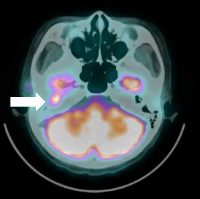
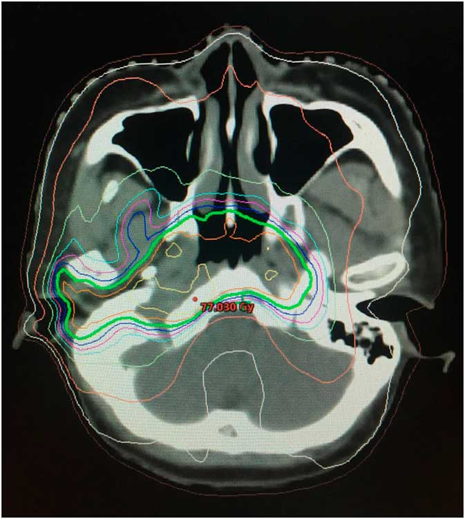
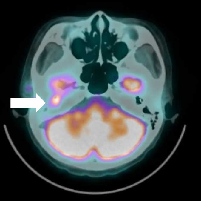
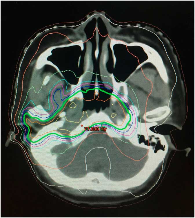
A 45-year-old Chinese woman presented with 1 year history of increasing right neck swelling and right-sided tinnitus. Clinically she had a palpable right level II lymph node and a right supraclavicular lymph node. There were no signs cranial nerve palsies. Flexible nasopharyngolaryngoscope showed fullness of the right fossa of Rosenmuller (FOR) with extension to right torus tubaris. Biopsy of the right FOR mass confirmed the diagnosis of undifferentiated nasopharyngeal carcinoma. Otoscopic examination showed retracted right tympanic membrane with hypervascularity. Computed tomography (CT) scan showed a mass in the right nasopharynx obliterating the right FOR and extending into right parapharyngeal space and posterior nasal space, and multiple bilateral cervical lymphadenopathy, largest at right level II and III measuring 3·8×3·4 cm. A staging positron-emission tomography (PET) scan, however, showed extension of the nasopharyngeal mass into the right external auditory meatus. She was staged as cT2N3bM1. She underwent six cycles of neoadjuvant cisplatin/5-fluorouracil, followed by radical concurrent chemoradiation that included the right external auditory meatus in the target volume and treated to 70 Gy in 33 fractions with intensity-modulated radiotherapy. Post-treatment clinical and radiological assessments showed complete response (Figures 1 and 2).
Positron-emission tomography scan showing fludeoxyglucose-avid disease in the right external auditory meatus (white arrow).
Isodose lines of radiotherapy plan showing 70 Gy coverage to the right external auditory meatus.
Case 2
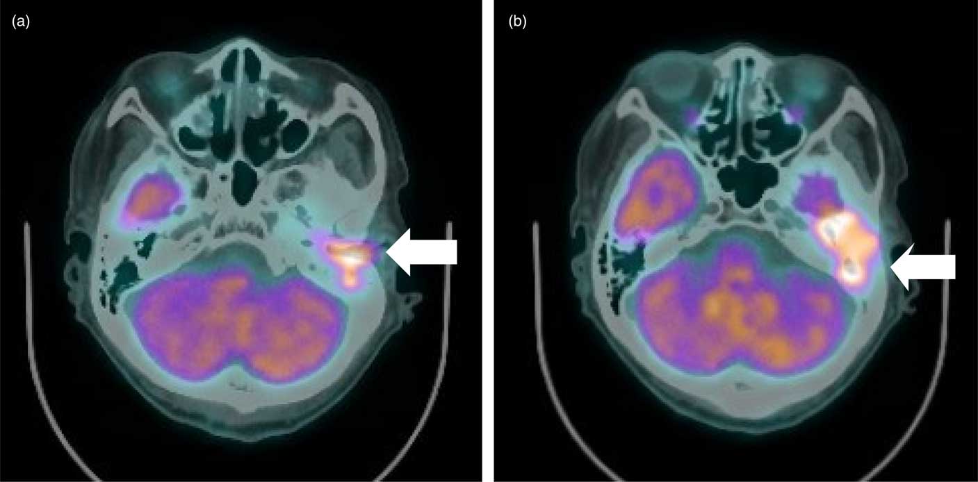
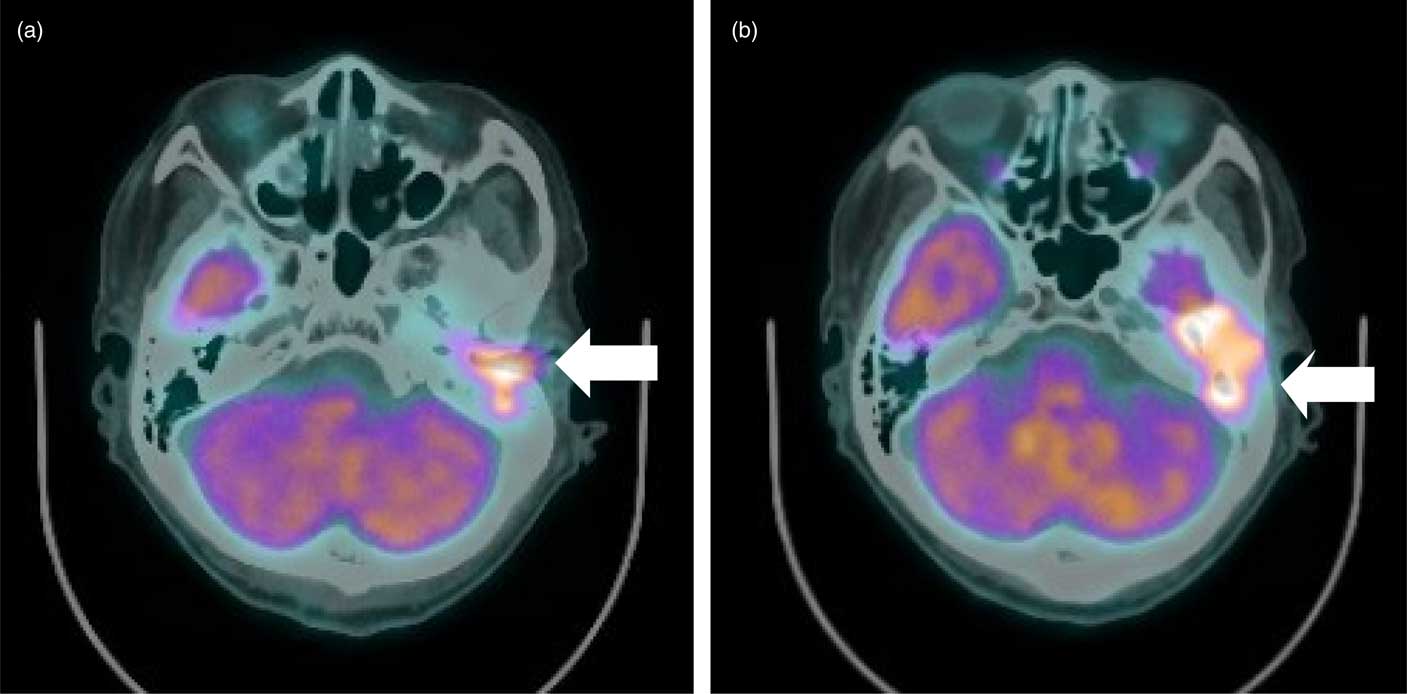
A 64-year-old Chinese man was initially diagnosed with NPC stage cT1N3aM0 1 year ago. He was treated with two cycles of neoadjuvant cisplatin and 5-fluorouracil followed by radical concurrent chemoradiation (up to 70 Gy). He was disease free for 1 year, then he was referred to our centre with complaint of facial asymmetry, left ear hearing loss and left ear discharge. On examination he had a left external auditory meatus mass and a left facial nerve palsy. Biopsy of the mass showed invasive carcinoma, suggestive of recurrence of NPC. PET scan showed 18F-fluorodeoxyglucose-avid disease in the left external auditory meatus, left petromastoid bone with intracranial extension into temporal lobe. He was commenced on cisplatin and 5-fluorouracil as palliative/potentially neoadjuvant chemotherapy before possible re-irradiation. Unfortunately he deteriorated after one cycle of treatment and passed away 6 months from the diagnosis of recurrent disease (Figures 3a and 3b).
(a, b) Positron-emission tomography scan showing fludeoxyglucose-avid disease in the left external auditory meatus (white arrow).
Discussion
Nasopharyngeal cancers typically spread via direct extension, perineural invasion, lymphatic and haematogenous modes. NPC commonly presents with cervical lymphadenopathy, while symptoms resulting from the primary tumour can be subtle. Direct invasion of NPC to external auditory meatus is rare.
Li et al.Reference Li, Sun and Chen 2 evaluated the locoregional extension patterns in 2366 patients with NPC. Anatomical sites with more than 30% incidence were classified as high risk and these include tensor veli palatine muscle, nasal cavity, base of sphenoid bone, pterygoid process, clivus, petrous apex, prevertebral muscle and foramen lacerum in descending order. No cases with inner ear, eustachian tube or external auditory meatus were reported.Reference Li, Sun and Chen 2
To date there are only 18 reported cases with NPC invading the EAM.Reference Cundy, Sando and Hemenway 3 – Reference Lai, Cheng, Tong and Yau 10 Among them, a Chinese case series reported nine cases of NPC with EAM involvement, three cases being at initial diagnosis; and six others at disease recurrence.Reference Wang, Yuan and Cheng 9 The similarities between these cases are presence of otologic symptoms such as hearing deficit, ear discharge and otalgia.Reference Wang, Yuan and Cheng 9
The mechanism of spread to EAM is unclear. Lai et al.Reference Lai, Cheng, Tong and Yau 10 suggested a possibility of a true mucosal spread of submucosal spread via the eustachian tube, rather than a locally advanced NPC spread via the skull bone.
Both our cases had at least one otologic symptom, that is, tinnitus in case one; hearing loss and ear discharge in case two. Incidentally both cases were also heavily node positive. Otoscopic examination and PET imaging were key to complete the diagnosis and staging in our experience. In the situation where radiotherapy is indicated (in case one), co-registration of PET images was particularly helpful in delineation of tumour volumes for intensity-modulated radiation therapy planning.
Conclusions
Invasion to external auditory meatus is a very unusual route of spread of NPC, both at initial presentation and at recurrence. But our two cases demonstrated this can occur, and should be kept in mind when patients present with otologic symptoms, as well as for heavily node positive patients. PET imaging is essential in detecting the extent of local invasion in this anatomical site, as well for radiotherapy planning. Covering this area in radiation field is potentially curative, and should not be missed.
Acknowledgements
The authors wish to thank both patient/next-of-kin for their kind permission to present these two cases.
Authors’ Contributions
D.W.L. wrote the work and G.F.H. contributed to the revision of the work and gave the final approval of the version to be published. All authors read and approved the final manuscript.
Financial Support
Neither author received any source of funding for this paper.
Conflicts of Interest
The authors declare they have no competing interests.
Ethical Standards
The authors declare they have observed appropriate ethical guidelines and legislation in writing the case report. Consent to participate was obtained from the patient/next-of-kin. Written informed consent was obtained from the patient/next-of-kin for publication of this Case Report. A copy of the written consent is available for review by the Editor-in-Chief of this journal.