Premature ventricular contractions in children without structural heart disease have generally been regarded as benign, regardless of premature ventricular contraction burden;Reference Conti 1 – Reference Rosenbaum 3 however, recent studies in adults with high premature ventricular contraction burdens have described a premature ventricular contraction-induced cardiomyopathy, characterised by left ventricular dilation and/or dysfunction,Reference Bogun, Crawford and Reich 4 – Reference Hasdemir, Kartal, Simsek, Yavuzgil, Aydin and Can 5 diastolic dysfunction,Reference Topaloglu, Aras and Cagli 6 and ventricular strain by speckle-tracking imaging.Reference Wijnmaalen, Delgado and Schalij 7 This cardiomyopathy appears reversible with reduction in ectopy burden.Reference Duffee, Shen and Smith 8 – Reference Zhong, Lee and Huang 9 Multiple adult studies have demonstrated rapid normalisation of left ventricular parameters following successful radiofrequency ablation of the premature ventricular contraction focus.Reference Bogun, Crawford and Reich 4 , Reference Hasdemir, Kartal, Simsek, Yavuzgil, Aydin and Can 5 , Reference Wijnmaalen, Delgado and Schalij 7 , Reference Baman, Lange and Ilg 10 – Reference Yokokawa, Good and Crawford 14 Further, this cardiomyopathy was reproduced in a model where dogs were paced in ventricular bigeminy; in all cases, ventricular changes resolved with termination of pacing, and no irreversible changes were found on pathology.Reference Huizar, Kaszala and Potfay 15 Despite the convincing adult literature, evidence of premature ventricular contraction-induced cardiomyopathy in children is limited,Reference Beaufort-Krol, Dijkstra and Bink-Boelkens 16 – Reference Tsuji, Nagashima and Hasegawa 20 with one abstractReference Patel, Ganley and Shah 17 showing decreased left ventricular fractional shortening in 7% of children with >20% ventricular ectopy burden at a single centre. The purpose of our study was to add to our understanding of the association between high premature ventricular contraction burden and cardiomyopathy in children. We also examined whether these high ectopy burdens persist through childhood or whether ectopy resolves at the latest follow-up. Finally, we sought to determine whether these children progress to having ventricular tachycardia as a significant fraction of total daily rhythm. Specifically, we hypothesised that (1) a sub-set of children with high premature ventricular contraction burden will have or develop left ventricular dilation or dysfunction, (2) high premature ventricular contraction burden will persist in most children through follow-up, and (3) few children with high premature ventricular contraction burden progress to having ventricular tachycardia as a significant portion of total daily rhythm.
Materials and methods
This study was approved by the Institutional Review Board of Seattle Children’s Hospital.
We retrospectively reviewed the records of children at Seattle Children’s Hospital from January, 1990, to November, 2013. Children between the age of 6 months and 18 years at presentation, with premature ventricular contractions comprising at least 20% of total daily rhythm on 24-hour Holter monitor, were included. Exclusion criteria included haemodynamically significant structural heart disease, a family history of cardiomyopathy (with the exception of family members with tachycardia-induced cardiomyopathy or premature ventricular contraction-induced cardiomyopathy), a major systemic disease associated with cardiomyopathy (for example, Duchenne muscular dystrophy), or ventricular tachycardia as >1% of total daily rhythm on baseline Holter monitor.
We collected demographic data, baseline and follow-up echocardiographic data, baseline and follow-up Holter monitor results, and surface electrocardiogram results, including bundle branch block pattern and polarity of R:S ratio in lead aVF during the ectopic beats. The echocardiogram database was reviewed for evidence of significant structural heart disease, left ventricular fractional shortening, left ventricular ejection fraction, left ventricular end-diastolic dimension, and qualitative description of ventricular function. Where appropriate, clinic notes and electrophysiology procedure narratives were reviewed as well.
Premature ventricular contraction-induced cardiomyopathy was defined by echocardiographic findings of left ventricular fractional shortening ⩽28%, left ventricular ejection fraction ≤54%, left ventricular end-diastolic dimension z-score ⩾2.2, or globally diminished function on qualitative interpretation. Resolution of high premature ventricular contraction burden was defined as a decrease in ventricular ectopy to <10% of total daily rhythm on the most recent Holter monitor. Data were initially obtained by review of echocardiogram reports; however, in order to check quality of assessments, and to avoid over-diagnosis of ventricular dilation or dysfunction, investigators reviewed images of those patients meeting the study criteria for cardiomyopathy. In three of these patients, measurements meeting criteria were taken on a sinus beat following a sinus beat. In one patient noted to be in trigeminy throughout the study, measurements were taken in M-mode on a sinus beat preceding a premature ventricular contraction. In two patients in bigeminy, measurements were taken on a sinus beat following a premature ventricular contraction. In one patient in whom diagnosis was based on provider’s qualitative assessment, only the echocardiogram report could be retrieved.
In examining outcomes for these children, values are expressed with 95% confidence intervals. In looking at the natural history of high premature ventricular contraction burden, we calculated the Kaplan–Meier product limit estimate of the survival curve. Patients were censored at the time of their first echocardiogram showing cardiomyopathy, at the time of radiofrequency ablation if for reasons other than cardiomyopathy, at the time of Holter monitor showing resolution of high ectopy burden, at age 21 years, if lost to follow-up, or at the end of the study period.
Results
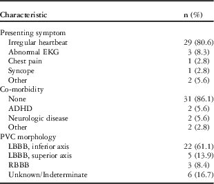
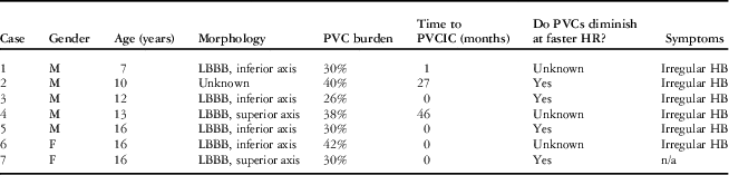
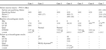
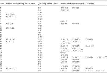
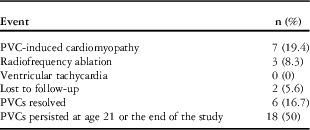
A total of 36 patients met the study criteria. Characteristics of the study population are detailed in Table 1. Mean age of the study population was 11 years (standard deviation 4 years); seven patients (19.4%, 95% CI 6.2–32.6%) met the criteria for premature ventricular contraction-induced cardiomyopathy, mostly at initial presentation. Features of these patients are detailed in Table 2. Table 3 shows Holter monitor results and echocardiographic parameters for this group of patients. The majority of these patients were male, and all of these children with identified premature ventricular contraction morphology by surface electrocardiogram had a left bundle branch block pattern to their ectopic beats. Where available, exercise testing showed suppression of ectopy at faster heart rates. No patient with echocardiographic criteria for cardiomyopathy was clinically symptomatic. Mean premature ventricular contraction burden in the cardiomyopathy group was 34.7% (standard deviation 6.3%). Mean premature ventricular contraction burden in the group without cardiomyopathy was 27.2% (standard deviation 5.1%). Holter monitor results for the group without cardiomyopathy are shown in Table 4.
Characteristics of study patients.

ADHD=attention deficit and hyperactivity disorder; EKG=electrocardiogram; LBBB=left bundle branch block; RBBB=right bundle branch block
Characteristics of patients with premature ventricular contraction-induced cardiomyopathy.

F=female; HB=heartbeat; LBBB=left bundle branch block; M=male; n/a=not applicable; PVC=premature ventricular contraction; PVCIC=premature ventricular contraction-induced cardiomyopathy
Holter monitor and echocardiogram results for the cardiomyopathy group.

BIG=bigeminy-sinus beat following a PVC; LV EF=left ventricular ejection fraction; LV FS=left ventricular fractional shortening; LVEDD=left ventricular end-diastolic dimension; Mos=months from qualifying Holter monitor; PVC=premature ventricular contraction; Qual=qualitative assessment of ventricular function by provider; RR=by report only; SS=sinus beat after a sinus beat; TRI=trigeminy-sinus beat preceding a PVC
Holter monitor results for patients without cardiomyopathy.

AB=underwent radiofrequency ablation of PVC focus following Holter monitor; Mos=time in months from qualifying Holter monitor; PVC=premature ventricular contraction
Among all study patients, three patients (8.3%) without premature ventricular contraction-induced cardiomyopathy underwent radiofrequency ablation. The age range for these patients was 8–12 years. Time from diagnosis of high premature ventricular contraction burden to ablation ranged from 2 to 30 months; one patient with 37% ectopy had ablation performed due to his palpitations being bothersome to him. The other two patients underwent ablation due to provider concern for degree of ectopy (26 and 37% ectopy).
In all, six patients (16.7%, 95% CI 4.3–29.1%) experienced resolution of their high premature ventricular contraction burden during the study. In those who experienced resolution of premature ventricular contractions, time from presentation until resolution of ectopy burden varied, ranging from 3 to 37 months from presentation. Burden decreased to <1% of total daily rhythm in five of six patients. Age ranged from 4 to 15 years. No predominant premature ventricular contraction morphology was identified in these patients.
After all censoring events were taken into consideration (Table 5), 50% of the patients remained with persistence of high ventricular ectopy burden and without cardiomyopathy, ablation, or loss to follow-up. Figure 1 shows the Kaplan–Meier product limit estimate of the survival curve for persistence of high premature ventricular contraction burden.
Kaplan–Meier product limit estimate of the survival curve. The x-axis gives time in months from baseline Holter monitor. The number of patients remaining uncensored at each time point is listed below. The y-axis gives the cumulative fraction of patients remaining with high premature ventricular contraction (PVC) burden.

Distribution of censoring events.

CI=confidence interval; PVC=premature ventricular contraction
No patient progressed to having ventricular tachycardia as greater than 1% of total daily rhythm on follow-up Holter monitor.
Discussion
This study demonstrates that a high premature ventricular contraction burden in children may not be benign, and there is a sub-set in whom associated left ventricular dilation and/or dysfunction is found. The significance of these echocardiographic changes is unclear, as none of the patients in our study were clinically symptomatic; however, it is possible that unrecognised and untreated, these patients could go on to develop the symptomatic heart failure that is seen in the adult population.
Significance of study results
In contrast to previous paediatric studiesReference Beaufort-Krol, Dijkstra and Bink-Boelkens 16 , Reference Sestito, Pardeo and Sgueglia 18 – Reference Tsuji, Nagashima and Hasegawa 20 – with the exception of the Patel abstractReference Patel, Ganley and Shah 17 – we have focussed our study on children with the highest premature ventricular contraction burdens, suspecting that the natural history of disease in these children may be different from those with lower burdens. We chose 20% of total daily rhythm as the cut-off for defining high premature ventricular contraction burden based on several previous studies.Reference Wijnmaalen, Delgado and Schalij 7 , Reference Baman, Lange and Ilg 10 , Reference Takemoto, Yoshimura and Ohba 12 Takemoto et alReference Takemoto, Yoshimura and Ohba 12 showed significant changes in left ventricular ejection fraction, end-diastolic dimension, end-systolic dimension, and mitral regurgitation above the 20% cut-off. Furthermore, Wijnmaalen et alReference Wijnmaalen, Delgado and Schalij 7 showed 30 times the ventricular strain at >20% ectopy when compared with controls. Baman,Reference Baman, Lange and Ilg 10 using a receiver operator curve, showed that focussing resources on patients with a premature ventricular contraction burden of about 24% gave the best balance of specificity and sensitivity for cardiomyopathy, although they found cases of cardiomyopathy in patients with lower ectopy burdens.
There are similarities between the trends noted in our study and findings in previous paediatric and adult studies. In terms of demographics, our population was fairly evenly split between males and females, but the majority of patients who had changes on echocardiogram were male. We found an overall high persistence of ectopy over time. This finding seems to be in line with the Beaufort–Krol study,Reference Beaufort-Krol, Dijkstra and Bink-Boelkens 16 which suggested that ventricular ectopy of left ventricular origin tended to resolve at follow-up, whereas ectopy of right ventricular origin tended to persist without significant change in burden at latest follow-up. As 75% of our patients had electrocardiograms suggesting right ventricular origin of premature ventricular contractions, it may be that we might have seen more resolution had we followed-up more patients with ectopy of left ventricular origin. It may also be that our population behaved differently, as in general our patients had much higher baseline ectopy burdens. In the small number of patients in whom high ectopy burden resolved, there was no trend noted in terms of origin of ectopic beats.
In looking at our cardiomyopathy group, similar to the Patel study,Reference Patel, Ganley and Shah 17 in all the patients in whom ectopic beat morphology was determined, a left bundle branch block pattern was present. The majority of patients already had ventricular changes at the time of baseline echocardiogram, but two patients with normal baseline echocardiograms later developed changes at the 27- and 46-month follow-up, respectively. Although too small a population to determine significance, the above findings, when taken in the context of the previous studies, suggest that perhaps premature ventricular contractions of right ventricular origin are more likely to lead to cardiomyopathy, and as this ectopy does not tend to resolve during childhood, it is especially important to follow-up these children over time. Of note, in our patients meeting criteria for cardiomyopathy in whom a determination was made, all had decrease or disappearance of ectopy at faster heart rates, and thus historically would likely have been considered to have benign ectopy. In addition, not all of our patients who developed a ⩾20% ventricular ectopy burden had such a high burden on initial Holter monitor, and thus a potentially significant finding would have been missed had they been dismissed from follow-up after their initial encounter. As such, we have made it a practice to at least follow-up children with ventricular ectopy burden between 10 and 20% of total daily rhythm with repeat Holter monitor.
There was no particular trend in the features of our patients who experienced resolution of high ectopy burden. Although our Kaplan–Meier curve (Fig 1) appears to represent our clinical findings well over the first 3 years of follow-up, it is less reliable beyond about 36 months due to a small remaining uncensored population. These findings might suggest that in patients without echocardiographic changes, ectopy burden has the potential to resolve without intervention, and thus initial conservative management favoured.
Limitations
There are several important limitations to our study. First, it is retrospective and examines the experience at only a single regional referral centre. The small study population may have a tendency to exaggerate the prevalence of cardiomyopathy among children with high premature ventricular contraction burden. This limitation also hampers our ability to look for significant differences among the sub-groups of our study population – for example, although there is a trend in the cardiomyopathy group towards primarily males and left bundle branch block morphology, a much larger population would need to be studied in order to determine whether there is a significant difference from the group without cardiomyopathy. A second limitation is the change in and variety of practice during the study period. Early on, if premature ventricular contractions diminished or disappeared at faster heart rates, and were not associated with structural heart disease, patients were often discharged from follow-up. It is only recently that more attention has been paid to this population of children. Thus, longitudinal data including serial echocardiographic assessment of function and change in ectopy burden over multiple Holter monitors have only recently become available. A third limitation deals with the difficulties in echocardiographic assessment of function during frequent ectopy. Ventricular mechanics and loading conditions change during ectopic beats and even on sinus beats directly following an ectopic beat. Although care can be taken to assess ventricular size and function on a sinus beat following a sinus beat, in higher ectopy burdens such as ventricular bigeminy, these opportunities are limited. We suggest that it may be worth looking at ventricular sphericity index and left ventricular posterior wall thickness to determine whether these changes are more consistent with mechanical re-modelling of the ventricle into a more spherical shape, or whether changes are more consistent with a dilated cardiomyopathy. Should the former be the case, then ejection fraction may be a better measure of function in these patients than shortening fraction. Should the latter be the case, then it may prove useful to follow a biomarker such as B-type natriuretic peptide. Nevertheless, further research in these areas is necessary to make these determinations. Ventricular strain may be a useful tool in these patients if examined prospectively as a technique mainly independent of loading conditions. Fourth, as many of these patients had their premature ventricular contractions picked up on routine physical examinations, it is impossible to know how long these children had high premature ventricular contraction burden before presentation, thus making it difficult to describe the actual duration of ectopy before development of cardiomyopathy or before resolution. For this reason, we decided to include those patients who had ventricular changes at the time of presentation, so as not to exclude the very patients we were trying to identify. Fifth, patients were primarily identified through an advanced search function in our echocardiogram database, thus some patients who did not undergo echocardiographic assessment of function may have been missed. Finally, the finding of ventricular dilation or dysfunction in children with high premature ventricular contraction burden is an association, and the present study cannot demonstrate that the high premature ventricular contraction burden caused the echocardiographic findings. It is possible that an underlying cause exists for both features that might be better delineated with more advanced imaging techniques.
Conclusion
High premature ventricular contraction burden in children may not be a benign condition. In light of the findings from our study and others, it would seem prudent to perform a baseline evaluation and provide ongoing follow-up for children with a premature ventricular contraction burden in excess of 20%. The initial evaluation should include history, physical examination, and performance of an electrocardiogram. It has been shown that two premature ventricular contractions on a 10-second surface electrocardiogram is predictive of high ectopy burden on a 24-hour ambulatory monitor,Reference Evenson, Welch, Cascio and Simpson 21 and has been recommended as a trigger for further work-up in adolescent athletes.Reference Drezner, Ackerman and Cannon 22 We have made it a practice to quantify ectopy by Holter monitor in patients thus presenting, and obtain a baseline echocardiogram in children with ventricular ectopy comprising ⩾20% of total daily rhythm. Based on the findings of diagnostic studies, we then schedule an initial follow-up at 6–12 months with Holter monitor and echocardiogram. Further follow-up is then determined based on findings at that time. Given the beneficial effects of catheter ablation in adults with premature ventricular contraction-induced cardiomyopathy, it seems reasonable to consider radiofrequency ablation of the ectopic ventricular focus for those patients with signs of ventricular dilation or dysfunction on echocardiogram. Given the limitations of our study, a larger, well-powered, multi-centre investigation is warranted to better characterise this population and determine factors predictive of development of ventricular dilation and dysfunction, and conversely, resolution of high ectopy burden.
Acknowledgements
The authors thank Terrence Chun, MD, Jennifer Keylon, RN, Jack Salerno, MD, and the Seattle Children’s Paediatric Electrophysiology Work Group for their assistance in this study.
Financial Support
This research received no specific grant from any funding agency, commercial, or not-for-profit sectors.
Conflicts of Interest
None.