Introduction
The end-Ordovician marked the first major extinction event of the Phanerozoic. There were significant changes in the brachiopod faunas during the extinction and subsequent recovery. The biotic event heralded the arrival of new groups of brachiopod communities assigned to the Hirnantia Fauna (Temple, Reference Temple1965). The Hirnantia Fauna is a worldwide, cool-water brachiopod fauna that originated and flourished in shallow-water marine environments at intermediate to low latitudes (e.g., Rong and Harper, Reference Rong and Harper1988, Reference Rong and Harper1999; Rong et al., Reference Rong, Chen and Harper2002). The uniqueness of the Hirnantia Fauna throughout geological time, its near global distribution, and its distinctive and limited stratigraphical range (Hirnantian, uppermost Ordovician) have made it a focus for evolutionary paleobiologists and biostratigraphers.
After almost half a century of intense investigation, the Hirnantia Fauna is now well known for its importance in the study of end-Ordovician macroevolutionary processes (e.g., Temple, Reference Temple1965; Wright, Reference Wright1968; Amsden, Reference Amsden1974; Lespérance and Sheehan, Reference Lespérance and Sheehan1976; Rong, Reference Rong1979; Nikitin et al., Reference Nikitin, Popov, Rukavishnikova, Apollonov, Bandaletov and Nikitin1980; Cocks, Reference Cocks1982; Benedetto, Reference Benedetto, Racheboeuf and Emig1986; Rong and Harper, Reference Rong and Harper1988, Reference Rong and Harper1999; Owen et al., Reference Owen, Harper, Rong, Barnes and Williams1991; Harper and Rong, Reference Harper and Rong1995, Reference Harper and Rong2001; Rong et al., Reference Rong, Chen and Harper2002; Jin and Zhan, Reference Jin and Zhan2008; Zhan et al., Reference Zhan, Liu, Ian, Jin and Li2010; Hints et al., Reference Hints, Parnaste and Gailite2012; Benedetto et al., Reference Benedetto, Halpern and Galeano Inchausti2013; Harper and Hints, Reference Harper and Hints2016). Detailed investigation of the Hirnantia Fauna on a global scale has already made a significant contribution to the recognition of the essence of the end-Ordovician mass extinction. There are more than 100 localities in nearly 30 countries and regions where the Hirnantia Fauna has been documented (Rong, Reference Rong1979; Rong et al., Reference Rong, Chen and Harper2002). In South China, Hirnantia faunas from 24 localities have been summarized and discussed, most of which are in the Guizhou, Sichuan, and Hubei provinces on the Upper Yangtze Platform (Rong et al., Reference Rong, Chen and Harper2002; Zhan et al., Reference Zhan, Liu, Ian, Jin and Li2010). Among them, the collections from Yichang, Hubei Province, where the Hirnantian Global Stratotype Section and Point (GSSP) is situated, were systematically described in detail (Rong, Reference Rong and Mu1984; Zeng et al., Reference Zeng, Chen, Wang, Zhang and Han2016). The brachiopod fauna is highly diverse (over 20 genera) and was assigned to Benthic Assemblage (BA) 3, the bathymetric zone of the deeper parts of the shallow-water platform (Rong, Reference Rong and Mu1984).
Fieldwork by the author group during the past several decades has dramatically increased collections of the Hirnantia Fauna in South China. Recently, we discovered two new associations of Hirnantia Fauna in Meitan County and on the east border of the Zunyi area (Fig. 1). The two associations dominated by Hirnantia sagittifera (M'Coy, Reference M'Coy1851) with moderate diversity (14 species) are assigned to BA 2–3, a typical shallower-water environment. The sufficiency of specimens of Hirnantia sagittifera in the study has permitted analysis of its population dynamics following the previous study (Li, Reference Li1996) and detection of intraspecific variation. In this paper, the Hirnantia Fauna in Meitan County is reported for the first time and its ecology discussed. One new species, Minutomena missa, is systematically described. For Hirnantia sagittifera, Temple (Reference Temple1965) noted that significant variation exists in populations of the species; however, this fact and its significance have largely been ignored. Here, on the basis of measurements of global representative specimens of species, together with related specimens from Yichang and this study, we quantatively analyze this variation.
Map showing the locations of the two sections where the nearshore Hirnantia brachiopod fauna was discovered.

Geological setting
The Kuanyinchiao Beds were also named the Kuanyinchaio Formation (e.g., Wang et al., Reference Wang, Zhan, Rong, Huang, Percival, Luan and Wei2018). However, due to its limited thickness, which is mostly less than 2 meters, we retain the term Kuanyinchiao Beds in this study. Graptolites from shales underlying and overlying the Kuanyinchiao Beds in many nearshore areas indicate that the unit lies mostly between the Metabolograptus extraordinarius Biozone and the M. persculptus Biozone. The Kuanyinchiao Beds in South China are commonly composed of dark gray argillaceous limestone containing abundant brachiopods, trilobites, corals, and a few other fossil groups (Rong, Reference Rong1979). However, lithological and faunal variations in the Kuanyinchiao Beds have been reported at different localities (e.g., Rong et al., Reference Rong, Chen and Harper2002). In many localities in the nearshore facies belt, the beds are split into two parts on lithological differences, such as those in Bijie, Renhuai, Tongzi, Fenggang, and Yinjiang, northern Guizhou, South China. The lower bed is commonly a gray-black mudstone whereas the upper bed is argillaceous limestone (Wang et al., Reference Wang, Zhan, Rong, Huang, Percival, Luan and Wei2018).
The majority of the specimens in this study were collected from the Hetaoba section, Meitan, northern Guizhou Province (Fig. 2). The shelly bed consists of calcareous mudstone with a thickness of only 20 cm, whereas the collection from the Baixiangyan section is confined to 15 cm of strata in calcareous mudstone.
The Hirnantia Fauna occurring in the Kuanyinchiao Beds at two sections from northern Guizhou, South China.

Paleoecological significance
Community ecology
The term ‘Hirnantia Fauna’ was replaced by ‘Hirnantia Community’ (Lespérance and Sheehan, Reference Lespérance and Sheehan1976) and assigned to BA 4, or even 5 (Sheehan, Reference Sheehan, Gray and Boucot1979), which belonged to relatively deeper-water environment settings; this was based principally on their understanding of the depth ranges of the Hirnantia Fauna from Pérce, Quebec. However, the Hirnantia Fauna may comprise more than one ecological community (Rong, Reference Rong1979; Harper, Reference Harper1979, Reference Harper1981). Wang et al. (Reference Wang, Boucot, Rong and Yang1987) recognized six communities, with four of them occupying BA 3, the other two occupying BA 2 and upper BA 4, respectively. Detailed studies of the paleogeographic framework for the region indicate that Hirnantia-bearing faunas may occupy a position equivalent to BA 3 (Rong, Reference Rong and Mu1984, Reference Rong1986) or lower BA 2 to BA 3 (Rong and Harper, Reference Rong and Harper1999), although the relative frequencies of the genera vary. On the basis of the data from South China, it is suggested that the commonly highly diverse Hirnantia Fauna (>15 genera) may have lived in a deeper-water environment than that of the more strictly defined Hirnantia Fauna (commonly less than 10 genera) and may occupy a position in low BA 3.
The brachiopod specimens collected from two sections in this study are all identified (Table 1). Although the relative abundance of common taxa such as Hirnantia sagittifera and Eostropheodonta hirnantensis (M'Coy, Reference M'Coy1851) differ between the two assemblages, the similarities in composition indicate that they may have belonged to a single paleoecologic community. Two communities dominated by Hirnantia and Aphanomena are assigned to upper BA 3 (Rong, Reference Rong1986; Wang et al., Reference Wang, Boucot, Rong and Yang1987). Rong (Reference Rong1986) recognized the Aphanomena–Hirnantia Community on the basis of the materials from Donggongsi section, Zunyi City, which is very close to the Baixiangyan section in this study. Since Aphanomena has been reassigned to Eostropheodonta (Cocks and Rong, Reference Cocks, Rong and Kaesler2000), the two assemblages are recognized as the Hirnantia–Eostropheodonta community, probably inhabiting a normal shallow-water environment and occupying an upper BA 3 position (Rong, Reference Rong1986; Wang et al., Reference Wang, Boucot, Rong and Yang1987). With the assumption that a community dominated by Paracraniops lived in a lower BA 3 environment (Wang et al, Reference Wang, Boucot, Rong and Yang1987), had lower diversity, and lacked the two craniid genera Petrocrania and Xenocrania, together with the fact that there is a great deal of biological debris in shell beds in the Baxiangyan section, implying deposition above normal wave base, it may be inferred that the environmental setting for the Hirnantia Fauna in the Hetaoba section is relatively deeper than that of Baixiangyan.
Taxonomic list at species level for this study with numbers of specimens from the two collections.

Population ecology and its significance
Since Hirnantia faunas immediately followed the first phase of the end-Ordovician mass extinction, the ecological effect of the event was a major driver of the adaption of the Hirnantia Fauna to its environment. The environmental effects of the extinction can be monitored by population analysis of the main components of the Hirnantia Fauna.
Although fossil populations cannot be defined in the same terms as those in modern biology because of the nature of the fossil record, and some attributes of fossil populations are difficult to study, paleoecologists have successfully applied their own approaches to population ecology, particularly the study of population structure or dynamics in analyzing fossil populations through time (see Dodd and Stanton, Reference Dodd and Stanton1990; Brenchley and Harper, Reference Brenchley and Harper1998). Many studies have revealed that survivorship patterns of time-averaged normal populations without selective destruction can also represent an accurate census of their former living populations (e.g., Cate and Evans, Reference Cate and Evans1992; Li, Reference Li1996). During the identification of the specimens from the Hetaoba section (GHH-02), we found that many delicate structures and even the juveniles are well preserved, which suggests that population dynamics in these fossil assemblages can be described and evaluated.
The brachiopods from Hetaoba are relatively abundant and assigned to a Hirnantia–Eostropheodonta community since specimens of two genera account for nearly 60% of the total. We measured complete specimens of the two taxa, followed the method of Levinton and Bambach (Reference Levinton and Bambach1970), recommended by previous studies (e.g., Li, Reference Li1996), and calculated and plotted size frequency and survivorship curves for the two dominant components (Fig. 3). The size-frequency distributions of the two populations with abundant juveniles are all left-skewed, especially the population of Hirnantia sagittifera, indicating that there were many young individuals and fewer older ones. These two populations, with a high juvenile mortality and a lower mortality later in life, are characterized by survivorship curves that are concave upward. Most of the brachiopods died young, with few individuals living to maturity. The high juvenile mortality of Hirnantia sagittifera indicates that an ecological threshold may exist during the early life of the population. Compared with the curve of Hirnantia sagittifera, there is a lag in the peak for the population of Eostropheodonta hirnantensis, suggesting better adaptation to the same environment. The adaptation of both populations indicates that they are eurytopic, or their habitat was not so severe as to affect the major components of the fauna, even in the shallow-water environment after the end-Ordovician mass extinction.
Size frequency histograms and survivorship curves for the two populations of the dominant components (Hirnantia and Eostropheodonta) in the Hetaoba section (GHH-02).

Materials and locations
The shelly beds of the Hirnantian Stage are widely distributed on the Yangtze Platform of South China. The brachiopod fauna in this study was collected from the lower part of the Kuanyinchiao Beds (for the division of the Kuanyinchiao Beds, see Wang et al., Reference Wang, Zhan, Rong, Huang, Percival, Luan and Wei2018) at two sections in Meitan County, and on the east border of Zunyi (Fig. 1) by the authors and their colleagues in 2011, 2017, and 2018. All specimens are preserved as internal and external molds.
The section previously investigated is located at Hetaoba, 6 km northwest of Tiancheng village, east of Meitan County Town (27°48′07.5″N, 107°34′22.9″E). Common fossils include abundant brachiopods, some trilobites, a few bryozoans, and rare gastropods, which are preserved in gray-yellow calcareous mudstone. There are 603 specimens and 13 species of brachiopods identified in our collections from the Kuanyinchiao Beds (GHH-02) in the section. More than 90 specimens (33.8%) of Hirnantia sagittifera were collected together with many Eostropheodonta hirnantensis (15.4%), Paromalomena macmahoni (Reed, Reference Reed1915) (10.3%), Dalmanella testudinaria (Dalman, Reference Dalman1828) (8.0%), Palaeoleptostrophia sp. (6.5%), Hindella crassa (Williams, Reference Williams1951) (6.5%), Leptaena rugosa (Dalman, Reference Dalman1828) (5.8%), Minutomena missa n. sp. (4.1%), Pseudopholidops partibilis (Rong, Reference Rong1979) (4.8%), Cliftonia cf. C. psittacina (Wahlenberg, Reference Wahlenberg1821) (2.5%) together with rare Fardenia (Coolinia) sp. (0.8%), Xenocrania haimei (Reed, Reference Reed1915) (0.5%) Petrocrania sp. (0.2%), and Discinidae gen. indet. sp. indet. (0.2%).
During recent fieldwork, a relatively low-abundance shallow-water shelly brachiopod association was collected from the Baixiangyan section (GMB-01), 7 km east of Yongle village, the east border of Zunyi area (27°51′51.8″N, 107°24′36.15″E). The fauna is dominated by brachiopods together with a few trilobites and much crinoidal debris. In total, 79 specimens were gathered, including Hirnantia sagittifera (35.4%), Eostropheodonta hirnantensis (31.6%), Leptaena rugosa (15.2%), Hindella crassa (7.1%), Cliftonia cf. C. psittacina (5.1%), Dalmanella testudinaria (4.3%), and Paromalomena macmahoni (2.9%).
Repository and institutional abbreviation
Types, figures, and other specimens examined in this study are deposited in the following institution: Nanjing Institute of Geology and Paleontology, Chinese Academy of Sciences (the catalog numbers are prefixed by NIGP).
Systematic paleontology
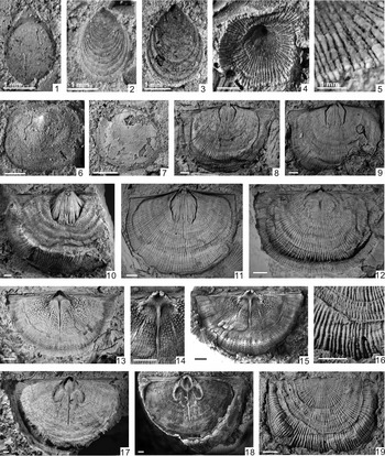
There are 13 species belonging to 13 genera together with an undeterminate species in the two collections under study. The list of species reported is as follows, with some representative specimens being illustrated of each: Discinidae gen. indet. sp. indet. (Fig. 4.7), Pseudopholidops partibilis (Fig. 4.1–4.3), Xenocrania haimei (Fig. 4.4–4.5), Petrocrania sp. (Fig. 4.6), Leptaena rugosa (Fig. 4.8–4.19), Minutomena missa n. sp. (Fig. 5), Paromalomena macmahoni (Fig. 6), Eostropheodonta hirnantensis (Fig. 7.10–7.15), Palaeoleptostrophia sp. (Fig. 7.1–7.9), Fardenia (Coolinia) sp. (Fig. 7.16–7.18), Hirnantia sagittifera (Figs. 8, 9), Dalmanella testudinaria (Fig. 10.1–10.4), Hindella crassa (Williams) (Fig. 10.5–10.8), and Cliftonia cf. C. psittacina (Fig. 10.9–10.12). The one new species and two most dominant species are discussed in detail. Data and some statistical information are also provided.
(1–3) Pseudopholidops partibilis: (1) a dorsal(?) internal mold (NIGP 170724); (2, 3) two dorsal(?) external molds (NIGP 170724, 170726). (4, 5) Xenocrania haimei: (4) dorsal external mold and (5) its local enlargement showing detail of ornamentation (NIGP 170727). (6) Petrocrania sp., dorsal internal mold (NIGP 170728). (7) Discinidae gen. indet. sp. indet. dorsal valve exterior (NIGP 170729). (8–19) Leptaena rugosa: (8–11) four ventral internal molds (NIGP 170730–170733); (12) dorsal internal mold (NIGP 170734); (13–15) dorsal internal mold: (13) its latex cast, (15) local enlargement, and (14) detail of cardinalia (NIGP 170735). (17) Dorsal internal mold and (18) its latex cast (NIGP 170736); (16) dorsal external mold, (19) its local enlargement, and (16) detail of ornamentation (NIGP 170737). All specimens from GHH-02 except (17) and (18) from GMB-01. Scale bars = 2 mm except where noted.

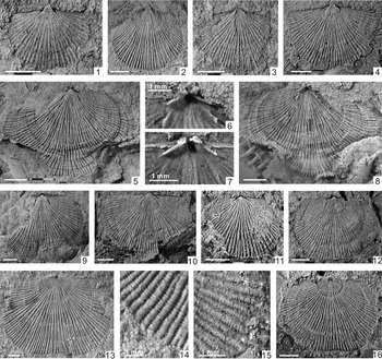
Minutomena missa n. sp. (1–4) Four ventral internal molds (NIGP 170738–170741); (5) dorsal internal mold, (8) its latex cast, and (6) local enlargement showing detail of cardinalia (NIGP 170742). (11) dorsal internal mold and (7) local enlargement of its latex showing detail of cardinalia (NIGP 170743); (9, 10) two dorsal internal molds (NIGP 170744, 170745); (12) dorsal internal mold, (16) its external mold, and (15) local enlargement of its latex showing detail of ornamentation (NIGP 170746); (13) dorsal external mold and (14) its local enlargement showing detail of ornamentation (NIGP 170747). All specimens from GHH-02. Scale bar = 2 mm except where noted.

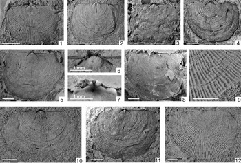
Paromalomena macmahoni (Reed). (1, 2) two ventral internal molds (NIGP 170748, 170749); (3, 4, 10, 11) four dorsal internal molds (NIGP 170750–170753); (5) dorsal internal mold, (8) its latex cast, (6) local enlargement of mold, and (7) cast showing detail of cardinalia (NIGP 170754); (12) dorsal external mold and (9) its local enlargement showing detail of ornamentation (NIGP 170755). All specimens from GHH-02. Scale bars = 2 mm except where noted.

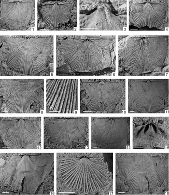
(1–9) Palaeoleptostrophia sp.: (1, 4) two ventral internal molds (NIGP 170756, 170757); (2) ventral internal mold and (3) its local enlargement showing absence of dental plates (NIGP 170758); (5) dorsal internal mold, (8) its external mold, and (9) local enlargement showing detail of ornamentation (NIGP 170759); (6, 7) two dorsal internal molds (NIGP 170760, 170761). (10–15) Eostropheodonta hirnantensis (M'Coy): (10–12) three ventral internal molds (NIGP 170762–170764); (13, 14) two dorsal internal molds (NIGP 170765, 170766); (15) local enlargement of a dorsal internal mold (NIGP 170767). (16–18) Fardenia (Coolinia) sp.: (16) ventral internal mold and (18) its external mold (NIGP 170768); (17) dorsal internal mold (NIGP 170769). All specimens from GHH-02. Scale bars = 2 mm.

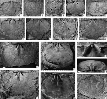
Hirnantia sagittifera: (1–3, 6) four ventral internal molds (from GHH-02, NIGP 170770–170773); (4, 5, 7–9, 14) seven dorsal internal molds (all from GHH-02 except 7 from GMB-01, NIGP 170774–170779); (10) dorsal internal mold and (16) its external mold (from GHH-02, NIGP 170780); (11) dorsal internal mold, (13) its posterior view, (15) its latex cast, and (12) local enlargement showing detail of cardinalia (from GMB-01, NIGP 170781). Scale bars = 2 mm.

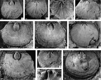
Hirnantia sagittifera: (1) ventral internal mold, (2) dorsal internal mold, and (3) its latex cast, from Huangtian section of Gongxian, Sichuan province (NIGP 170782, 170783); (4) ventral internal mold and (7) a dorsal internal mold, from Wangjiawan section of Yichang area (the GSSP of Hirnantian Stage) (NIGP 170784, 170785); (5) ventral internal and (6) its latex cast (from AAE-080, NIGP 170786); (9) dorsal internal mold and (10) its latex cast (from AAE-080, NIGP 170787); (8) ventral internal mold and (11) dorsal internal mold (AAE-080); the two specimens were designated as paratype and holotype of Hirnantia magna Rong et al., Reference Rong, Xu and Yang1974 separately (NIGP 22275, 22276). Scale bars = 2 mm.

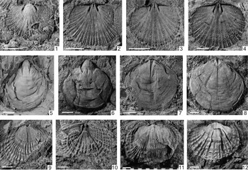
(1–4) Dalmanella testudinaria: (1) ventral internal mold (NIGP 170788); (2–4) three dorsal internal molds (NIGP 170789–170791). (5–8) Hindella crassa: (5, 6) two ventral internal molds (NIGP 170792, 170793); (7, 8) two dorsal internal molds (NIGP 170794, 170795). (9–12) Cliftonia cf. C. psittacina: (9) ventral internal mold (NIGP 170796); (10) ventral external mold (NIGP 170797); (11, 12) two dorsal internal molds (NIGP 170798, 170799). All from GHH-02 except 11 and 12 from GMB-01. Scale bars = 2 mm.

Class Strophomenata Williams et al., Reference Williams, Carlson, Brunton, Holmer and Popov1996
Order Strophomenida Öpik, Reference Öpik1934
Superfamily Strophomenoidea King, Reference King1846
Family Glyptomenidae Williams, Reference Williams and Moore1965
Genus Minutomena Zeng et al., Reference Zeng, Chen, Wang, Zhang and Han2016
- Reference Zeng, Chen, Wang, Zhang and Han2016
Minutomena Zeng et al., p. 21.
Type species
Minutomena yichangensis Zeng et al., Reference Zeng, Chen, Wang, Zhang and Han2016, p. 21, pl. 2, figs. 1–5; Kuanyinchaio Beds (Hirnantian), Yichang, western Hubei, Central China.
Minutomena missa new species
Figure 5
Type specimens
Holotype: NIGP 170742 (Fig. 10.5). Paratypes: NIGP 170738–170741, NIGP 170743–170746. All from Kuanyinchiao Beds (Hirnantian, uppermost Ordovician) of Hetaoba section, Meitan County, northern Guizhou, South China.
Diagnosis
Larger Minutomena with coarser ornamentation and relatively small cardinalia.
Occurrence
The new species is known only from the Kuanyinchiao Beds (Hirnantian, uppermost Ordovician) of Meitan County, Guizhou Province, South China.
Description
Shells small to medium sized, 3.5–11.8 mm long, 4.3–13.6 mm wide, transversely semicircular; ventral valve slightly convex with maximum convexity near umbo, dorsal valve nearly flat to weakly concave, lateral profile rarely resupinate.
Ornamentation of multicostellae, unequally round ribs increasing mostly by branching, about 3–4 per 2 mm at 10 mm from the umbo (Fig. 5.13); fine and prominent concentric growth lines well developed. Substance of shell coarsely pseudopunctate.
Ventral interior with thin and short dental plates, divergent at an angle of about 70°–110°; muscle scars small, broad, faintly visible, rarely extending beyond ends of dental plates anteriorly; diductor scars subtriangular. Pedicle callist broad and well developed (Fig. 5.2, 5.4).
Dorsal interior with small bilobed cardinal process situated in the depressed posterior end of notothyrial platform; socket ridges short, straight or slightly concave posteriorly, widely divergent about 110°–135°; paired low and conspicuous ridges well developed in front of socket plates (Fig. 5.5–5.10), divergent about 60°–100°; muscle field not seen.
Etymology
Latin, missus, with regard to having overlooked this species during the previous study.
Materials
Eight ventral internal molds, 14 dorsal internal molds, and three external molds.
Remarks
The genus was published for the first time in a Chinese book on the basis of the specimens collected from the GSSP area of the Hirnantian Stage (Zeng et al., Reference Zeng, Chen, Wang, Zhang and Han2016). Its close relationship with Paromalomena was not noticed in the original study. However, the specimens in this study were mistakenly assigned to species of Paromalomena by the first author during the earlier study because of a lack of sufficiently informative materials. On the basis of detailed systematic study, the differences between the two genera can be summarized as follows: (1) Ornamentation of Paromalomena is of fine costellae, and increasing by intercalation; concentric wrinkles or rugae commonly irregularly developed; weak and wavy (Fig. 6.2, 6.9). Costae of Minutomena are coarser and increasing mostly by branching, the wrinkles or rugae rarely found on the genus (Fig. 5.13). (2) More important, side septa or paired low ridges are absent or are inconspicuous in Paromalomena (Fig. 6.10) but are essentially developed in front of socket plates in Minutomena (Fig. 5.11).
The new species is different from the type species mainly in two aspects: (1) The size of Minutomena yichangensis Zeng et al., Reference Zeng, Chen, Wang, Zhang and Han2016 is commonly less than 8 mm in width; whereas the specimens of M. missa n. sp. are mostly larger than 10 mm in width. (2) For the type species, a pair of lateral ridges are often very prominent, with its anterior end even reaching half of the valve length (Zeng et al., Reference Zeng, Chen, Wang, Zhang and Han2016, pl. 2, fig. 4a); however, the lateral ridges of M. missa n. sp. are always weaker, with their anterior ends at one-fourth to one-fifth of valve length.
Order Strophomenida Öpik, Reference Öpik1934
Superfamily Strophomenoidea King, Reference King1846
Family Leptostrophiidae Caster, Reference Caster1939
Genus Eostropheodonta Bancroft, Reference Bancroft and Lamont1949
Type species
Orthis hirnantensis M'Coy, Reference M'Coy1851; Hirnant Formation (Hirnantian), Upper Ordovician, Aber Hirnant, near Bala, Wales, UK.
Eostropheodonta hirnantensis (M'Coy, Reference M'Coy1851)
Figure 7.10–7.15
Type specimens
Lectotype: internal mold of pedicle valve, A 28831, from Aber Hirnant, North Wales (M'Coy, Reference M'Coy and Sedgwick1852, pl. 1H, fig. 11c; Temple, Reference Temple1965, pl. 19, fig. 1).
Remarks
The ornamentation of species assigned to Eostropheodonta has been considered to be of taxonomic significance. However, it is highly variable even in specimens from the same population (see discussion in Rong et al., Reference Rong, Aung, Zhan, Huang, Harper, Chen, Zhou and Zhang2019). Furthermore, the development of dental plates is also variable. During the early study of the fauna, all related specimens were assigned to Eostropheodonta. After detailed analysis of the variation, we consider that specimens with vestigial or absent dental plates, together with the triangular and weakly impressed muscle field, are better assigned to Palaeoleptostrophia rather than Eostropheodonta. On the basis of the dorsal internal molds of the species of the two genera, it is difficult to differentiate the two (Rong and Cocks, Reference Rong and Cocks1994). Since species of Palaeoleptostrophia have numerous coarse pseudopunctae in the posterolateral parts of both its ventral and its dorsal internal molds, we assign the dorsal internal molds with the developed coarse pseudopunctae to Palaeoleptostrophia. The genus Palaeoleptostrophia was thought to originate in the Rhuddanian, and the oldest Leptostrophiidae lacked dental plates (Rong and Cocks, Reference Rong and Cocks1994). There are some specimens of that family recorded as leptostrophiids in the Hirnantian rocks of Latvia and Estonia (Hints and Harper, Reference Hints and Harper2015). Thus, it could be regarded as a progenitor taxon evolved during the crisis interval (Rong et al., Reference Rong, Aung, Zhan, Huang, Harper, Chen, Zhou and Zhang2019). The evolutionary relationship between Eostropheodonta and Palaeoleptostrophia together with other genera of Leptostrophiidae will be discussed in detail elsewhere.
Order Orthida Schuchert and Cooper, Reference Schuchert and Cooper1932
Suborder Dalmanellidina Moore, Reference Moore, Moore, Lalicker and Fischer1952
Superfamily Enteletoidea Waagen, Reference Waagen1884
Family Draboviidae Havlíček, Reference Havlíček1950
Subfamily Draboviinae Havlíček, Reference Havlíček1950
Genus Hirnantia Lamont, Reference Lamont1935
Type species
Orthis sagittifera M' Coy, Reference M'Coy1851; Hirnant Formation (Hirnantian), Upper Ordovician, Aber Hirnant, near Bala, Wales, UK.
Hirnantia sagittifera (M’Coy, Reference M'Coy1851)
Figures 8, 9
Here only the data used in the analysis are listed; see appendix of Rong et al., Reference Rong, Aung, Zhan, Huang, Harper, Chen, Zhou and Zhang2019 for a detailed list.
- Reference M'Coy1851
Orthis sagittifera M'Coy, p. 398.
- Reference Lamont1935
Hirnantia sagittifera; Lamont, p. 313, pl. 7, figs. 20–22.
- Reference Temple1965
Hirnantia sagittifera; Temple, p. 349, pl. 11, fig. 8; pl. 12, figs. 1–10; pl. 13, figs. 1–10; pl. 14, figs.1–8.
- Reference Bergström1968
Hirnantia sagittifera; Bergström, p. 10, pl. 3, fig. 13; pl. 4, figs. 1, 2.
- Reference Rong, Xu and Yang1974
Hirnantia magna Rong, Xu, and Yang, p. 196, pl. 92, fig. 25, 26.
- Reference Lespérance and Sheehan1976
Hirnantia sagittifera; Lespérance and Sheehan, p. 723, pl. 109, figs. 3–11.
- Reference Havlíček1977
Hirnantia sagittifera; Havlíček, p. 266, pl. 29, figs. 15–23.
- Reference Rong1979
Hirnantia sagittifera fecunda Rong, pl. 1, figs. 10, 14, 15.
- Reference Nikitin, Popov, Rukavishnikova, Apollonov, Bandaletov and Nikitin1980
Hirnantia sagittifera; Nikitin, Popov, and Rukavishnikova, p. 43, pl. 11, figs. 1–4.
- Reference Cocks1982
Hirnantia sagittifera; Cocks, p. 763, pl. 78, figs. 6–10; pl. 79, figs. 1–3; pl. 80, fig. 8.
- Reference Mergl1983
Hirnantia sagittifera; Mergl, p. 343, pl. 4, figs. 14–16.
- Reference Rong and Mu1984
Hirnantia sagittifera; Rong, p. 139, pl. 6, figs. 1–17; pl. 7, figs 1–8; pl. 12, fig. 15; text-figs 10, 11.
- Reference Benedetto, Racheboeuf and Emig1986
Hirnantia sagittigera; Benedetto, p. 444, pl. 1, figs. 11–15; pl. 2, figs. 1–3.
- Reference Rong and Xu1987
Hirnantia sagittifera; Rong and Xu, p. 5, pl. 1, figs. 9, 12, 13, 16, 17, 20–22, 24, 26.
- Reference Villas, Lorenzo and Gutierrez-Marco1999
Hirnantia sagittifera; Villas, Lorenzo, and Gutierrez-Marco, p. 193, fig. 4 (a–i).
- Reference Hints, Parnaste and Gailite2008
Hirnantia sagittifera; Jin and Zhan, p. 52, pl. 36, figs. 1–16.
- Reference Hints, Parnaste and Gailite2012
Hirnantia sagittifera; Hints, Parnaste, and Gailite, p. 67, fig. 2A–J.
- Reference Zeng, Chen, Wang, Zhang and Han2016
Hirnantia sagittifera morph. Bohemia Havlíček (Reference Havlíček1977), Zeng et al., p. 69, pl. 35, figs. 1–12; pl. 36, figs. 1, 2.
- Reference Zeng, Chen, Wang, Zhang and Han2016
Hirnantia sagittifera morph. Poland Temple (Reference Temple1965), Zeng et al., p. 70, pl. 37, figs.1–11.
- Reference Zeng, Chen, Wang, Zhang and Han2016
Hirnantia magna; Zeng et al., p. 71, pl. 36, figs. 3–12.
- Reference Zeng, Chen, Wang, Zhang and Han2016
Hirnantia septumis Zeng et al, p. 72, pl. 38, figs. 1–12.
- Reference Zeng, Chen, Wang, Zhang and Han2016
Hirnantia fecunda (Rong), Zeng et al., p. 74, pl. 34, figs. 6–12.
Type specimens
Lectotype: internal mold of brachial valve, A 41217, from Aber Hirnant, North Wales (M'Coy, Reference M'Coy and Sedgwick1852, pl. 1H, fig. 17; Temple, Reference Temple1965, pl. 11, fig. 8).
Remarks
H. sagittifera has been described in detail by many different authors (e.g., Temple, Reference Temple1965; Havlíček, Reference Havlíček1977; Cocks, Reference Cocks1982; Villas et al., Reference Villas, Lorenzo and Gutierrez-Marco1999; Hints et al., Reference Hints, Parnaste and Gailite2012, and so on). The population variation in the species was noted mainly in size and outline (Temple, Reference Temple1965; Cocks, Reference Cocks1982; Cocks and Torsvik, Reference Cocks and Torsvik2002). Its adult shell width can reach 50 mm (Bergström, Reference Bergström1968; Hints et al., Reference Hints, Parnaste and Gailite2012) or be less than 15 mm in different populations. The variation of its internal characters is also significant. Rong (Reference Rong and Mu1984) initially discussed the morphologic variation of the cardinal process and considered it could be both simple and bilobate with peculiar crenulations on its top and sides. The variation in H. sagittifera is so considerable that some specimens in the same population could be easily identified as separate species.
Recently, Zeng et al. (Reference Zeng, Chen, Wang, Zhang and Han2016) monographed a Hirnantia Fauna from Yichang, where the GSSP of the Hirnantian Stage is situated. With abundant and well-preserved specimens of Hirnantia from the same beds, they identified five species (or morphotypes): H. sagittifera morph. Bohemia, H. sagittifera morph. Poland, H. magna, H. septumis, and H. fecunda. The main criteria for discrimination were shell size and the development of muscle fields. However, these key differences are not clear with reference to their specimens.
To assess the value of the differences between the ‘five species,’ we measured most of the taxa from published figures. We also compared several specimens from other collections from South China: (1) Donggongsi section (AAE-080) of Zunyi city, Guizhou province; (2) Tianbatou section of Gongxian, Sichuan province; (3) Wangjiawan section of Yichang area, Hubei Province (the GSSP of the Hirnantian). Seven parameters were selected for dorsal internal molds, such as length, width, length of cardinalia (from anterior margin of brachiophore bases to the apex of shell), width of cardinalia (the maximum distance between two brachiophore bases), muscle field position (distance from anterior margin of muscle fields to the apex of the shell), width of muscle fields, and angle of brachiophore supporting plates. Six parameters were available for the ventral internal molds: length, width, length of muscle field, width of muscle field, width of adductor muscle scar, and angle of dental plates (Appendices 1, 2). Using principal components analysis (PCA) to interrogate these measurements, most of the variation in these specimens can be analyzed and evaluated. Since related specimens from a previous study (Rong, Reference Rong1979, Reference Rong and Mu1984) were collected from the same locality, we also included them in the analysis. The results (Fig. 11.1) displayed a clear pattern of variation. No single morphological group can be clearly recognized from the scatters, indicating that the variation of each ‘species’ is significant but continuous for both outline and internal structures.
Principal components analysis of data for six parameters for seven ‘species’ of (1) Hirnantia and (2) globally distributed Hirnantia sagittifera to show the variation.

Zeng et al. (Reference Zeng, Chen, Wang, Zhang and Han2016) considered that ‘H. sagittifera (M'Coy) morph.’ Bohemia is different from ‘H. sagittifera (M'Coy) morph.’ Poland mainly in its larger size. However, as the specimens were collected from the same beds, we consider that many of ‘H. sagittifera (M'Coy) morph. Poland’ are immature specimens lacking the development of the dorsal muscle field. Since the material from Bohemia and Poland are both assigned to H. sagittifera, this morphotype is not necessarily present. Similarly, H. magna was erected mainly by virtue of its larger size (it can reach 40 mm) and the development of cardinalia and muscle fields according to specimens from very shallow-water (BA 2) populations and is represented only by large adult shells rather than different species. Most of the shells were significantly larger than those collected from other localities at that time (Rong et al., Reference Rong, Xu and Yang1974). However, large and even larger specimens H. sagittifera have been reported from Sweden (Bergström, Reference Bergström1968), Oslo (Cocks, Reference Cocks1982), Argentina (Benedetto, Reference Benedetto, Racheboeuf and Emig1986), and Estonia (Hints et al., Reference Hints, Parnaste and Gailite2012), which undermines the validity of the species H. magna. Here, we figure the holotype of this species (Fig. 9.11) and note the similarity between the holotype of H. magna and the large specimens from this study (Fig. 8.11), suggesting H. magna can be treated as a junior synonym of H. sagittifera.
H. fecunda (Zeng et al., Reference Zeng, Chen, Wang, Zhang and Han2016) was first erected as H. sagittifera fecunda (Rong, Reference Rong1979) mainly on the basis of its shorter and narrower cardinalia and ventral muscle fields. Both the measurements and the results of the PCA analysis indicate that the variation of H. sagittifera encompasses that of H. fecunda. In this study, specimens collected from Meitan with short brachiophore bases and muscle fields are also similar to ‘H. sagittifera fecunda.’ However, we consider that the subspecies should be suppressed and assigned to H. sagittifera. The typical character of H. septumis is a short and narrow medium septum developed in the ventral valves. We consider that this character is an indicator of adult or gerontic individuals; these specimens are quite large with a well-developed muscle field, and some of the ventral valves even have muscle-bounding ridges. Similar but weaker muscle-bounding ridges are also identified on the specimens collected from Guizhou (Fig. 9.5, 9.6).
We suggest that the ‘five species’ of Zeng et al. (Reference Zeng, Chen, Wang, Zhang and Han2016) together with H. magna (Rong et al., Reference Rong, Xu and Yang1974) and H. sagittifera fecunda (Rong, Reference Rong1979) should be reassigned to H. sagittifera sensu stricto. The ‘unique characters’ described for each of these taxa are part of wide-ranging, continuous intraspecific variation rather than criteria for the establishment of different species. Besides the data from Yichang of China presented in the preceding, representative specimens of H. sagittifera from different regions and paleoplates reveal further variation in these species. We measured the figured specimens from Wales (Temple, Reference Temple1965; Walmsley et al., Reference Walmsley, Boucot and Harper1969; Brenchley et al., Reference Brenchley, Marshall, Harper, Buttler and Underwood2006), Sweden (Bergström, Reference Bergström1968), Quebec, Canada (Lespérance and Sheehan, Reference Lespérance and Sheehan1976), Bohemia (Havlíček, Reference Havlíček1977), Kazakhstan (Nikitin et al., Reference Nikitin, Popov, Rukavishnikova, Apollonov, Bandaletov and Nikitin1980), Oslo (Cocks, Reference Cocks1982), Algeria (Mergl, Reference Mergl1983), Argentina (Benedetto, Reference Benedetto, Racheboeuf and Emig1986), Tibet (Rong and Xu, Reference Rong and Xu1987), Spain (Villas et al., Reference Villas, Lorenzo and Gutierrez-Marco1999), and Estonia (Hints et al., Reference Hints, Parnaste and Gailite2012). Most of the specimens are preserved as internal molds, with the exception of those from Estonia with calcareous shells, mainly from drill cores. Using the specimens from Yichang as a template, on the basis of their measurements (Appendices 1, 2), the results from PCA clearly indicate that the variation of the global species can be captured in the data from Yichang, China (Fig. 11.2). Some outliers, such as the data from Estonia and Sweden, may be due to different modes of preservation, which introduce a bias into the measurements. The differences between specimens from other regions were emphasized (e.g., Zeng et al., Reference Zeng, Chen, Wang, Zhang and Han2016). However, we consider that given the wide variability of the Yichang material, these differences may be accommodated within the intraspecific variability of H. sagittifera.
Specimens of Hirnantia sagittifera collected from Guizhou, South China, display a marked range of variation. Dorsal valves can be relatively flat or occasionally very convex (Fig. 8.13). The cardinal process can be simple (Fig. 8.15) or bilobed with crenulations (Fig. 9.10) in adult specimens. Dorsal muscle fields also vary from weak to well developed, with different outlines. The anterior pairs of dorsal muscle scars can be equal to (Fig. 8.10) or smaller than (Fig. 9.11) the posterior pairs. The angle between the brachiophore supporting plates or dental plates is clearly varied. The ventral adductor muscle field can be wide to very narrow. The anterior extremity of the adductor field on a few specimens can form a short septum as described in the different species (Zeng et al., Reference Zeng, Chen, Wang, Zhang and Han2016). With sufficient sampling of populations, more variation will be discovered.
Acknowledgments
The authors thank G.X. Wang, Y. Wang, R.B. Zhan, P. Tang, X.C. Luan, F.Y. Gong (NIGPAS), and Z.Q. Zhou (Xi'an Center of China Geological Survey) for help during fieldwork. The comments of I. Percival and R. Cocks together with those of the editor greatly improved the clarity of the manuscript. Financial support for this study was provided by the Strategic Priority Research Program of the Chinese Academy of Sciences, Grant No. XDB26000000; the National Natural Science Foundation of China (NSFC) 41530103. D.A.T. Harper acknowledges support from the Leverhulme Trust (UK) and IGCP Project 653 ‘The Onset of the Great Ordovician Biodiversification Event.’
Measurements (in mm except angle in degree) of ventral valves of representative specimens of Hirnantia sagittifera and related species, L. Mus. = length of muscle field, W. Mus. = width of muscle field, W. ad. Mus = width of adductor muscle field, Ang. D. P. = angle of dental plates.

Measurements (in mm except angle in degree) of dorsal valves of representative specimens of Hirnantia sagittifera and related species. L. Card = length of cardinalia, W. Card. = width of cardinalia, D. Mus-Apex = distance from anterior margin of muscle field to the apex of shell, Ang. BSP. = angle of brachiophore supporting plates.