Introduction
Nasopharyngeal carcinoma (NPC) is a lethal malignancy with a high prevalence in South-East Asia, particularly in the Guangdong and Guangxi provinces of Southern China.Reference Lee, Poon, Foo, Law, Cheung and Chan1, Reference Ahmad and Stefani2 In Southern China, the annual incidence of NPC is approximately 20 per 100 000 population; in the Guangdong province, it is estimated to be 80 per 100 000 population.Reference Cho3, Reference Glastonbury4 In addition, men are twice as likely as women to develop NPC, and the incidence increases with age.Reference Cho3
There are marked differences in disease manifestation and long-term prognosis for the three histological NPC types (I, II, and III). Type I NPC is the least common form and has the worst prognosis. It is analogous to pharyngeal squamous cell carcinoma (SCC) of the pharynx and is associated with cigarette smoking and alcohol use. Type I is further classified into poor, moderate and well-differentiated forms, with the latter being the least common. Type II NPC refers to non-keratinising carcinoma; it is similar to the more common type III, which is undifferentiated carcinoma. Indeed, types II and III NPC are considered to comprise a spectrum within the same disease. NPC type III was previously known as a lymphoepithelioma because it is associated with a combination of undifferentiated epithelial and non-malignant T lymphocytes. Types II and III NPC are more common than type I in both endemic and non-endemic regions. They are also more radiosensitive, thus having a better prognosis compared with type I NPC.Reference Glastonbury4
Most NPC tumour cells are poorly differentiated and biologically active in terms of cellular proliferation, infiltration and distant metastases. Although early-stage NPC is generally radiocurable, patients in Southern China with late-stage NPC have a poor prognosis, largely because of local treatment failure (19.8–36 per cent) and distant metastasis (21.2–41.8 per cent). Ahmad and Stefani reported the distant metastasis rate for NPC patients in Southern China to be as high as 38–81 per cent based on post-mortem examinations.Reference Ahmad and Stefani2 Many factors have been implicated in NPC metastasis. An increasing number of reports show that cancer cells specifically express chemokine receptors and that activation of these receptors plays an important role in distant metastasis.
Chemokines are chemotactic cytokines induced by inflammatory cytokines, growth factors and pathogenic stimuli and are involved in the directed migration of leukocytes.Reference Balkwill5 Chemokines are classified as C, C-C, C-X-C, and CX3C types according to the position of the cysteine residues that form two disulphide bonds. Chemokines mediate their effects through several G protein-coupled transmembrane domain receptors, which are currently divided into four families according to the type of chemokine to which they bind. These receptors include C-X-C chemokine receptor types 1 and 5 (CXCR1 and CXCR5), C-X-C chemokine receptor type 5 (CXCR5), C-C chemokine receptor types 1 and 9 (CCR1 and CCR9), and CX3C chemokine receptor 1 (CX3CR1). Chemokines were initially identified by their ability to transport inflammatory cells directly to areas of infection. Most chemokines are recognised by several receptors, and a single receptor can recognise more than one chemokine. However, some receptors, such as CCR9, recognise only a single chemokine. The CCR9 chemokine receptor binds to C-C motif chemokine 25 (CCL25; also known as thymus-expressed chemokine or TECK).Reference Papadakis, Prehn, Nelson, Cheng, Binder and Ponath6 CCR9 and its ligand, CCL25, are reported to be expressed only in the thymus, lymph nodes, spleen, haematopoietic stem cells, and both differentiated and undifferentiated T cells.Reference Wright, Bowman, Wagers, Butcher and Weissman7, Reference Zaballos, Gutiérrez, Varona, Ardavín and Márquez8
Functional CCR9 is selectively expressed on human small intestinal lymphocytes, while CCL25 is constitutively expressed in both murine and human small intestinal epithelial cells. The latter suggests a potential role for this chemokine receptor–chemokine complex in lymphocyte localisation to the small intestine.Reference Svensson, Marsal, Ericsson, Carramolino, Brodén and Márquez9 Furthermore, CCL25 is constitutively expressed in the epithelium and mucosal vessels of the small intestine, where it interacts with gut-homing, CCR9-expressing B and T cells.Reference Eksteen, Grant, Miles, Curbishley, Lalor and Hübscher10 CCR9 and CCL25 are known to mediate lymphocyte homing to the healthy small intestine, but the identity of chemokine receptors involved in intestinal inflammation are unknown.Reference Connor, Paraskevopoulos, Newman, Cuan, Hampartzoumian and Lloyd11
CCL25 has been demonstrated to play an essential role in the intestinal homing of IgA antibody secreting cells, primarily by mediating their migration into the intestinal lamina propria.Reference Hieshima, Kawasaki, Hanamoto, Nakayama, Nagakubo and Kanamaru12 There are some reports that CCR9 expression in leukaemic cells contributes to leukaemic relapse in the intestinal tract. Childhood acute lymphoblastic leukaemia is often associated with extramedullary leukaemic cell infiltration upon disease diagnosis or relapse. To understand the mechanisms behind T-cell acute lymphoblastic leukaemia cell dissemination, a recent study investigated homing receptor expression on the blast cells of 11 paediatric T-cell acute lymphoblastic leukaemia patients at diagnosis. This study revealed a unique profile: high levels of CCR9 and cluster of differentiation 103 (CD103; also known as integrin α-E) expression on T-cell acute lymphoblastic leukaemia cells. Both molecules are specifically associated with intestinal tract homing. This finding was clinically significant because the patient later relapsed with disease confined to the intestinal tract. Immunohistochemical analysis revealed that the intestinal leukaemic cells expressed CCR9 that co-localised with strong CCL25 expression.Reference Annels, Willemze, van der Velden, Faaij, van Wering and Sie-Go13
This study aimed to measure CCR9 and CCL25 expression in NPC tissues of different tumour–node–metastasis (TNM) stages to use as a reference for basic NPC research.
Materials and methods
Tissue samples
Nasopharyngeal carcinoma (NPC) tissue samples were collected from 42 patients between February 2005 and December 2005. Both in-patients and out-patients who were not administered radiotherapy at Zhongnan Hospital and Renmin Hospital of Wuhan University, Wuhan, China, were included. These comprised 27 men and 15 women in the patient group, with an average age of 49.7 ± 12.0 years. Staging was assessed according to the 6th Union for International Cancer Control TNM staging criteria (2002).Reference Sobin and Wittekind14 In the patient group, 12, 19 and 11 patients were classified as having stage I–II, stage III and stage IV cancer, respectively. In addition, 18, 17 and 7 patients were classified as having T1–2, T3 and T4 tumours, respectively. Lymph node metastases were found in 31 out of 42 patients, among whom 1 patient had a T1 tumour, 9 had T2 tumours, 14 had T3 tumours and 7 had T4 tumours. The major tumour type was poorly differentiated SCC. In the control group, all 18 out-patients (12 men and 6 women) had normal nasopharyngeal tissue biopsies. The average patient age in the control group was 35.1 ± 13.2 years. All tissue samples were divided into two parts and stored at –80 °C until being used for immunohistochemical analysis and to perform quantitative reverse transcription polymerase chain reaction and Western blotting. This study was approved by the ethics committee at the Medical School of Wuhan University.
Reverse transcription polymerase chain reaction
Total RNA was extracted from 50 mg NPC and normal tissue samples using Trizol reagent according to the manufacturer's instructions (Invitrogen, Carlsbad, California, USA). After treatment with RNase-free DNAse I (Takara Biotechnology, Dalian, China), RNA concentration and purity were determined by spectrophotometry and the concentration was adjusted to 1 µg/μl. Total RNA was diluted in diethyl pyrocarbonate treated water and stored at –80 °C. For quantitative reverse transcription polymerase chain reaction, single-strand complementary DNA (cDNA) was prepared using 2 μg RNA with an Exscript RT kit (Takara Biotechnology, Dalian, China), according to the manufacturer's protocol. Primer sequences and amplification conditions are shown in Table I. Relative standard curves were constructed for target genes using the corresponding reverse transcription polymerase chain reaction products isolated using a DNA extraction kit (Hangzhou Aisijin Biotechnology, Hangzhou, China) with concentrations of 10–10 000 pg per reaction. Polymerase chain reactions were performed in 48-well optical reaction plates using an RG3000 Rotor-Gene four-channel multiplexing system (Corbett Research, Sydney, Australia). Reverse transcription polymerase chain reaction kits (containing Premix Ex Taq and SYBR Green I) were purchased from Takara Biotechnology (Dalian, China). The reaction mixture (25 µl volume) contained 2 µl 0.1 µg/μl cDNA template, 0.5 µl each primer at 10 μmol/l, 12.5 µl 2× Premix Ex Taq, 0.5 µl 20× SYBR Green I and 9 µl diethyl pyrocarbonate treated water. For precise quantification, target gene expression was normalised to β-actin expression. Polymerase chain reaction cycling conditions were: pre-denaturation at 95 °C for 10 seconds, followed by 35 cycles of denaturation at 95 °C for 5 seconds and a combined annealing and extension step (specific conditions for each gene are shown in Table I).The final elongation step was conducted at 72 °C for 15 seconds. Duplicate samples were used for all assays.
Primer sequences and polymerase chain reaction annealing conditions

s = seconds
Western blotting
Western blot analysis was used to measure CCR9 and CCL25 protein expression. For this, tissue samples were homogenised in a lysis buffer, and protein extracts (100 µg per sample) were separated by 10 per cent sodium dodecyl sulfate polyacrylamide gel electrophoresis (Bio-Rad, Hercules, California, USA) and electrotransferred onto polyvinylidene difluoride membranes (Amersham Pharmacia Biotech, Piscataway, New Jersey, USA). Membranes were then incubated with an anti-CCR9 or anti-CCL25 monoclonal antibody (1:2000 dilution for both; Santa Cruz, California, USA) overnight at 4 °C with constant shaking. Antibody-bound proteins were detected using enhanced chemiluminescence (Amersham Pharmacia Biotech).
Immunohistochemical analysis
Tissue samples were fixed in 10 per cent formaldehyde for 24 hours and then stained with anti-CCR9 and anti-CCL25 polyclonal antibodies (Jingmei BioTech, Shanghai, China). Antibody binding was detected using a streptavidin–biotin–peroxidase complex method, and CCR9 and CCL25 expression were indicated by the presence of brown cytoplasmic granules. The mean optical density of five random fields in each section was determined at a magnification of ×400 and data were analysed using the Medical Colour Image Analysis System (HMIAS-2000; Guangzhou Longest Technology, Guangzhou, China).
Statistical analysis
All statistical analyses were performed using IBM SPSS Statistics software version 15.0 for Windows (Chicago, Illinois, USA). For categorical variables, a χ2 or Fisher's exact test was used to compare differences between groups. Messenger RNA (mRNA) expression data are expressed as the mean ± standard deviation. Significant differences between the mean values of various groups were determined using one-way analysis of variance followed by Dunnett's post hoc test. Statistical significance was set at less than or equal to 0.05 for two-sided tests.
Results
RNA and protein expression
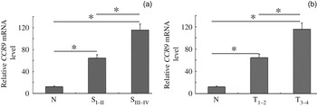
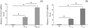
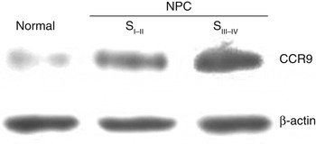
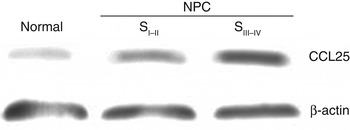
Compared with normal tissue, there was a significant increase in the CCR9 and CCL25 mRNA levels in both the early nasopharyngeal carcinoma (NPC) group (stage I–II or T1–2; p < 0.05) and the advanced NPC group (stage III–IV or T3–4; p < 0.05; Figures 1 and 2). In addition, CCR9 and CCL25 mRNA levels were significantly higher in advanced-stage tumours than in early stage tumours (p < 0.05). CCR9 and CCL25 protein expression were significantly higher in both stage I–II and stage III–IV tumours than in normal tissue (Figures 3 and 4).
Graph showing CCR9 messenger RNA (mRNA) levels in normal nasopharyngeal and nasopharyngeal carcinoma tissues, normalised to ACTB mRNA levels. Data are presented as the mean ± standard deviation. *p < 0.05 compared with the normal group (one-way analysis of variance followed by Dunnett's post hoc test). SI–II = clinical stage I–II; SIII–IV = clinical stage III–IV; N = normal; T1–2 = tumour–node–metastasis stage 1–2; T3–4 = tumour–node–metastasis stage 3–4

Graph showing CCL25 messenger RNA (mRNA) levels in normal nasopharyngeal and nasopharyngeal carcinoma tissues of various tumour–node–metastasis and clinical stages, normalised to ACTB mRNA levels. Data are presented as the mean ± standard deviation. *p < 0.05 as compared with the normal group (one-way analysis of variance followed by Dunnett's post hoc test). SI–II = clinical stage I–II; SIII–IV = clinical stage III–IV; N = normal; T1–2 = tumour–node–metastasis stage 1–2; T3–4 = tumour–node–metastasis stage 3–4

Western blot showing C-C chemokine receptor type 9 (CCR9) expression in normal nasopharyngeal tissue and stage I–II and III–IV nasopharyngeal carcinoma (NPC) tissue. β-actin is the loading control. SI–II = clinical stage I–II; SIII–IV = clinical stage III–IV

Western blot showing C-C motif chemokine 25 (CCL25) expression in normal nasopharyngeal tissue, stage I–II nasopharyngeal carcinoma tissue, and stage III–IV nasopharyngeal carcinoma tissue. β-actin is the loading control.

Immunohistochemical analysis
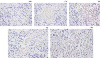
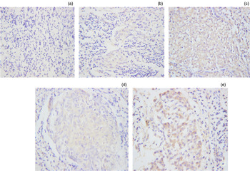
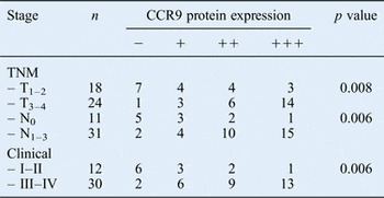
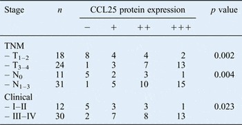
CCR9 and CCL25 protein expression was mainly localised to the cytoplasm of the NPC cells, but weak expression was also seen in stromal tumour cells (Figures 5 and 6). Expression of the CCR9–CCL25 complex was significantly increased in NPC tissues as compared with normal nasopharyngeal tissue (P < 0.05), and was significantly higher in advanced tumour tissues (stage III–IV or T3–4) than in early tumour tissues (stage I–II or T1–2; p < 0.05). CCR9 and CCL25 expression was higher in patients with cervical lymph node metastases than in those without lymph node metastases (Tables II and III).
Photomicrographs showing C-C chemokine receptor type 9 expression in (a) normal nasopharyngeal tissue and in clinical stage (b) I–II and (c) III–IV and tumour–node–metastasis stage (d) T1–2 and (e) T3–4 nasopharyngeal carcinoma tissue (×400).

Photomicrographs showing C-C motif chemokine 25 expression in (a) normal nasopharyngeal tissue and in clinical stage (b) I–II and (c) III–IV and tumour–node–metastasis stage (d) T1–2 and (e) T3–4 nasopharyngeal carcinoma tissue (×400).

CCR9 protein expression level by TNM and clinical stage in 42 NPC patients

CCR9 = C-C chemokine receptor type 9; TNM = tumour–node–metastasis; NPC = nasopharyngeal carcinoma; –, no expression; +, weak expression; ++, moderate expression; +++, strong expression
CCL25 expression level by TNM and clinical stage in 42 NPC patients

CCL25 = C-C motif chemokine 25; TNM = tumour–node–metastasis; NPC = nasopharyngeal carcinoma; –, no expression; +, weak expression; ++, moderate expression; +++, strong expression
Discussion
Many of the abundant stromal cells in solid tumours are infiltrating leukocytes.Reference Balkwill and Mantovani15–Reference Pollard19 The predominant stromal cell type in cancers are macrophages, lymphocytes, endothelial cells and fibroblasts. Eosinophils, granulocytes, natural-killer cells and B cells are also found in ovarian cancer, breast cancer and gastric cancer.Reference Brigati, Noonan, Albini and Benelli16–Reference Pollard19
Many human cancers have a complex chemokine network that influences the extent and phenotype of the infiltration, as well as tumour cell growth, survival, migration and angiogenesis.Reference Balkwill20 The number and type of cells involved in leukocyte infiltration into solid tumours are linked to local chemokine production by both tumour and stromal cells.Reference Brigati, Noonan, Albini and Benelli16, Reference Kidd, Caldwell, Dietrich, Samudio, Spaeth and Watson21 For example, CC chemokines determine macrophage and lymphocyte infiltration into human breast cancer, cervical carcinoma, pancreatic cancer, sarcoma and neuroglioma.Reference Balkwill and Mantovani22, Reference Bottazzi, Polentarutti, Acero, Balsari, Boraschi and Ghezzi23 In contrast, C-C and C-X-C chemokines regulate T-cell surface glycoprotein CD8 glycoprotein positive T lymphocyte infiltration into epithelial ovarian cancer and ascites.Reference Negus, Stamp, Hadley and Balkwill18 Abnormal chemokine and receptor expression and signalling has been reported in tumour cells.Reference Dhawan and Richmond24 In addition, the CCR9–CCL25 complex is involved in both acute and chronic leukaemic cell invasion and in non-Hodgkin's lymphoma metastasis.Reference Dhawan and Richmond24–Reference Arai, Yasukawa, Yakushijin, Miyazaki and Fujita28 Further, CCR9 and its CCL25 ligand are overexpressed in prostate cancer cells of epithelial origin; the CCL25–CCR9 complex is important for prostate cancer invasion and metastasis.Reference Singh, Singh, Stiles, Grizzle and Lillard29–Reference Fusi, Liu, Kummerlen, Nonnemacher, Jeske and Keilholz31 For example, neutralisation of the CCL25–CCR9 interaction impaired the migration and invasion potential of the LNCaP and PC3 cell lines. In addition, CCL25 overexpression differentially modulated the expression of collagenase-1 and matrix metalloproteinase (MMP) 1, 2, 10, 11 and 13 in prostate cancer cells.Reference Singh, Singh, Stiles, Grizzle and Lillard29 The CCL25–CCR9 complex is also expressed in ovarian cancer, breast cancer and melanoma cells.Reference Johnson, Singh, Singh, Johnson-Holiday, Grizzle and Partridge32–Reference Kuhnelt-Leddihn, Muller, Eisendle, Zelger and Weinlich34
In the present study, CCR9 and CCL25 protein localisation and expression, and CCR9 and CCL25 gene expression were determined in various nasopharyngeal carcinoma (NPC) stages using immunohistochemistry, Western blotting and quantitative reverse transcription polymerase chain reaction, respectively. Both CCR9 and CCL25 proteins mainly localised to the cytoplasm of NPC cells, with weak expression in stromal cells. In addition, protein expression was significantly associated with clinical staging, TNM staging and cervical lymph node metastasis. CCR9 and CCL25 expression was significantly higher in stage III–IV and T3–4 NPC than in stage I–II and T1–2NPC. In addition, NPC tissue from patients without lymph node metastasis was generally negative for CCR9 and CCL25 protein expression, whereas NPC tissue from patients with lymph node metastases showed intense staining of both proteins. Similarly, NPCs expressing CCR9 and CCL25 protein and mRNA (as determined by Western blotting and quantitative reverse transcription polymerase chain reaction, respectively) showed progression and were more advanced.
• Nasopharyngeal carcinoma is most common in South-East Asia and Southern China
• In most tumours, a complex chemokine network influences immune-cell infiltration and tumour migration
• CCL25–CCR9 upregulation correlates with tumour–node–metastasis stage
The promotion of tumour metastasis by the CCR9–CCL25 axis shows that a homing signal can form an integral part of the ‘seed and soil’ events of metastasis. Several elements in this pathway are likely to enable specific organs to attract tumour cells.
Conclusion
This study confirms that CCR9 and CCL25 expression is upregulated in NPC, especially in advanced disease.