Introduction
The diversity of lichenicolous fungi in Africa is largely underexplored and the limited information available is scattered in the literature (e.g. Bock et al. Reference Bock, Hauck and Fischer2007; Ertz Reference Ertz2009; Fryday Reference Fryday2015). Thus, we can expect to find a plethora of taxa among the lichenized and lichen-inhabiting fungi that live in the many habitat types unique to Africa. The Taita Hills in south-eastern Kenya form the northernmost section of the Eastern Arc Mountains which, together with nearby coastal forests, represent a hotspot of global biodiversity (Myers et al. Reference Myers, Mittermeier, Mittermeier, da Fonseca and Kent2000; Burgess et al. Reference Burgess, Butynski, Cordeiro, Doggart, Fjeldsa, Howell, Kilahama, Loader, Lovett and Mbilinyi2007; Dimitrov et al. Reference Dimitrov, Nogués-Bravo and Scharff2012). The moist and relatively cool conditions support a high diversity and considerable biomass of cryptogamic epiphytes (e.g. Enroth et al. Reference Enroth, Nyqvist, Malombe, Pellikka and Rikkinen2013; Malombe et al. Reference Malombe, Matheka, Pócs and Patiño2016; Stam et al. Reference Stam, Enroth, Malombe, Pellikka and Rikkinen2017). Even among macrolichens, many local taxa cannot be readily identified with the keys currently available for East Africa (Swinscow & Krog Reference Swinscow and Krog1988).
In the framework of lichenological exploration of tropical montane forests in Kenya, a remarkable lichenicolous fungus was repeatedly found growing out of the thalli of the tripartite cyanolichen Crocodia cf. clathrata (Peltigerales, Lecanoromycetes). Species of Peltigerales are known to host a large diversity of lichenicolous fungi of different phylogenetic origins (e.g. Kondratyuk & Galloway Reference Kondratyuk and Galloway1995; Hawksworth & Miadlikowska Reference Hawksworth and Miadlikowska1997; Lawrey & Diederich Reference Lawrey and Diederich2016) but none of the available descriptions correspond with our new fungus. To clarify the systematic position of the unknown fungus, we analyzed its morphology as well as slow-evolving ribosomal and mitochondrial markers, and describe a new genus and species based on the results.
Material and Methods
Sampling sites
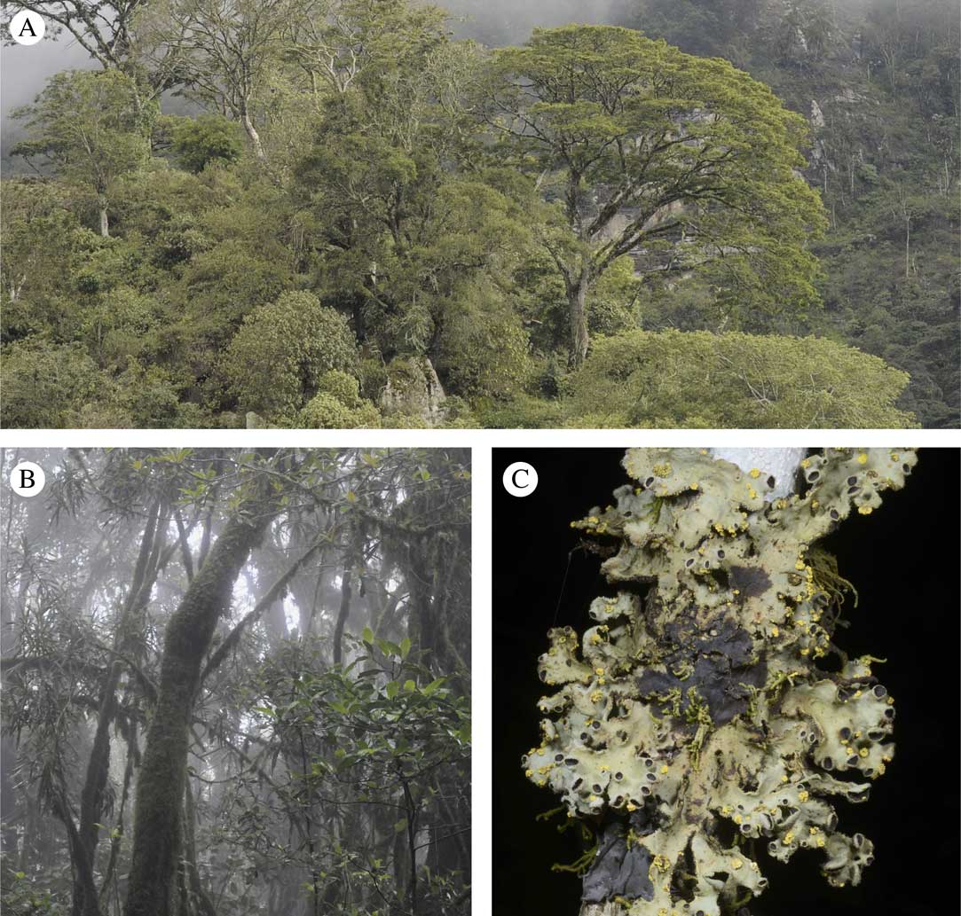
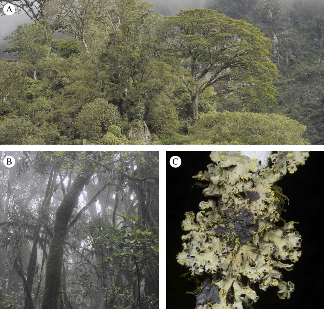
Cyanolichen specimens infected by the new fungus were collected from montane forests in the Taita Hills and the western part of the Rift Valley. The Taita Hills rise abruptly from the surrounding dry plains at c. 600–1000 m to a series of mountain ridges, reaching 2208 m at the highest peak Vuria. The upper slopes of East African mountains benefit from moisture brought by the trade winds and capture enough of this to sustain moist evergreen montane forests. The moist and relatively cool climate provides favourable conditions for the development of species-rich bryophyte and lichen communities and considerable epiphyte biomass (Fig. 1). The type locality of the new fungus is located close to the summit of Vuria, in a dense indigenous forest dominated by Maesa lanceolata, Nuxia congesta and Dracaena afromontana.
Habitat of Taitaia aurea in the Taita Hills, south-eastern Kenya. A, indigenous montane forest on the north-facing slope of Vuria, part of the Dawida massif; B, moss-rich forest at the type locality; C, Crocodia cf. clathrata with yellow outgrowths of T. aurea on the upper surface and thallus margins (holotype, Rikkinen 15258). In colour online.
Morphology
Light microscopy was carried out using hand-cut sections mounted in either tap water, a 10% aqueous solution of potassium hydroxide (KOH; K), Cresyl blue (CRB), Congo red or in Lugol’s solution (I). The apical apparatus of the ascus was observed in Lugol’s solution (I) pretreated with K (denoted as K/I). The measurements were made in water and the sizes are presented as minimum–(mean±1SD)–maximum value. The close-up photographs of ascomata and thallus cross-sections were taken using a microscope equipped with a Canon 60D digital camera. For the images, a series of up to 57 photomicrographs at different focal planes were stacked using HeliconFocus 4.45. The fluorescence microscopy, used to observe the pigments in the host thallus and in ascomata of the lichenicolous fungus, was carried out with a Zeiss Axioskop 40 FL microscope, an AxioCam MRc camera and the AxioVision 1.6. For the scanning electron microscopy (SEM), pieces were fixed onto an SEM-mount, sputter-coated with a 12 nm thick coating of Pt/Pd using an Automatic Sputter Coater (Canemco Inc.), and examined and imaged with a Carl Zeiss LEO 1530 Gemini field emission scanning electron microscope.
The material studied is deposited in the herbarium of the National Museums of Kenya (EA) and in the lichen herbarium of the Natural History Museum at the University of Tartu (TU).
DNA extraction, PCR amplification and sequencing
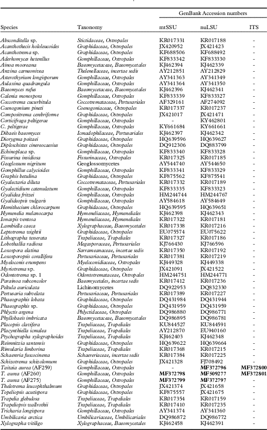
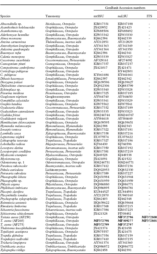
Genomic DNA was extracted from ascomata of specimens using High Pure PCR Template Preparation Kit (Roche Applied Science®) and following the protocol provided by the manufacturer with minor modifications. We amplified three gene loci: the internal transcribed spacer (ITS), the large subunit nuclear ribosomal RNA gene (nuLSU), and the mitochondrial small subunit ribosomal RNA gene (mtSSU). The primers used for amplification and sequencing were ITS0F, LA-W (Tedersoo et al. Reference Tedersoo, Jairus, Horton, Abarenkov, Suvi, Saar and Kõljalg2008), ITS4 and ITS5 (White et al. Reference White, Bruns, Lee and Taylor1990) for ITS, LROR and LR5 (Vilgalys & Hester Reference Vilgalys and Hester1990) and CTB6 (Garbelotto et al. Reference Garbelotto, Lee, Slaughter, Popenuck, Cobb and Bruns1997) for nuLSU, and mrSSU1 and mrSSU3R (Zoller et al. Reference Zoller, Scheidegger and Sperisen1999) for mtSSU. The PCR reaction mix (25 μl) consisted of 5 μl 5× HOT FIREPol Blend Master Mix (Solis BioDyne, Tartu, Estonia), 0·5 μl of 20 µM of both primers, 1–8 μl of target-DNA and distilled water. The PCR products were visualized in a 1% agarose gel stained with ethidium bromide. For the purification of PCR products, 1 μl of FastAP and 0·5 μl of Exonuclease I (Thermo Scientific, Waltham, MA, USA) were added to each tube per 20 μl of product. Both complementary strands were sequenced by Macrogen Inc. (Amsterdam, The Netherlands). Sequencher 4.10.1 (GeneCodes Corp.®, Ann Arbor, MI, USA) was used to check, assemble and manually adjust the resulting sequence fragments. The consensus sequences were compared with those publicly available in NCBI (https://www.ncbi.nlm.nih.gov/genbank) using a BLAST search to confirm their identity. For each gene, the top-scoring match was a member of Ostropomycetidae, even if the similarity did not exceed 90%. The newly generated sequences are available in NCBI (https://www.ncbi.nlm.nih.gov/genbank) under Accession numbers MF372796–MF372801 and MF509277 (Table 1).
GenBank Accession numbers for sequences used in this study. Newly generated sequences are in bold. Numbers in brackets are laboratory codes. The taxonomy of families follows Index Fungorum (http://www.indexfungorum.org/)
Phylogenetic analyses
The newly generated nuLSU and mtSSU sequences were aligned with sequences downloaded from NCBI (Table 1) using MUSCLE (Edgar Reference Edgar2004). We did not use ITS sequences in the analyses due to their high variability at higher taxon levels. The alignments were manually checked and trimmed with SeaView v.4.6 (Gouy et al. Reference Gouy, Guindon and Gascuel2010). The first analysis included representatives of major classes of Ascomycota and subclasses of Lecanoromycetes, but focusing on Ostropomycetidae (data not shown). After the first analysis, we excluded sequences with ambiguous alignment and reduced the dataset, but left a representation of families of Ostropales. The online version of Gblocks v.0.91b (Talavera & Castresana Reference Talavera and Castresana2007) run at http://molevol.cmima.csic.es/castresana/Gblocks_server.html was used to eliminate poorly aligned positions and divergent regions of the alignment while allowing gap positions within the final blocks. For each gene, the best-fit nucleotide substitution model was calculated and selected based on the lowest value of AIC criterion with jModelTest v.2.1.6. (Darriba et al. Reference Darriba, Taboada, Doallo and Posada2012). The best-fit model for nuLSU was TIM+I+G and for mtSSU it was TVM+I+G. Each gene locus was aligned and analyzed separately with a maximum likelihood (ML) approach using PHyML (Guindon et al. Reference Guindon, Dufayard, Lefort, Anisimova, Hordijk and Gascuel2010) and, as no topological conflict was detected in supported clades (bootstrapping over 100 replicates) by visual inspection (data not shown), nuLSU and mtSSU alignments were concatenated. The Markov chain Monte Carlo (MCMC) approach using MrBayes v.3.2.1 (Ronquist et al. Reference Ronquist, Teslenko, van der Mark, Ayres, Darling, Höhna, Larget, Liu, Suchard and Huelsenbeck2012) was applied to reconstruct phylogeny. For the final partitioned analysis, two parallel simultaneous runs with four chains run were applied over 7 M generations until convergence of the chains was confirmed by the standard deviation of split frequencies reaching 0·01. The first 25% of saved data was discarded as burn-in, and the 50% majority-rule consensus tree and posterior probabilities (PP) were calculated from the rest. As an alternative, a maximum likelihood (ML) analysis using GTR+G as the substitution model was implemented with RAxML v.8.1.10 (Stamatakis Reference Stamatakis2014) at the CIPRES Science Gateway (Miller et al. Reference Miller, Pfeiffer and Schwartz2010). Bootstrap support (BS) was calculated from over 500 pseudoreplicates. The outgroup included Loxosporopsis corallifera Brodo et al., Dibaeis baeomyces (L. f.) Rambold & Hertel (Pertusariales, Lecanoromycetes), Peltula auriculata Büdel et al. (Lichinomycetes) and Geoglossum nigritum (Pers.) Cooke (Geoglossomycetes).
The phylogenetic tree was visualized with FigTree v.1.4.2 (http://tree.bio.ed.ac.uk/) and Adobe Illustrator CS3® was used for artwork.
Results and Discussion
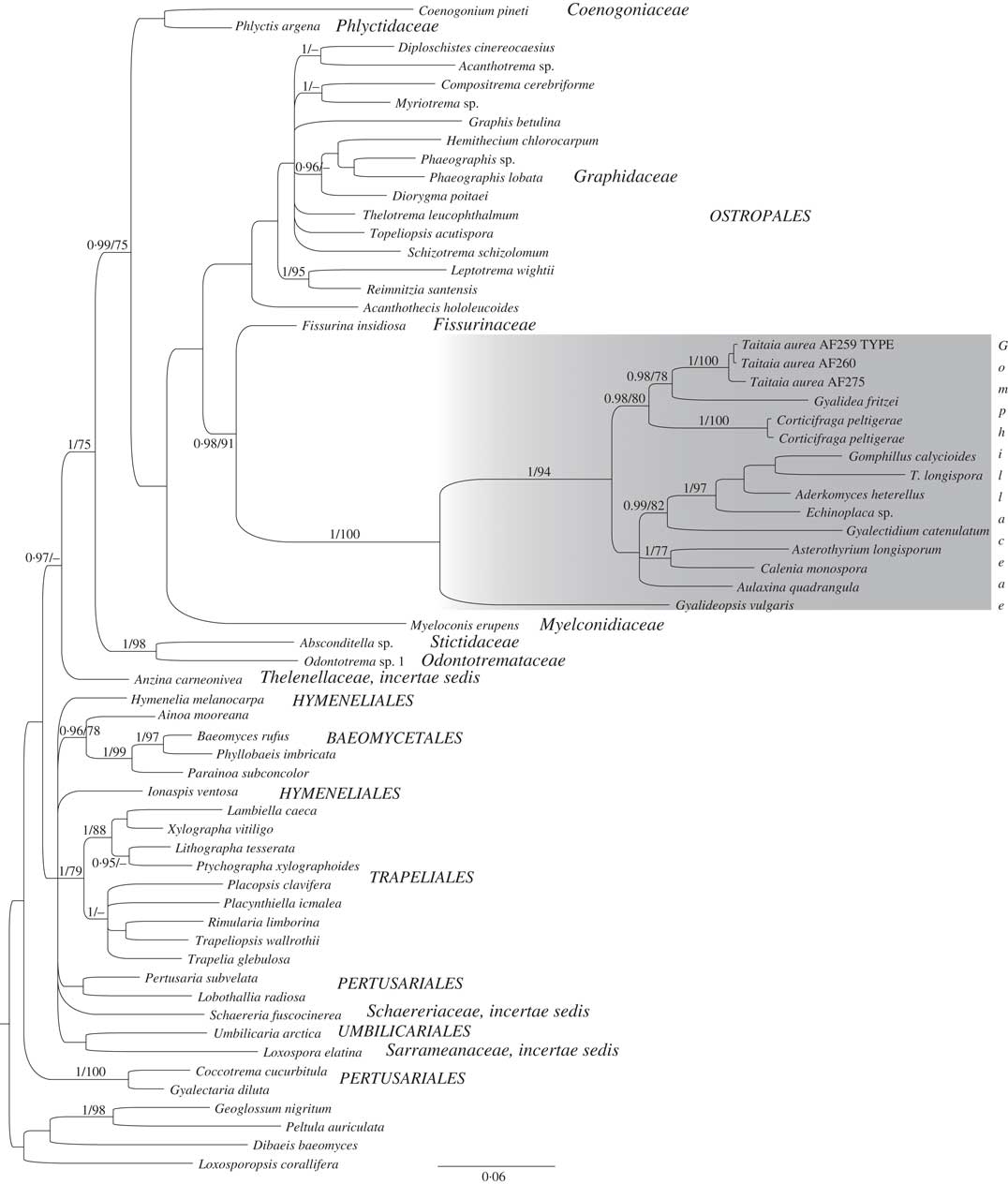
The combined nuLSU+mtSSU dataset consisted of 912 characters, 366 of which were nuLSU and 530 mtSSU. The single-gene and concatenated nuLSU+mtSSU analyses placed the sequences within Gomphillaceae, Ostropales (PP=1, BS=100; Fig. 2). The combination of phenotypic characters (hemiangiocarpic ascomata with non-amyloid asci without distinct apical apparatus; see Taxonomy, Fig. 4) supports the phylogenetic placement within Ostropales (Lumbsch et al. Reference Lumbsch, Schmitt, Lücking, Wiklund and Wedin2007; Jaklitsch et al. Reference Jaklitsch, Baral, Lücking and Lumbsch2016), an ascomycete order of various lifestyles, including symbiotic, optionally lichenized and lichen-dwelling fungi (Baloch et al. Reference Baloch, Lücking, Lumbsch and Wedin2010).
50% majority-rule consensus tree using two loci (nuLSU+mtSSU) based on Bayesian approach showing the position of Taitaia aurea within Gomphillaceae, Ostropales (grey box). Numbers at nodes indicate phylogenetic relationships which are considered to be supported when posterior probabilities (PPs) are ≥0·95 (before slash) and bootstrap values (BS)≥75 (after slash).
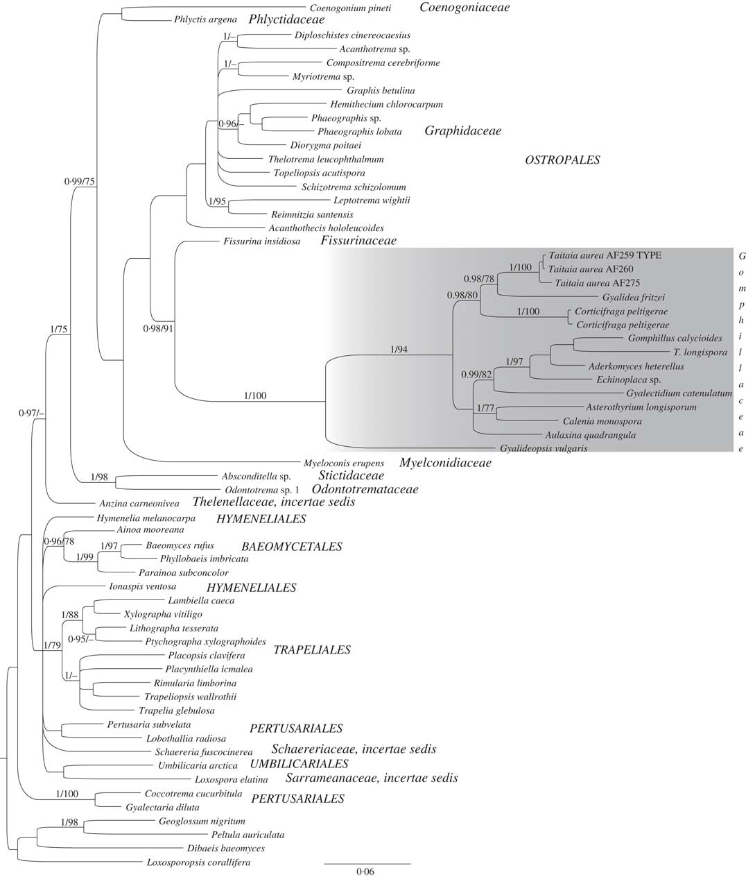
The family Gomphillaceae includes mainly lichen-symbiotic species distributed in the tropics and variously growing on, for example, living leaves, tree bark, bryophytes and soil or rock (Lücking et al. Reference Lücking, Stuart and Lumbsch2004; Baloch et al. Reference Baloch, Lücking, Lumbsch and Wedin2010; Jaklitsch et al. Reference Jaklitsch, Baral, Lücking and Lumbsch2016). In addition to the new fungus, only two other genera of lichen-inhabiting fungi are currently known in the family, Gyalideopsis Vězda (Etayo & Diederich Reference Etayo and Diederich2001; Etayo Reference Etayo2010) and Corticifraga D. Hawksw. & R. Sant. (Pino-Bodas et al. Reference Pino-Bodas, Zhurbenko and Stenroos2017). The phylogenetic concept of Gomphillaceae itself has remained obscure and unresolved (Lücking et al. Reference Lücking, Stuart and Lumbsch2004). The family includes taxa previously placed in a separate family, Asterothyriaceae, distinguished by simple, unbranched paraphyses in the hamathecium and by the lack of hyphophores (Vězda Reference Vězda1987; Henssen & Lücking Reference Henssen and Lücking2002). Both of these characters are also observed in our new fungus (see Taxonomy). Considering the proportion of publicly available sequences (Table 1) and the number of species and genera currently assigned to Gomphillaceae (Jaklitsch et al. Reference Jaklitsch, Baral, Lücking and Lumbsch2016), a re-evaluation of these two families can be expected in the future.
The new fungus, represented in the phylogeny by three specimens, showed the closest relationship to Gyalidea fritzei (Stein) Vězda (PP=0·98, BS=78; Fig. 2). The two fungi have different ecologies, with the latter species being lichenized and growing on lime-rich stone (Gilbert et al. Reference Gilbert, James and Woods2009). Both species in turn are sister to Corticifraga peltigerae (PP=0·98, BS=80; Fig. 2), another lichenicolous fungus which was recently shown to be a member of Gomphillaceae (Pino-Bodas et al. Reference Pino-Bodas, Zhurbenko and Stenroos2017). Similar to the new fungus, the species of Corticifraga live on cyanolichens of the Peltigerales (e.g. Hawksworth & Santesson Reference Hawksworth and Santesson1990; Zhurbenko Reference Zhurbenko2007). All three genera share similar ascoma development, ascus arrangement, simple, septate paraphyses and colourless, ellipsoid to fusiform septate ascospores (Hawksworth & Santesson Reference Hawksworth and Santesson1990; Gilbert et al. Reference Gilbert, James and Woods2009). Both the new fungus and Corticifraga have a similar type of exciple consisting of more or less globular cells (i.e. textura globularis-type) (Hawksworth & Santesson Reference Hawksworth and Santesson1990); no information is provided about the exciple type of Gyalidea (e.g. Gilbert et al. Reference Gilbert, James and Woods2009). Both Corticifraga and Gyalidea differ from the new fungus by having ascomata that arise singly, that are immersed, pale cream to almost black and with discs that are deeply concave (urceolate) to plane when mature (Hawksworth & Santesson Reference Hawksworth and Santesson1990; Gilbert et al. Reference Gilbert, James and Woods2009).
Taxonomy
Taitaia Suija, Kaasalainen, Kirika & Rikkinen gen. nov.
MycoBank No.: MB 821739
Type species: Taitaia aurea Suija, Kaasalainen & Rikkinen.
Taitaia aurea Suija, Kaasalainen & Rikkinen sp. nov.
MycoBank No.: MB 821740
DNA barcode/reference sequence (rDNA ITS): MF372800 (from holotype: Rikkinen 16258, DNA sample: AF259)
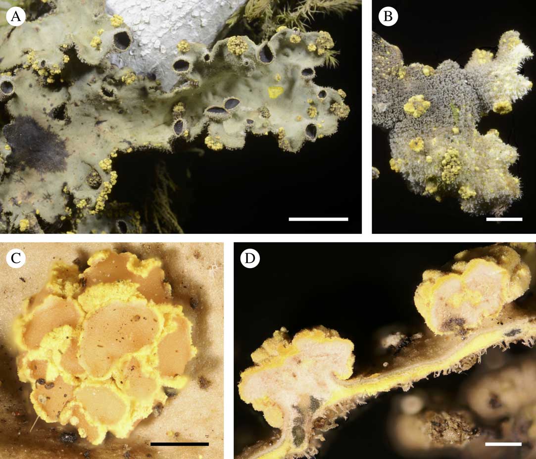
Lichenicolous fungus. Ascomata apothecioid, hemiangiocarpic, breaking through cortex of host thallus, sometimes with ten or more ascomata arising from a single base, margins yellow and disc salmon red. Hymenium with unitunicate non-amyloid asci and simple, septate paraphyses. Ascospores fusiform and 1-septate.
Type: Kenya, Taita-Taveta Province, near summit of Vuria, dense moist forest with Maesa lanceolata, Nuxia congesta and Dracaena afromontana, UTM 421443, 9623094, 2072 m, on thallus of Crocodia cf. clathrata growing on stem of woody climber, 15 January 2015. J. Rikkinen 16258 (TU56326—holotype; DNA sample: AF259)
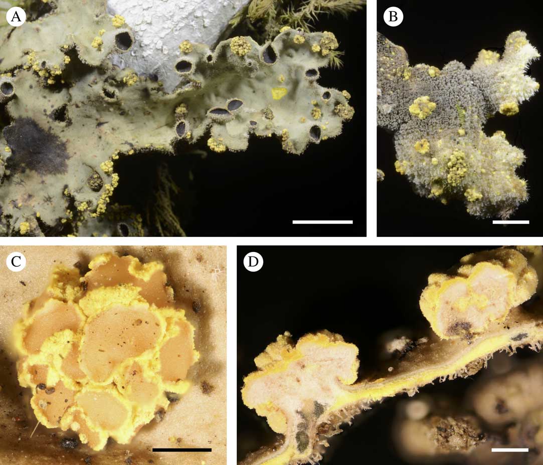
Taitaia aurea. A, ascomata developing on the upper surface and thallus margins of Crocodia cf. clathrata; B, ascomata developing through pseudocyphellae on the lower surface of the host; C, group of mature ascomata; D, cross-section through two ascomata and the host thallus. A–C, Rikkinen 15258 (holotype), D, Rikkinen 15260. Scales: A=5 mm; B=2 mm; C & D=0·5 mm. In colour online.
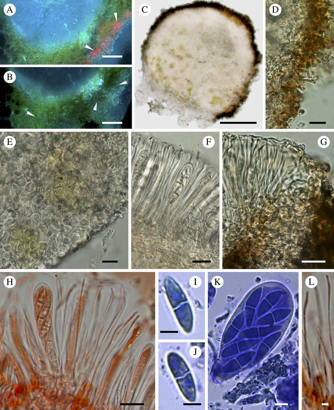
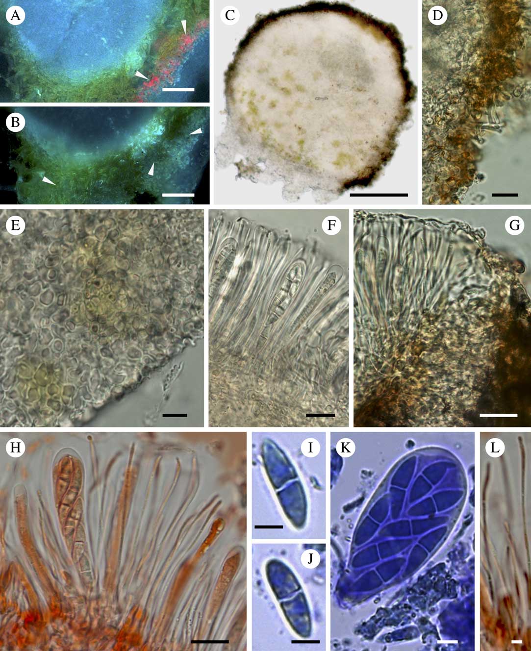
Taitaia aurea (Kirika 5103). A, lower part of ascoma immersed within the host thallus (note fluorescing pulvinic acid crystals using blue fluorescence filter (arrows)); B, same as A, but showing lichenicolous fungal hyphae (arrows) intruding between the hyphae of host fungus; C, cross-section through immature ascoma; D, section of exciple showing protruding hyphal tips; E, section of hyaline hypothecium of textura globulosa type; F, section of hymenium with asci and paraphyses; G, section of borderline between hymenium and exciple; H, section of hymenium with asci and unbranched paraphyses; I & J, ascospores; K, ascus; L, simple septate paraphyses. A–F, in water; G, H & L, in Congo red; I–K, in Cresyl blue. Scales: A, B, D, F–H=10 μm; C=40 μm; E, I–K=5 μm; L=2 μm. In colour online.
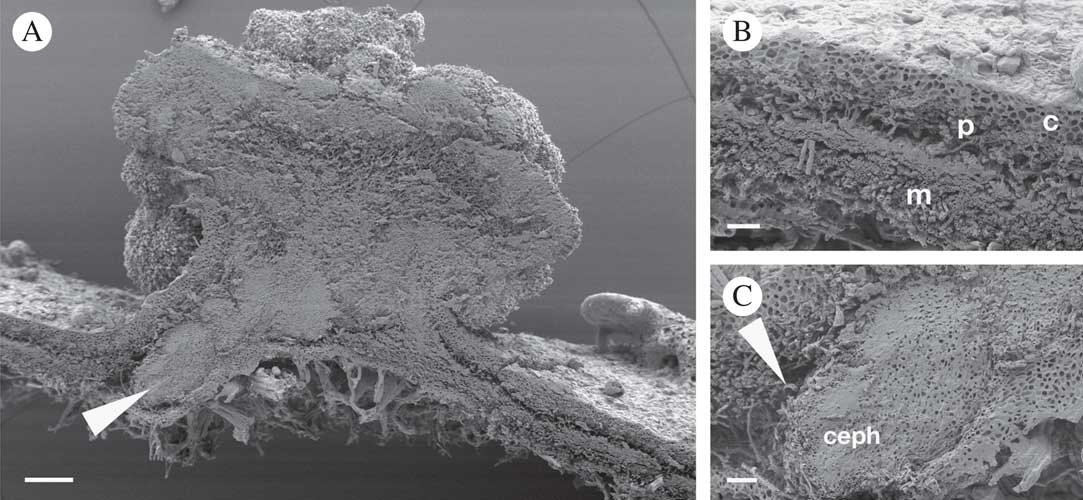
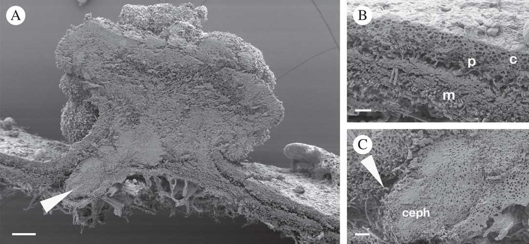
SEM of Taitaia aurea (Rikkinen 15260; see Fig. 3D). A, cross-section through young ascomata and host lichen. Note that the ascomata of Taitaia associate with internal cephalodia (Nostoc) of the host (arrow) and develop through its upper cortex; B, cross-section of the host showing upper cortex (c), green algal photobiont layer (p), medulla (m) and lower cortex; C, cross-section of internal cephalodium (ceph) showing superficial hyphae (arrow) which do not penetrate into the central parts of the cephalodium. Scales: A=100 μm; B & C=20 μm.
Lichenicolous ascomycete, producing ascomata through the upper and lower surface and margins of the thallus of Crocodia cf. clathrata (Figs 1C, 3A & B).
Ascomata apothecioid, sessile, the developing fruiting bodies bursting through the cortex of the host thallus; grouped, sometimes ten or even more ascomata starting from a single base, basally constricted (Fig. 3C & D); diameter of aggregations of immature ascomata 0·4–(0·7±0·3)–0·8 mm (n=19), and aggregations of mature, opened ascomata 0·6–(1·4±0·4)–2·4 mm (n=16; Fig. 3C); hemiangiocarpic, at first closed (Fig. 4C), then opening with the edge rolling outwards exposing a salmon red disc (Fig. 3C), 0·2–(0·3±0·1)–0·7 mm (n=20). True exciple well developed, more-or-less concolorous with the yellow medulla of the host lichen (Fig. 3D), textura globularis-type (Fig. 4E), c. 10–15 µm wide. Exciple surrounding hymenium and hypothecium impregnated with dark yellowish to brownish pigment grains (Fig. 4C, D & G), this pigment does not dissolve in either K or N, and does not autofluoresce, (Fig. 4A), and is distinct from pulvinic acid that gives the yellow colour to the host thallus (pinkish fluorescence in Fig. 4A); the hyaline hyphal tips of the external excipular cells extend out of the margin (Figs 4D & 5A) revealing the mealy appearance of the ascomatal margin (Figs 3D & 5A). Hymenium c. 35 µm high, hyaline (Fig. 4F), without hymenial gel, the upper part covered by an amorphous hyaline layer, K−, K/I−, CRB+ dark blue; subhymenium slightly yellowish, textura intricata (Fig. 4F). Hypothecium hyaline, c. 160 µm (until the pigmented rim), similar in structure to exciple (i.e. textura globularis-angularis type) (Fig. 4E), K−, K/I−, CRB+ pale blue; sometimes with clumps of yellow pigment grains (Fig. 4C & E). Paraphyses irregularly septate, unbranched (Fig. 4H & L) or only dichotomously branched at the base, lax (no hymenial gel), c. 1 µm wide, not or only very slightly widened at tips, remaining hyaline, but tips surrounded by a hyaline amorphous substance (forming a layer over the hymenium), CRB+ dark blue; contents of paraphyses in Congo red+ reddish (Fig. 4H & L), CRB+ blue. Asci functionally unitunicate, clavate, 40–(44·8±5·6)–55×10–(13·2±3·5)–20 µm (n=10); apical ascus wall not widened or slightly widened, non-amyloid (K/I−) without distinct apical apparatus; the ascus base distinctly narrowed; 8 biseriate spores (Fig. 4H & K). Ascospores 2-celled, hyaline, fusiform, spore tips attenuated, not constricted or only slightly constricted at septum, symmetrical or sometimes asymmetrical (one cell longer than the other), 14–(15·8±1·5)–19×4–(4·8±0·8)–7 µm, l/w=2·1–4·5 (n=20), with perispore <1 µm, which is Congo red– and CRB− (Fig. 4I & J); intracellular matrix of ascospores Congo red+, CRB+ (Fig. 4I–K). Vegetative hyphae hyaline, non-amyloid, I−, K/I−, protruding between the medullary hyphae of the host (Fig. 4B). Asexual morph not observed.
Etymology. The genus name refers to the type locality, Taita Hills in Kenya; the epithet ‘aurea’ refers to the golden yellow colour of the ascoma margin.
Taxonomy. Monotypic genus in Gomphillaceae (Ostropales, Ostropomycetidae, Lecanoromycetes, Ascomycota).
Ecology and distribution. Obligately lichenicolous, growing on thalli of Crocodia cf. clathrata (Peltigerales). The ascomata can burst through the host thallus but no obvious damage to mycobiont or photobiont was detected by microscopic inspection. On the lower surface of the host the ascomata usually develop through pseudocyphellae (pores); however, on the upper surface and thallus margins they might develop preferentially at locations where the cortex of the host has been damaged. Inside the host, the vegetative hyphae of Taitaia are slightly narrower than those of the host (Fig. 5A & B) and can be followed for some distance between the medullary hyphae of the host (Fig. 4B). The ascomata of Taitaia are closely associated with the internal cephalodia of the host (Figs 3D & 5A) that contain a nitrogen-fixing cyanobacterium (Nostoc). In cephalodiate lichens, the green-algal photobiont typically produces most photosynthate and the cephalodial cyanobiont mainly fixes nitrogen (Cornejo & Scheidegger Reference Cornejo and Scheidegger2013; Rikkinen Reference Rikkinen2017). While vegetative hyphae of the fungus are seen on cephalodial surfaces, they do not appear to penetrate into these structures.
The new taxon is known from two localities in tropical lower-mountain forests in Kenya (East Africa).
Additional specimens examined. Kenya: Taita-Taveta Province: near summit of Vuria, dense moist forest with Maesa lanceolata, Nuxia congesta and Dracaena afromontana, UTM 421443, 9623094, 2072 m, on thallus of Crocodia cf. clathrata growing on stem of woody climber, 2015, J. Rikkinen 16260 (TU 56327; DNA sample: AF260). Rift Valley Province: Cherangani Hills, Kapcherop, along Chepkait River, riverine in disturbed montane forest, 01°02'N, 35°19'E, 2200 m, on Crocodia cf. clathrata, on bark, 2016, P. Kirika 5103 (EA; DNA sample: AF275).
We thank the staff of the Taita Research Station of the University of Helsinki. The fieldwork in Taita Hills was carried out under the National Council for Science and Technology, Kenya, permit no. NCST/RCD/17/012/33. Rasmus Puusepp is thanked for molecular work, Bellis Kullman (Tartu) for help with fluorescence microscopy, Alexander R. Schmidt and Dorothea Hause-Reitner (Göttingen) for the collaboration and assistance with light microscopy and SEM imaging, and Raquel Pino-Bodas for supplying sequences from Corticifraga peltigerae. We are grateful to the reviewers for their criticism of the manuscript. The financial support for AS was provided by IUT 20-30 and by the European Regional Development Fund (Centre of Excellence EcolChange). The project has also received funding from the European Union’s Horizon 2020 research and innovation programme under grant agreement no. 705777. We also gratefully acknowledge financial support from the Academy of Finland (TAITAWATER 2012–2016) and the Department of Biosciences, University of Helsinki.