Tracheal intubation and mechanical ventilation are life-saving interventions to address respiratory failure, but adverse events occur during the course of mechanical ventilation that can result in prolonged critical illness and increased risk of death. Traditional surveillance has focused on infectious causes of adverse events, particularly ventilator-associated lower respiratory tract infection (VA-LRTI), including ventilator-associated pneumonia (VAP) and tracheobronchitis. But other adverse events, such as congestive heart failure or septic shock, can also result in the need for increased ventilator support.
In 2013, the Centers for Disease Control and Prevention (CDC) National Healthcare Safety Network (NHSN) implemented a new surveillance strategy for adverse ventilator-associated events (VAEs) that advanced a broader, more objective definition of ventilator-associated complications (VACs).Reference Klompas1–Reference Magill, Rhodes and Klompas3 We considered VACs in 3 tiers: (1) VACs without signs of infection; infection-associated VACs (IVACs), which are accompanied by leukocytosis or leukopenia or fever or hypothermia as well as altered antibiotic treatment; and (3) possible VAP (ie, PVAP), which are IVACs with sputum studies suggestive of VA-LRTI. The NHSN definitions of VAE have been well validated to predict ventilator-associated 'morbidity and mortality.Reference Kobayashi, Uchino, Takinami and Uezono4–Reference Klompas, Li, Menchaca and Gruber6 However, current VAE surveillance definitions capture a different population than clinically defined VA-LRTIs, which are typically diagnosed by physical examination findings such as sputum production, laboratory test results, radiographic findings, and microbiologic test results.Reference Fan, Gao, Wu, Zhang, Zhu and Xiong7–Reference Vaewpanich, Akcan-Arikan, Coss-Bu, Kennedy, Starke and Thammasitboon18
Sputum production is a physical examination finding that is often used in the clinical diagnosis of a VA-LRTI, but its relationship to a VAE defined by CDC NHSN criteria is unknown. It is challenging to quantify sputum production during mechanical ventilation, but an indicator of sputum production is the frequency with which subglottic suction is performed. Subglottic suction is a procedure performed on an as-needed basis by nurses, respiratory therapists, and physicians to clear lower airway secretions via the endotracheal tube and to reduce the risk of a VA-LRTI.Reference Damas, Frippiat and Ancion19–Reference Mao, Gao and Wang21 We sought to evaluate the relationship between the quantity of sputum produced, measured by the frequency of subglottic suctioning, and VAEs. Specifically, we sought to define how subglottic suction frequency in the immediate pre-VAE period relates to subglottic suction frequency in the preceding period. We also sought to determine whether sputum quantity measured by subglottic suction frequency discriminates between VACs and IVACs or VAPs. Here, we report the results of a case-crossover study designed to address these questions.
Materials and methods
Study design
We performed a case-crossover study to define the relationship between the quantity of sputum production (primary exposure), measured as the frequency with which nurses and respiratory therapists chose to perform as-needed subglottic suctioning, and the occurrence of VAEs (primary outcome). We also analyzed whether each VAE was categorized as a VAC without suspected infection or as an IVAC or PVAP. For the primary analysis, the day of the VAE served as the case period. The days prior to the VAE during which the patient could have been diagnosed with a VAE [ie, after at least 2 days of stable or decreasing positive end-expiratory pressure (PEEP) and fraction of inspired oxygen (FiO2) requirements] served as the control period. As sensitivity analyses, we treated the day before the VAE as the case period, or 2 days before the VAE as the case period, with the antecedent days during which a VAE might have been diagnosed as the control period.
Study setting
We evaluated 90 consecutive VAEs that occurred across 87 unique patients over 3.5 years in the medical intensive care unit (MICU) of the Hospital of the University of Pennsylvania (HUP), a tertiary-care academic medical center. All patients who depend on mechanical ventilation at HUP were monitored for VAEs, and all VAEs were reported to the NHSN. VACs and IVACs were captured by a review of the HUP NHSN reporting records. The study was approved by the University of Pennsylvania Institutional Review Board (protocol no. 824613) with a waiver of informed consent.
Study population
Patients were eligible for inclusion if they had been admitted to the HUP MICU, had required mechanical ventilation for >2 calendar days (ie, at risk for VAE), and had experienced a VAE event within the enrollment period. We chose a case-crossover design and mixed-effects modeling approach to account for the heterogeneity of lung disease and other medical comorbidities in this critically ill population.
Clinical data collection
The VAE dates and VAC versus IVAC categorization were obtained from NSHN reporting records. Patient demographics, medical diagnoses, laboratory, and microbiology data were obtained from the Penn Data Store, a repository of electronic health record data. Subglottic suction events were extracted from daily flow sheets that report all activity related to mechanical ventilation.
Definition of exposures
The primary exposure was the quantity of sputum production, defined as the number of as-needed subglottic suction events performed per calendar day. Calendar days were used because VAE (the outcome of interest) are adjudicated per calendar day.
Definition of outcomes
The primary outcome was VAE, and we compared the frequency of subglottic suction on the day of VAE and in the preceding 48 hours to the pre-VAE period. To evaluate whether quantity of sputum discriminates between VAC and IVAC, we similarly compared subglottic suction frequency across VAE categories.
Precision and sample size
We determined the necessary sample size by specifying the desired precision with which to detect the effect of sputum quantity to discriminate the pre-VAE and VAE risk periods.Reference Rothman and Greenland22 We anticipated that, with 85 patients, we could detect an absolute risk difference attributable to sputum quantity of 15% with posterior certainty of 4%–26%. With the same number of patients, we anticipated detecting a 25% absolute risk of IVAC versus VAC attributable to sputum quantity, with 95% posterior certainty of 4%–45%.
Statistical methods and models
We organized data using R version 3.6.1 statistical software, and we generated plots using the “ggplot2” package.23 We evaluated potential confounders by comparing clinical variables of interest across exposure groups (ie, high vs low frequency of subglottic suction at onset of mechanical ventilation) using Wilcoxon rank-sum testing for continuous variables and the Fisher exact test for categorical variables. For the primary analysis, we performed Bayesian multilevel logistic regression with random, patient-level slopes and intercepts to address the possibility of confounding by patient-level factors. Like conditional logistic regression, this approach accounts for patient-level strata in the case-crossover design.Reference Maclure24–Reference Rice27 To permit partial pooling of parameter estimates and rigorous evaluation of model fit, Bayesian models were fit using Stan Hamiltonian Monte Carlo (HMC) version 2.19 via the “rethinking,” “rstan,” and “brms” packages.Reference Bürkner28–Reference McElreath30 Models were fit with 4 chains of 2,000 iterations and were then confirmed using HMC diagnostics (no divergent iterations, no iterations saturating maximum tree depth, Rhat statistic < 1.1 for all parameters, and E-BFMI > 0.2) and by examining the posterior distributions.Reference Gabry, Simpson, Vehtari, Betancourt and Gelman31 We characterized posterior credibility intervals and type S error.Reference Gelman and Tuerlinckx32
Availability of data
The data, model code, and code used to produce the figures are available online at https://github.com/bjklab/Subglottic-suction-frequency-and-adverse-events-during-mechanical-ventilation.
Results
Sputum quantity at onset of mechanical ventilation does not predict time to VAE
We evaluated 90 consecutive VAEs, which occurred across 87 unique patients. For patients with >1 VAE, we considered only the first VAE, so 87 VAEs (47 VACs without associated infection; 40 IVACs, including 12 PVAPs) were included in the final analysis. The median number of subglottic suction events at onset of mechanical ventilation (ie, first full calendar day on ventilator) was 7. We first compared clinical characteristics across 39 patients with high (ie, greater than median) sputum quantity and 48 patients with low (ie, less than or equal to median) sputum quantity at the onset of mechanical ventilation (Table 1). We noted no significant differences in patient demographics, medical history, or features of critical illness at onset of mechanical ventilation. We did note a disparity in bacterial growth from respiratory culture in the 7 days prior to mechanical ventilation onset: only 1 (2.1%) of the patients with low sputum quantity had had recent bacterial growth from respiratory culture, versus 5 (13%) of patients with high sputum quantity. But this difference was not statistically significant. We next evaluated the sputum quantity at onset of mechanical ventilation as a predictor of time to VAE. A Weibull parametric survival model showed no significant differences in time to VAE attributable to sputum quantity (Supplementary Fig. 1 online).
Comparison of Clinical Characteristics Among Patients With High or Low Sputum Production at Onset of Mechanical Ventilation

Note. COPD, chronic obstructive pulmonary disease; MV, mechanical ventilation; VAE, ventilator-associated event; IVAC, infection-associated ventilator-associated complication; PVAP, possible ventilator-associated pneumonia; WBC, white blood cell count; Cr, creatinine; AST, aspartate aminotransferase; ALT, alanine aminotransferase; PEEP, positive end-expiratory pressure; IV, intravenous; IQR, interquartile range.
a Continuous variables are described as median (IQR).
b Categorical variables were compared using the χ2 test or the Fisher exact test; continuous variables were compared using the Wilcoxon rank-sum test.
Sputum quantity increases in the immediate pre-VAE period
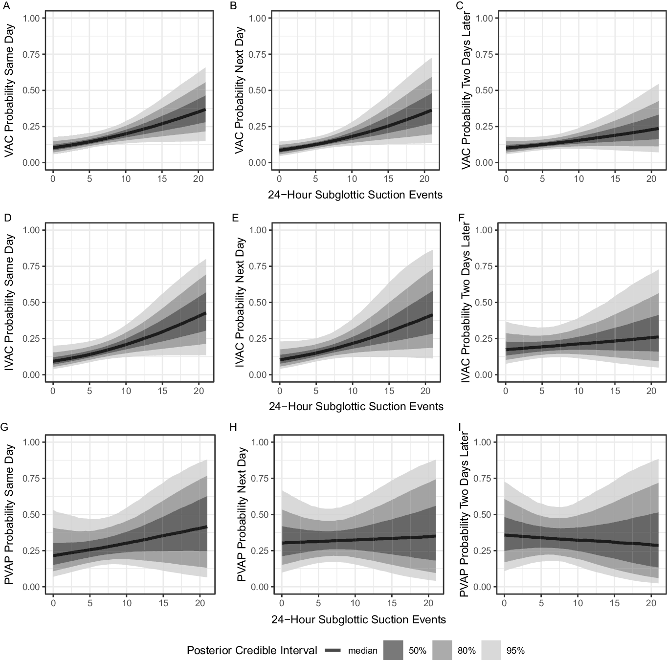
Among the 87 patients in this study, we evaluated 848 ventilator days at risk for VAE (ie, not including the first 2 days of mechanical ventilation). Although sputum quantity at onset of mechanical ventilation was not associated with time to VAE, we did observe an association between increased subglottic suction events and increased risk of both VAC and IVAC. Subglottic suction events across the pre-VAE period and across VAE types are summarized in Supplementary Table 1 (online). Figure 1 shows the binomial model predicted probability of VAC or IVAC based on the number of observed subglottic suction events across all ventilator days at risk for VAE. With subglottic suctioning at 10 events versus 5 events over a 24-hour period, the absolute probability of VAC increased by 3.07% (type S error < 0.024) the same day, by 2.97% (type S error < 0.018) the next day, and by 1.71% (95% CI included 0) the day after. With subglottic suctioning at 10 events versus 5 events over a 24-hour period, the absolute IVAC probability increased by 3.57% (type S error < 0.035) the same day, by 3.43% (type S error < 0.018) for increased risk of IVAC the next day, and by 1.25% (95% CI included 0) the day after. We also evaluated the impact of 15 versus 5 subglottic suction events over a 24-hour period and found that VAC probability increased by 6.75% (type S error < 0.024) the same day, by 6.67% (type S error < 0.018) the next day, and by 3.67% (95% CI included 0) 2 days later. With the same increase in subglottic suction events, IVAC probability increased by 8.1% (type S error < 0.035) the same day, by 7.67% (95% CI included 0) the next day, and by 2.57% (95% CI included 0) 2 days later. Given the small number of observed PVAP events, the posterior credibility intervals were wide. However, we detected a positive relationship between subglottic suction events and same-day PVAP risk, which increased by 2.82% (95% CI includes 0) as subglottic suction events increase from 5 to 10 and by 5.86% (95% CI includes 0) as events increased from 5 to 15.
Increased sputum quantity is associated with increased risk for a ventilator-associated event (VAE). Panels depict the results of random effects binomial regression models relating VAE to the frequency of subglottic suctioning. Panels A, B, and C show the association between increased sputum quantity and absolute probability of a ventilator-associated complication (VAC) on the same day, a VAC on the next day, and a VAC 2 days later, respectively. Panels D, E, and F show the between infection-associated ventilator-associated complication (IVAC) and subglottic suctioning. Panels G, H, and I the association between possible ventilator-associated pneumonia (PVAP) and subglottic suctioning, in the same order. The best estimates of the association are represented with a dark line surrounded by bands of posterior certainty. The left panels show the primary analysis (case period defined as the day of the VAE), and the center and right panels show sensitivity analyses with the case period defined as 1 or 2 days prior to the VAE.

Sputum quantity alone does not discriminate IVAC from VAC
Greater sputum quantity was associated with increased probability of VAC, IVAC, and PVAP. To determine whether sputum quantity could be used to discriminate IVAC from VAC or PVAP from VAC/IVAC (not including PVAP), we evaluated a model comparing subglottic suction events in the periods before IVAC versus VAC, and we evaluated a model comparing subglottic suction events in the periods before PVAP versus VAC/IVAC. No level of subglottic suction events reliably discriminated IVAC from VAC. Furthermore, >15 subglottic suction events was associated with greater probability of IVAC, but this threshold did not have high posterior certainty (Fig. 2). Subglottic suction events did not discriminate PVAP from VAC/IVAC. In an exploratory secondary analysis, we evaluated a multivariable model including subglottic suctioning and other clinical variables that may contribute to respiratory failure to determine features not included in the IVAC definition (ie, excluding temperature and white blood cell count) that discriminate IVAC from VAC. In our cohort of medical intensive care patients, we found that serum liver enzymes, alanine aminotransferase (ALT) and aspartate aminotransferase (AST) best discriminated IVAC and VAC and that elevated enzymes favoring VAC. The odds ratio of IVAC per standard deviation increase in ALT was 0.51 (95% CI, 0.26–0.92). The odds ratio of IVAC per standard deviation increase in AST was 0.56 (95% CI, 0.26–0.996). The odds ratio of IVAC per standard deviation increase in the sum of AST and ALT was 0.54 (95% CI, 0.27–0.98). The number of pressors and the number of antibiotics favored VAC and IVAC, respectively. But unlike AST and ALT, these alone did not discriminate well (Supplementary Fig. 2 online).
Subglottic suction events do not distinguish infection-associated ventilator-associated complications (IVACs) from ventilator-associated complications (VACs). Panels A, B, and C show the results of random effects binomial regression comparing the subglottic suctioning in the pre-IVAC period to the period before a noninfectious VAC. Panels D, E, and F show the pre-PVAP period to the period before the VAC and IVAC (excluding PVAP). The best estimates of the association are represented with a dark line surrounded by bands of posterior certainty. Very frequent subglottic suctioning is associated with IVAC, but no number of observed suction events discriminates IVACs from VACs with high certainty.

The clinical decision to send sputum for bacterial culture depends on fever more than sputum quantity
The association between sputum quantity and VAE risk prompted us to examine the relationship between sputum quantity and the behavior of clinicians. In a multivariable model utilizing all mechanical ventilation days (in contrast to the analyses above, which utilized only the pre-VAE period), we examined the relationship between subglottic suction events, fever, and the decision to order a sputum culture. Whereas fever was associated with the probability of a sputum culture order (odds ratio 1.9; 95% CI, 1.5–2.5), sputum quantity was not (Supplementary Fig. 3 online).
Discussion
Using a case-crossover design, we investigated the relationship between sputum production during mechanical ventilation, measured by subglottic suction frequency, and adverse ventilator-associated events (VAEs) with the goal of understanding whether sputum quantity reliably reflects underlying lung disease that will result in increased oxygen (FiO2 and PEEP) requirement. We quantified sputum production as the number of subglottic suction events per calendar day, and we categorized VAEs using the CDC NHSN definitions of VAC and IVAC. In this cohort of medical intensive care patients with VAEs, sputum quantity measured by subglottic suction frequency was associated with both IVAC and VAC. Although the association with IVAC was stronger, sputum quantity did not discriminate well between infection-associated and noninfectious VAEs. Secondary analyses revealed other variables, prominently AST and ALT, that better discriminated IVACs from VACs in this cohort. The fact that a IVAC was less likely in patients with evidence of livery injury suggests that pulmonary edema may have driven the VAE in these cases. We detected no association between sputum quantity at onset of mechanical ventilation and time to VAE; this finding supports VAE as a sign of new lung pathology that develops during mechanical ventilation rather than progression of pathology already present at time of intubation.
The relationship between sputum quantity and VAE is of interest because sputum quantity is often clinically interpreted as a sign of VA-LRTI, and the relationship between VAE and VA-LRTI remains unclear.Reference Fan, Gao, Wu, Zhang, Zhu and Xiong7–Reference Stevens, Silva and Gillis10 The finding that sputum quantity, measured by subglottic suction frequency, is higher at the time of VAE and immediately before VAE, relative to the earlier pre-VAE period, validates sputum quantity as a physical examination sign associated with risk for worsening respiratory failure. However, sputum quantity did not discriminate well between IVAC and VAC. Greater sputum quantity was associated with VAC without fever or leukocytosis, as well as with IVAC.
There are several possible explanations for the discordance between sputum quantity and the NHSN criteria used to discriminate infection-associated adverse events. One possible explanation is that the number of subglottic suction events does not consistently represent the quantity of sputum produced. Misclassification of sputum production could result in poor discrimination between IVAC and VAC. Another possible explanation is that NHSN IVAC criteria, which are known to exclude a large proportion of clinically diagnosed VA-LRTI,Reference Klompas11 exclude true VA-LRTI events that would drive a stronger association with sputum quantity. A third possible explanation is that sputum production, though commonly interpreted as a clinical sign of infection, is nonspecific, or that sputum quality (eg, purulence) is more specific than sputum quantity. Indeed, we observed that clinician orders for respiratory bacterial culture were not associated with sputum quantity, whereas culture orders were associated with fever. This likely reflects the clinical suspicion for noninfectious etiologies (eg, pulmonary edema) that can also drive increased sputum quantity. Further complicating efforts to use subglottic suction frequency to discriminate IVAC and VAC is the potential protective effect against infection exerted by suctioning.Reference Damas, Frippiat and Ancion19–Reference Mao, Gao and Wang21 Subglottic suctioning may help to prevent some VAEs that it would otherwise herald.
Our study has several limitations. The case-crossover design was chosen because VAEs are rare outcomes, and the exposure of interest—sputum quantity—is dynamic and most likely to impact VAE risk acutely. However, our exclusive focus on VAE cases leaves open important questions about the generalizability of our findings across the broader population of patients who depend upon mechanical ventilation. Likewise, our cohort was enrolled from the medical intensive care unit of a single, academic medical center, and medical histories of patients were limited to International Classification of Disease, Tenth Revision (ICD-10) codes associated with their intensive care unit admission. Sputum quantity may differ significantly in surgical, neurological, or pediatric intensive care patients, and practices for the management of tracheal secretions may differ across institutions. Our findings must be validated with studies of sputum quantity across a broader population of patients dependent on mechanical ventilation.
Nevertheless, our study demonstrates a significant positive association between subglottic suction events and VAE risk, supporting the validity of subglottic suction frequency ascertained from electronic medical records as a means to quantify sputum production. The relationships between the respiratory tract microbiome, lung immune activation, and sputum production during mechanical ventilation warrant further study, as does the impact of sputum production on clinical behavior. The frequency of subglottic suction events appears to serve as a valuable marker of sputum quantity for these future analyses.
In conclusion, the frequency of subglottic suction events, which are performed on an as-needed basis for patients dependent on mechanical ventilation, may serve as a valuable marker of sputum quantity, which is associated with risk for adverse VAEs. However, the relationship between subglottic suction events, sputum quantity, and VAE must be validated in a broader population of mechanically ventilated patients and intensive care settings.
Supplementary material
To view supplementary material for this article, please visit https://doi.org/10.1017/ice.2020.1298
Acknowledgments
We acknowledge the ARES Research Collaborative at the University of Pennsylvania for scientific discussions and support.
Financial support
B.J.K. is supported by the National Institute for Allergy and Infectious Diseases (grant nos. K23 AI121485 and L30 AI120149) as well as the Centers for Disease Control and Prevention (CDC) (contract award nos. BAA 200-2016-91964 and 200-2018-02919). B.J.K. and D.P. are supported by the CDC Healthcare-Associated Infection Prevention Epicenters Program (grant no. U54CK000485).
Conflicts of interest
All authors report no conflicts of interest relevant to this article.