Introduction
The pork tapeworm, Taenia solium, completes its life cycle between pigs (the intermediate host) and humans (the definitive host). This parasite matures exclusively in the human intestine after a human eats uncooked pork contaminated with the T. solium larval stage, cysticercus/cysticerci. Eggs released from mature adult tapeworm(s) in the human intestine can either infect pigs or humans, including the tapeworm carriers themselves. Cysticercosis develops following the accidental ingestion of eggs by pigs or humans. Neurocysticercosis (NCC) is potentially one of the most lethal parasitic zoonoses spreading worldwide. However, most NCC cases are asymptomatic until the parasite becomes damaged by the host defence mechanisms (Schantz et al., Reference Schantz, Wilkins and Tsang1998; Garcia et al., Reference Garcia2016; Pawlowski, Reference Pawlowski2016; Wandra et al., Reference Wandra2016; WHO, 2016).
In working towards the control or prevention of NCC, it is important to conduct field work with a ‘one health’ approach that integrates medical, veterinary and environmental research, i.e. research on pigs for cysticercosis, on humans for both taeniosis and cysticercosis, and on transmission ecology in the affected communities, along with appropriate education for community members. Local people may not realize that their pigs could harbour numerous T. solium cysticerci, which can cause taeniosis in humans. Eggs from an adult tapeworm in the human intestine can cause NCC either in the tapeworm-carriers themselves or in other people who have some intimate contact with the tapeworm-carriers (Schantz et al., Reference Schantz, Wilkins and Tsang1998; Kobayashi et al., Reference Kobayashi2013; Ito and Budke, Reference Ito and Budke2014; Pawlowski, Reference Pawlowski2016; Davaasuren et al., Reference Davaasuren2017). One highly endemic area for this parasite is Asia, including Thailand (Ito et al., Reference Ito, Nakao and Wandra2003, Reference Ito and Budke2014, Reference Ito, Yanagida and Nakao2016; Li et al., Reference Li2006, Reference Li2007; Anantaphruti et al., Reference Anantaphruti2007, Reference Anantaphruti2010; Kobayashi et al., Reference Kobayashi2013; Ito and Budke, Reference Ito and Budke2014; McCleery et al., Reference McCleery2015; Wandra et al., Reference Wandra2016; Davaasuren et al., Reference Davaasuren2017; Wu et al., Reference Wu2017; Sato et al., Reference Sato2018).
In cysticercosis endemic areas, pigs act as intermediate hosts for taeniid cestodes; these animals are often infected with the cysticerci of not only T. solium (definitive host: humans) but also Taenia hydatigena (definitive host: canids) and, less commonly, Taenia asiatica (definitive host: humans). Although humans and domestic animals, i.e. pigs and dogs, cohabit in NCC-endemic communities, especially in remote or rural areas (Ito et al., Reference Ito2002), most reports dealing with swine cysticercosis have ignored this point and failed to account for the possibility of coinfection with T. solium and T. hydatigena and/or infection with T. hydatigena (fig. 1). It is possible that higher numbers of pigs may be infected with T. hydatigena than previously reported (Dermauw et al., Reference Dermauw2016), particularly in areas where local people have ceased defecating outdoors. Any serological detection performed without complementary necropsy evidence could produce a false-positive bias, overestimating the T. solium seroprevalence in instances where some or all of the seropositive cases are caused by T. hydatigena infection. This risk of bias and the importance of necropsy data were previously highlighted by Ito (Reference Ito2013) and Lightowlers et al. (Reference Lightowlers2016).
Model of routes of pig infection with cysticerci of T. solium and/or T. hydatigena in remote or rural areas of developing countries. (1) Humans (H) and dogs (D) have free access to pigs (P) or vice versa, which occurs in the majority of endemic areas, such that pigs become infected with both T. solium and T. hydatigena. (2) Humans, but not dogs, have free access to pigs and vice versa, resulting in pig infection with only T. solium. (3) Dogs, but not humans, have free access to pigs and vice versa, resulting in pig infection with only T. hydatigena.

Based on naked-eye enzyme-linked immunosorbent assays (ELISAs) conducted in the field in Bali, Indonesia, Swastika et al. (Reference Swastika2016) reported that strongly seropositive pigs harboured many T. solium cysticerci or cysticerci from both T. solium and T. hydatigena, whereas weakly seropositive pigs were infected with only T. hydatigena. Here, we applied the same serology evaluation method for screening the swine cysticercosis in backyard pigs from Karen minority villages at the Thai–Myanmar border, with subsequent necropsies of a subset of pigs to confirm our findings with direct evidence of infection.
Materials and methods
Collection of pig sera
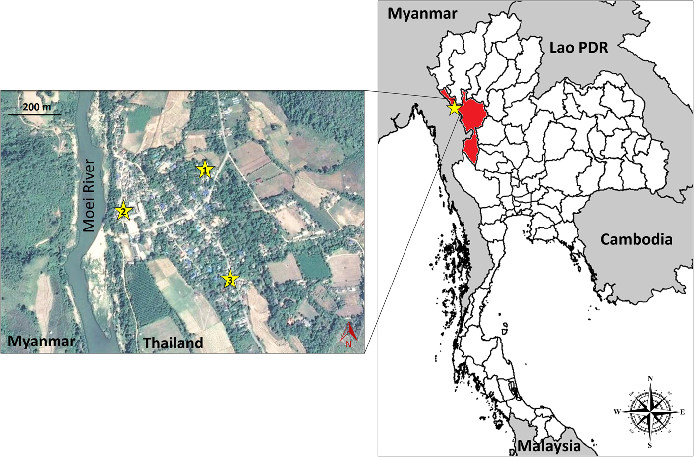
During three visits, in January, April and December 2012, pig serum samples were collected from three hill-tribe (Karen) villages: Bann Nong Bua, Bann Tala Orka and Bann Khue Kho in Tha Song Yang district, Tak province, Thailand (17°20′23.291″N, 98°6′32.579″E) (fig. 2). Blood samples were collected from each pig (c. 1 ml per sample) by jugular or ear vein puncture and left at room temperature for c. 1 h prior to centrifugation for serum separation. All pigs were marked with gentian violet, and detailed information was recorded, including the pig's name, owner's name, and house number, to avoid re-sampling. Attribute data, such as animal sex and age (ranging from 6 months to 3 years old) estimated by owners, were also recorded. Based on age, pigs were categorized as either juvenile (≤ 8 months old) or adult (> 8 months old) (Swastika et al., Reference Swastika2016).
Location of swine cysticercosis sampling sites in Tha Song Yang district, Tak province, Thailand. The surveys of cysticercosis seroprevalence were conducted in three villages: Nong Bua (star 1), Khue Kho (star 2), and Tala Orka (star 3).

Naked-eye and quantitative ELISAs
ELISAs were performed to detect antibody responses against the partially purified low-molecular-weight glycoproteins (LMW-GPs) of T. solium cyst fluid. LMW-GPs were purified by cation exchange chromatography (Sako et al., Reference Sako2013). A brief protocol for the antigenic preparation was described in Swastika et al. (Reference Swastika2016). The protein antigen (100 μl per well of 1 μg/ml stock) was used to coat 96-well microplates; the plates were incubated at 4°C overnight, and then excess antigen was removed by washing with phosphate-buffered saline (PBS) containing 0.1% Tween 20 (PBS-T). Blocking buffer (1% casein in PBS-T) was applied to each well, and the plates were incubated at 37°C for 1 h prior to washing twice with PBS-T. Diluted pig sera (100 μl per well of 1 : 100 serum in blocking buffer) were added to the respective wells and incubated at 37°C for 1 h, after which the wells were washed five times with PBS-T. Protein-G–peroxidase conjugate (used as the secondary antibody) (100 μl per well of 1 : 4000 conjugate in blocking buffer) was added, and the plates were then incubated at 37°C for 1 h before being washed five times with PBS-T followed by a final rinse with PBS without Tween 20. Substrate solution (0.5 mg/ml 2, 2-azino-di (3-ethyl-benzthiazoline-6-sulfonate) (ABTS)) in 50 mm phosphate–citrate buffer (pH 5.0) was added, and the plates were incubated at room temperature for 30 minutes. The reactions were stopped by adding 100 μl per well of 1% sodium dodecyl sulphate (SDS).
In the field, ELISA readings were performed by the naked-eye method. Based on these results, a subset of seropositive (with different ELISA well colour intensities) and seronegative (colourless ELISA wells) pigs were purchased so that we could perform necropsies to obtain direct evidence of infection. Quantitative ELISA readings at an optical density of 405 nm (OD405) that produced absorbance values were subsequently performed at the Department of Helminthology, Faculty of Tropical Medicine, Mahidol University (Bangkok, Thailand). Negative control serum samples from eight pigs raised on a private commercial farm in Nakhon Pathom province (closed-system farming, free of any helminthic infection by faecal examination methods: simple smear and simple flotation techniques) were used for setting the cut-off value, which was defined as the mean absorbance value plus seven times the standard deviation of control sera. Here, the cut-off OD405 was 0.183.
Immunoblot
Serum samples from the 16 necropsied pigs were checked for the presence of antibody responses to the LMW-GPs used for the ELISA (Sako et al., Reference Sako2013). Polyvinylidene difluoride (PVDF) sheets, used as immunoblotting membranes, were prepared with the LMW-GPs following a method previously described by Sako et al. (Reference Sako2013). The membrane was cut into individual strips, rinsed briefly in methanol for 30 s, and washed for 10 minutes in fresh PBS-T. The strips were blocked in blocking solution (1% casein in PBS-T) for 1 h prior to the addition of serum samples diluted 1 : 20 with blocking solution. The samples and membranes were then incubated together at room temperature for 2 h. After washing the strips four times in PBS-T, horseradish peroxidase–protein G conjugate (Thermo Fisher Scientific, Eugene, OR, USA) diluted 1 : 500 in blocking solution was added, and the reaction was allowed to proceed for 1 h at room temperature. The strips were again washed five times in PBS-T and then exposed to the substrate solution (0.5 mg/ml 4-chloro-1-naphthol in 0.015% H2O2) for 30 minutes at room temperature for the development of visible protein bands.
Pig necropsy for confirmation of parasitic infection
A subset of the pigs with positive serological results, with the corresponding ELISA-well colours spanning a range of intensities, (n = 10) as well as some pigs with negative ELISA results (n = 6) were selected for necropsy. With the permission of the pig owners, a total of 16 pigs were bought and subsequently necropsied at the local Primary Health Care Unit. The animal carcasses were inspected intensively for the presence of cysticerci throughout the whole body, including muscle, tongue, eyes and other visceral organs, i.e. brain, diaphragm, mesentery, lung, liver and kidney. Those visceral tissues and striated muscle from several parts of the body were sliced into approximately 5-mm-thick sections to facilitate the investigation process. We spent at least 1 h, and often several hours (depending on the pig size), inspecting each pig and estimating the total cyst number. The gastrointestinal tracts were also examined for intestinal helminth infection.
Statistical analysis
The swine cysticercosis seroprevalences, with confidence intervals of 95%, in the three different villages, were computed using Quantitative Parasitology software, version 3.0 (Rózsa et al., Reference Rózsa, Reiczigel and Majoros2000). The effect of sex (male vs female) and maturity (juvenile vs adult) on the proportion of seropositive pigs was examined using the non-parametric chi-squared function ‘prop.test’ that is embedded in R (R Core Team, 2015).
Results
Serological screening of pigs using ELISAs and immunoblot assays
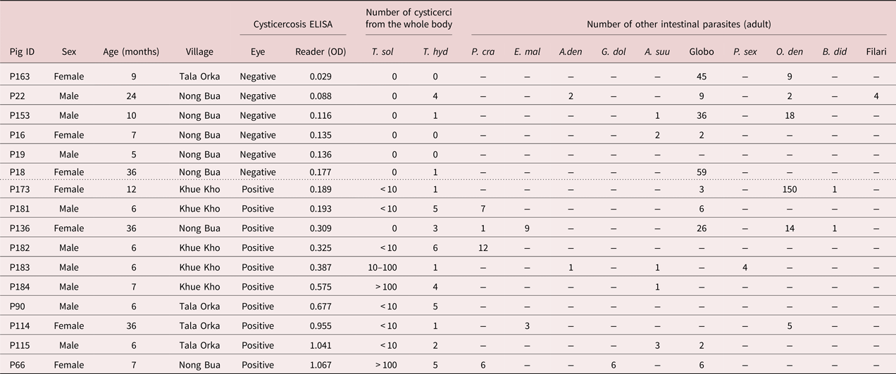
Serological screening of 188 pigs from three hill-tribe villages in Tak province, Thailand revealed that 19.7% of the tested animals had antibody responses to the LMW-GPs. The pigs from two of the villages, Nong Bua (star 1 in fig. 2) and Tala Orka (star 2 in fig. 2), had similar seroprevalence rates, 15.9% and 19.3%, respectively; in contrast, the pigs from Khue Kho village (star 3 in fig. 2) exhibited a much higher seropositivity, 38.9% (table 1). Of the 188 screened pigs, 16 were selected for use in subsequent necropsies to obtain evidence of the presence of cysticerci and to conduct further assessments of their serological data (fig. 3, table 2). Antibody responses to the LMW-GPs were much stronger in pigs that were coinfected with T. solium and T. hydatigena.
Scatter plot of the swine cysticercosis serological screening results. ELISAs to detect swine cysticercosis were performed on serum samples from 188 pigs from hill-tribe villages in Tak province, Thailand. The plot markers with open circles indicate non-necropsied pigs, whereas the results from the 16 necropsied pigs are presented with various black shapes. The dashed line indicates the ELISA cut-off value (OD405 = 0.183), which was calculated from eight parasite-free pigs.

Seroprevalence of swine cysticercosis among three hill-tribe villages in Tha Song Yang district, Tak province, Thailand (fig. 2).

Swine cysticercosis and serological test results of the 16 necropsied pigs. The data are presented from the smallest to largest ELISA OD405 value. Data on other gastrointestinal helminth infections in pigs is also included (Chaisiri et al., Reference Chaisiri2017). The dashed line indicates the ELISA cut-off value (OD405 = 0.183). T. sol = Taenia solium; T. hyd = Taenia hydatigena; P. cra = Pseudanoplocephala crawfordi; E. mal = Echinostoma malayanum; A. den = Ascarop dentata; G. dol = Gnathostoma doloresi; A. suu = Ascaris suum; Globo = Globocephalus sp.; P. sex = Physocephalus sexalatus; O. den = Oesophagostomum dentatum; B. did = Bourgelatia diducta; Filari = Filariidae; Eye = naked-eye ELISA in the field study; reader = quantification of optical density value by an ELISA machine in the laboratory.

In terms of the effects of age and sex on cysticercosis seropositivity, we found no significant difference in proportion of ELISA-based seropositivity between juvenile (24.6%) and adult (14.8%) pigs (χ2 = 1.589, P = 0.207) or between male (17.2%) and female (25%) pigs (χ2 = 0.826, P = 0.363).
To compare the antigen patterns recognized by pigs, either Taenia-infected or non-infected, immunoblot assays were performed on serum samples from the 16 necropsied pigs (fig. 4). Pigs coinfected with T. solium and T. hydatigena clearly showed much stronger banding patterns compared with those infected with only T. hydatigena or with pigs with no cysts. Only one pig (P136) had different results between the ELISA absorbance OD405 and the immunoblot assay. However, a second ELISA analysis revealed P136 as exclusively negative. Overall, the samples with higher ELISA OD405 values tended to have darker bands on the immunoblot strip.
Immunoblot analysis of the 16 necropsied pigs. Immunoblots were performed to access the antibody responses of the 16 necropsied pigs against the T. solium antigens used for the ELISAs. The ELISA absorbance OD405 value corresponding to each pig is in brackets above the blot.

Necropsy
Based on exhaustive work to inspect the whole body of the pig carcasses, examining the presence of cysticerci, ten seropositive pigs were confirmed to be infected with at least T. hydatigena. Among them, nine pigs with OD405 values greater than 0.189 were confirmed to be infected with both T. hydatigena and T. solium, whereas the remaining pig (P136) was infected exclusively with T. hydatigena. However, the sample from P136 showed no bands on the immunoblot strip (fig. 4). The number of T. hydatigena cysts in each pig ranged from one to six. The pig (P136, OD405 = 0.309) infected with three T. hydatigena cysts showed higher antibody responses than only two of the pigs coinfected with T. solium and T. hydatigena (P173, OD405 = 0.189 and P181, OD405 = 0.193). Additionally, two of the three mature pigs, each approximately 3 years old, were free from T. solium cysts, but all three of them were infected with T. hydatigena. The pig with the greatest number of T. hydatigena cysts (P182, OD405 = 0.325) was confirmed to harbour 6 T. hydatigena cysts and < 10 T. solium cysts. The pig with the highest antibody response (P66, OD405 = 1.067) harboured five T. hydatigena cysts and > 100 T. solium cysts. Regarding the six seronegative pigs, none of them showed any evidence of T. solium infection, but three of them (P18, P22 and P153) were confirmed to be infected with T. hydatigena.
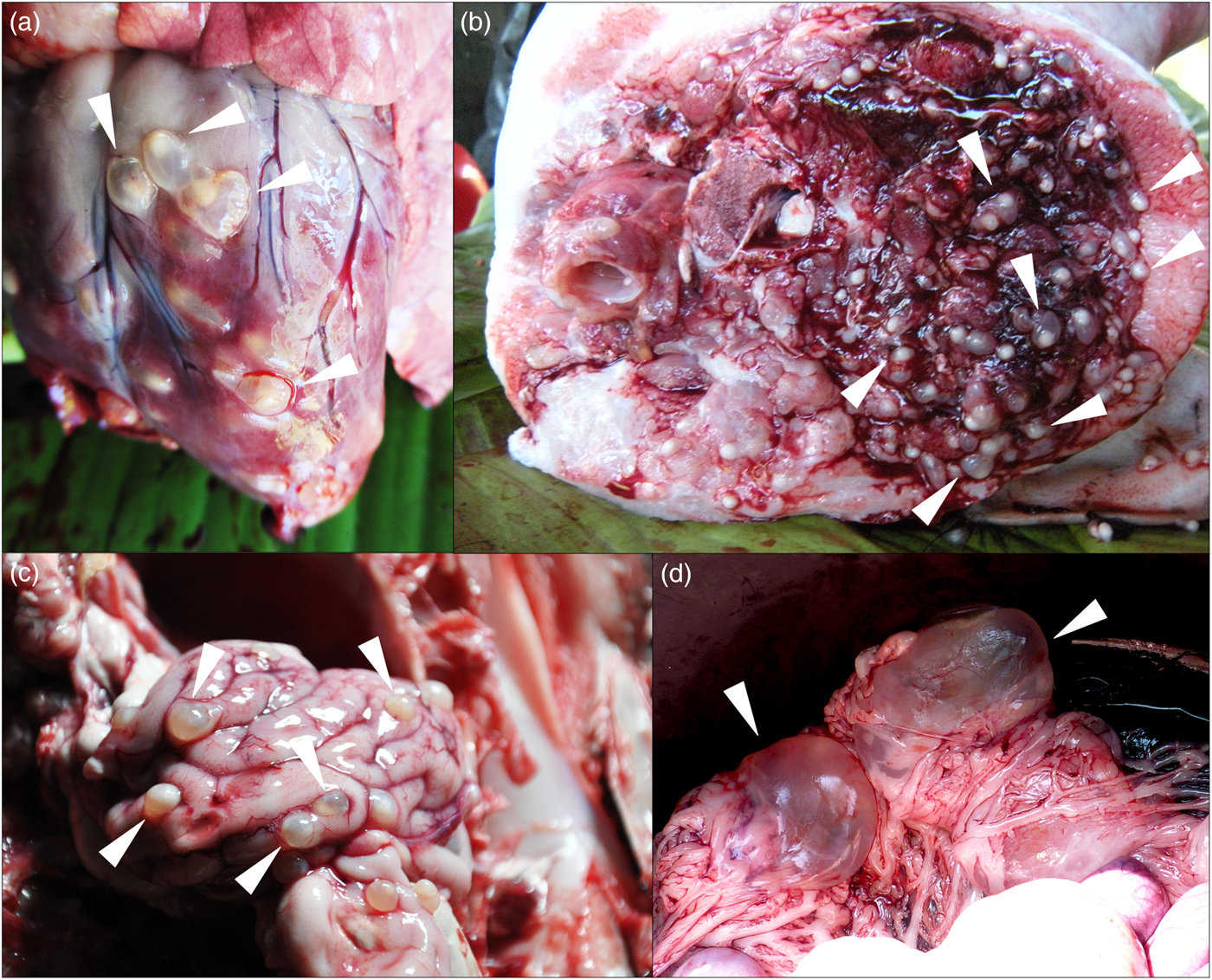
Gross aspects of T. solium and T. hydatigena found in the necropsied pigs are presented in fig. 5. The presence of intestinal parasites was also examined, and the details of the worm recovery for each necropsied animal are presented in table 2.
Cysticerci of T. solium and T. hydatigena in infected pigs. Representative images of cysticerci of T. solium (a: heart, b: muscle, c: brain) and of T. hydatigena (d: mesentery) from the necropsied pigs. Cysticercus of T. solium is transparent cylinder shape < 10 mm in length, as shown in a, b, c. The size of invaginated scolex inside is variable according to the maturity or age of the cysticercus. In contrast, the size of cysticercus of T. hydatigena is generally much bigger but highly variable according to age. It often becomes large, transparent and spherical in shape (2–5 cm in diameter). However, if it is small and similar in size to T. solium, it has no scolex and looks like a transparent round ball.

Discussion
We conducted serological detection of swine cysticercosis in three ethnic minority villages (Karen ethnic group) in a remote area at the Thai–Myanmar border (fig. 2). Our serological screenings indicated the presence of pigs infected with T. solium cysticerci, and this result was confirmed through subsequent necropsy. All pigs confirmed to be infected with T. solium were also coinfected with T. hydatigena cysticerci.
A recent report of a cross-sectional abattoir study in Burkina Faso by Dermauw et al. (Reference Dermauw2016) indicated that 8.8% (40/452) of pigs were infected with T. hydatigena, whereas 2.9% (13/452) were infected with T. solium, and one pig was coinfected with both cestode species. This result contrasts with our finding that all seropositive pigs except one were coinfected with both species. The dissimilarity in the coinfection rates of these two Taenia species between our study and theirs could be explained by several possible factors. For example, the pigs examined in Burkina Faso might have been raised with less free access to dog and human faeces (potentially due to educational level and hygienic practices) compared with the pigs surveyed in the present study. In addition, there may also be some factor of difference in the host specificity between Asian and African T. hydatigena (Braae et al., Reference Braae2015; Nguyen et al., Reference Nguyen2016).
Like our survey, a study by Conlan et al. (Reference Conlan2012) conducted in Lao People's Democratic Republic also found that T. hydatigena was predominant compared with T. solium. Based on observations of naturally Taenia-infected pigs in endemic areas, which suggest that the prevalence of T. solium in pigs can sometimes be much less than that of T. hydatigena (Sikasunge et al., Reference Sikasunge2008; Conlan et al., Reference Conlan2012), there may be some cross-immunity between Taenia spp. (Ito et al., Reference Ito, Takami and Itoh1991; Lightowlers, Reference Lightowlers2010, Reference Lightowlers2013; Lightowlers et al., Reference Lightowlers2015). However, our present observation found that all seropositive pigs except one (P136) were coinfected with both T. solium and T. hydatigena. Therefore, even if there is some cross-immunity induced by infection with a Taenia species, it is unable to protect against infection, at least in the case of T. solium versus T. hydatigena.
Regardless of the immunity effect, the occurrence of coinfection or a single infection may be based on the environmental contamination dynamics with either only a single species (either humans or dogs) or multiple species, including at least humans and dogs (fig. 1). Additional data, specifically direct evidence of infection with Taenia spp., such as that obtained via necropsy, paired with environmental observations concerning whether pigs have free access to human and/or dog faeces (fig. 1), are needed to better address this question (Ito, Reference Ito2013; Ito et al., Reference Ito, Yanagida and Nakao2016).
Meat inspections that focus on the detection of only T. solium in abattoirs have limited value (Phiri et al., Reference Phiri2002) when the potential contamination with T. hydatigena is taken into account. Even in places where some meat inspection is available, no examination of the abdominal cavity and/or viscera for the confirmation of T. hydatigena cysts has been recommended because T. hydatigena has no economic importance.
Although mature pigs (n = 3) were expected to have higher T. solium infection rates based on their greater opportunity to become infected due to their age, only one of the three mature pigs infected with T. hydatigena was coinfected with T. solium. It is possible to explain that those pigs were kept in pig pens where they had no opportunity for free roaming and less opportunity to access human faeces. This suggests that pigs of all ages should always be kept in pens to limit the chance of parasitic exposure and block the parasitic life cycle.
There have been many serological reports on swine cysticercosis in endemic areas, but most were based only on serology, without direct evidence of parasitic infection from necropsy confirmation (Dorny et al., Reference Dorny2004; Krecek et al., Reference Krecek2008; Deckers and Dorny, Reference Deckers and Dorny2010; McCleery et al., Reference McCleery2015; Dermauw et al., Reference Dermauw2016; Kungu et al., Reference Kungu2017). Furthermore, most studies on swine cysticercosis have ignored the notable factor of pigs and dogs typically being free roaming in endemic areas, therefore those pigs are frequently coinfected with both T. solium and T. hydatigena (Ito, Reference Ito2013; Swastika et al., Reference Wandra2016). Thus, an evaluation of the endemic area environment must be prerequisite for any field work on swine cysticercosis. Notably, these coinfections and/or infection with other Taeniid species may complicate the serology of swine cysticercosis in rural or remote areas because there is currently no available method of conclusively confirming parasitic infection without necropsy (Ito, Reference Ito2013; Ito et al., Reference Ito2014; Lightowlers et al., Reference Lightowlers2016; Swastika et al., Reference Swastika2016; Muro et al., Reference Muro2017).
Compared with a previous study in Indonesia (Swastika et al., Reference Swastika2016), our ELISA and immunoblot results show an improved ability to identify infection, but it is still not perfect, as confirmed by the necropsy results. The ELISA OD450 values of pigs coinfected with T. solium and T. hydatigena cysticerci were generally higher and could be used to discriminate the coinfected pigs from the cyst-negative or T. hydatigena-infected pigs, except for the case of P136, which was exclusively infected with T. hydatigena but had one positive ELISA result (fig. 3 and table 2). Additionally, antibody responses against the cysticerci were prominently detected by the immunoblot assay. The sera of pigs coinfected with T. solium and T. hydatigena exhibited strong antibody–protein reactions; in contrast the sera of pigs infected with only T. hydatigena produced no clear bands on the immunoblot membrane, even sera from P136, which produced one positive ELISA result (fig. 4). The immunoblot result for P136 is our first experience of a serum sample with a reasonably high ELISA OD405 value from a necropsy-confirmed T. hydatigena-positive case showing no bands in an immunoblot. It is possible that this result may be a product of some technical error.
Our serological results contrast with the serology reported for a study in Bali in which all pigs harbouring T. hydatigena had positive ELISA results (Swastika et al., Reference Swastika2016). The pigs confirmed to be infected with T. hydatigena in Bali may have been coinfected with T. solium but were falsely judged not to be infected with this species due to harbouring only a very small number of cysts and/or due to the < 1 mm size of T. solium immature cysticerci, which have not yet developed an anlagen or scolex, and are difficult to detect with the naked eye even during a thorough necropsy. To avoid contradictory or inaccurate results, the molecular detection of T. solium and/or T. hydatigena using pig meat without any macroscopic evidence of T. solium infection may be necessary in future studies. To our knowledge, there remains no truly species-specific serology available for cysticercosis in pigs (Sciutto et al., Reference Sciutto1998; Ishida et al., Reference Ishida2003; Ito and Craig, Reference Ito and Craig2003; Decker and Dorny, Reference Deckers and Dorny2010; Abuseir et al., Reference Abuseir2013; Ito et al., Reference Ito, Yanagida and Nakao2016; Swastika et al., Reference Swastika2016; Muro et al., Reference Muro2017). A big limitation of the present work is that we did not detect any pigs that were infected exclusively with T. solium; all the studied pigs that were infected with T. solium were also coinfected with T. hydatigena.
Based on our present study, serum samples from pigs infected with T. hydatigena may produce negative immunoblot results when using LMW-GPs purified by cation exchange chromatography (Sako et al., Reference Sako2013). A similar analysis was previously carried out using LMW-GPs (10–26 kDa) prepared by preparative isoelectric focusing (PIEF) (Ito et al., Reference Ito1998, Reference Ito1999; Yang et al., Reference Yang1998; Sato et al., Reference Sato2003). Ito et al. (Reference Ito1999) showed the usefulness of LMW-GPs for the screening of pigs in endemic areas in Mexico, China and Indonesia where pigs and dogs were living outdoors together. Additionally, Subahar et al. (Reference Subahar2001) and Ito et al. (Reference Ito2002) used LMW-GPs (10–26 kDa) to confirm T. solium infection in pigs and dogs that were naturally infected in Papua, Indonesia. Therefore, further studies using LMW-GPs from these two species are crucial for the evaluation of species specificity in serology. Our previous work and the results of this study indicate that if pig samples produce an OD405 value of > 0.4 in an ELISA based on LMW-GPs, the corresponding pigs are expected to harbour T. solium cysticerci, at least in Tak or in Bali (Swastika et al., Reference Swastika2016).
Pigs from three villages in Tak were estimated to be highly contaminated with both Taenia cysticerci and other intestinal helminths (table 2) (Chaisiri et al., Reference Chaisiri2017). However, intestinal parasites, including Pseudanoplocephala crawfordi (adult cestode in the small intestine) and Echinostoma malayanum (adult trematode in the small intestine) are unlikely to have affected the antibody responses measured by our ELISAs.
Given the difficulty of looking for T. solium cysts in whole pig carcasses, particularly when those pigs are infected with a very small number of early-developmental-stage parasites as discussed above, there can be technical issues with the necropsy procedure. Lingual examination is currently considered to be the most reliable screening method for swine cysticercosis without necropsy (Sikasunge et al., Reference Sikasunge2008). Positive serological results in pigs with nodules on their tongues may be reasonably reliable; however, one report indicates that many cysticercotic pigs lack nodules on the tongue (Sikasunge et al., Reference Sikasunge2008). The most concerning problem is the uncertainty of how to assess pigs in which the tongue examination was negative. There are a huge number of publications dealing with Ag-ELISAs and tongue examinations that stress the usefulness of Ag-ELISAs. However, it is now very clear that many pigs are infected with T. hydatigena rather than T. solium or are coinfected with both species. Because the available serological tests are probably not species specific, Ag-ELISA positive results may be caused by T. hydatigena rather than T. solium in environments where dogs have free access to pig farming (Sciutto et al., Reference Sciutto1998; Ishida et al. Reference Ishida2003; Ito and Craig, Reference Ito and Craig2003; Abuseir et al., Reference Abuseir2013; Ito, Reference Ito2013; Ito et al., Reference Ito2014, Reference Ito, Yanagida and Nakao2016; Swastika et al., Reference Swastika2016; Muro et al., Reference Muro2017). Pigs infected with T. hydatigena, other Taenia spp., or even Echinococccus spp. may produce positive results in these Ag-ELISA tests. Therefore, we would like to reemphasize that it is impossible to conclude anything about the swine cysticercosis status of an animal with certainty in the absence of direct evidence from necropsy (Dorny et al., Reference Dorny2004; Dermauw et al., Reference Dermauw2016) and that results from environments potentially contaminated with T. solium (human faeces) and/or T. hydatigena (dog faeces) in endemic areas must be considered with caution (fig. 1).
Prior to the application of widespread serological detections of swine cysticercosis, studies confirming parasite infection by necropsy are crucially important, particularly to assess any serological cross-reactivity of T. solium and other Taenia spp. in an endemic area. Further studies aimed at improving the screening of pigs infected with T. solium are necessary to determine means of differentiating them from pigs infected with T. hydatigena. It may be possible by introduction of ELISA using cyst fluid of T. hydatigena.
Acknowledgements
The authors would like to thank the local administrations in Tha Song Yang sub-district, Tak province (i.e. Nong Bua Health Promoting Hospital, Tha Song Yang District Health Office, and Tha Song Yang District Livestock Office) for their kindness and facilitation in the field work and data collection. We also sincerely thank the local health volunteers and villagers for their cooperation and responses. We thank Katie Oakley, PhD, from Edanz Group (www.edanzediting.com/ac) for editing a draft of this manuscript.
Financial support
This study was supported by Grants-in-Aid for Scientific Research from the Japan Society for the Promotion of Science (JSPS) (24256002 for A. Ito; 24406011 for M. Okamoto), the Asia/Africa Science Platform Fund from JSPS, and the Special Cooperation Fund for Promoting Science and Technology, Ministry of Education, Japan for A. Ito.
Conflict of interest
None.
Ethical standards
All experimental procedures involving pigs, including permission for animal restraint, blood collection, serological screening procedures, and necropsy, complied with the Institutional Animal Care and Use Committee (IACUC) guidelines and were approved by the district livestock office (document number TMHM 0517.116/00442). Animal euthanasia strictly followed the traditional method of the local Karen minority.