Introduction
In the UK, prostate cancer is the most common cancer in men, accounting for 47,200 new cases in 2015.1 In the UK at present, one in eight men will be diagnosed with prostate cancer during their lifetime, and between 2014 and 2035 the incidence of prostate cancer is predicted to rise by 12%.1 At the end of 2010, an estimated 280,500 men in the UK who had previously been diagnosed with prostate cancer were alive.1 It is estimated that all men with early-stage prostate cancer will survive their disease for 5 years or more.1 However, this level of successful treatment means more people are now living with the side effects of treatment. When limiting analysis to inverse intensity-modulated radiotherapy (IMRT) planned patients (337) from the large-scale study of prostate patients, who were assessed as being low-risk (LR) of prostate radiotherapy CHHiP (conventional or hypofractionated high-dose intensity-modulated radiotherapy for prostate cancer) trial conducted across the UK, showed acute bowel toxicity ≥grade 2 in 21% and acute bladder toxicity ≥grade 2 in 46% of patients.Reference Naismith, Griffin and Syndikus2
The challenge involved with radical prostate radiotherapy is delivering high dose to the clinical target volume (CTV), while minimising irradiation to the bladder and rectum due to their close proximity to the prostate. Rectal spacers have been developed to increase the distance between the prostate and the rectum, which in theory allows for reduced radiotherapy dose received to the rectum. Several studies reported on the use of a polyethylene glycol (PEG) hydrogel spacer (HS) to reduce radiation toxicity to the rectum. HS remains in the body throughout the radiotherapy course for approximately 3–6 months, gradually liquefying, via hydrolysis, to be absorbed and cleared via renal filtration.3 It is inserted through a targeted aspiration needle mid-gland in the perirectal fat space (PRS), image-guided by transrectal ultrasound. HS is approved by the US Food and Drug AdministrationReference Hamstra, Mariados and Sylvester4 and is now established globally into multiple national guidelines, including the National Institute for Health and Care Excellence guidelines5 for the use of biodegradable spacers for prostate cancer radiotherapy including both external beam radiotherapy (EBRT) and brachytherapy.
HS is most commonly used in prostate cancer patients with LR cancer receiving a dose and fractionation of 79·2 Gy in 44 fractions, as demonstrated in the phase 3 randomised controlled PIVATOL trial in the United States.Reference Hamstra, Mariados and Sylvester4 However, there are other risk factors which influence adverse effects from radiotherapy. This can be through the form of other patient-specific risk factors (such as diabetes, previous abdominal surgery, use of anticoagulants, anti-hypertensives), or for high-risk (HR) patients when radiotherapy is combined with androgen deprivation therapy (ADT) or when pelvic lymph-node irradiation or dose-escalation is used.Reference Vanneste, Hoffman and van Lin6 Extending the use of spacers beyond LR patients, there are published results for HS insertions with non-conventional dose-escalation radiotherapy techniques such as stereotactic ablative body radiotherapy (SABR), intensity-modulated proton therapy (IMPT) and brachytherapy. The Royal College of Radiologists acknowledges sufficient evidence supports the theory that prostate cancer has a low alpha/beta ratio and therefore, hypofractionation of >2 Gy per fraction may be radiobiologically advantageous.7 However, the national comprehensive cancer network (NCCN) in the United States recommends prohibiting the use of HS for prostate cancer malignancies which have obvious rectal invasion or visible T3 tumours and posterior extensions.8
The aim of this literature review is to evaluate the quality of rectal (HS) insertions in patients undergoing radical radiotherapy for prostate cancer. The secondary aim is to assess the benefit of HSs in patients with risk factors more likely to have rectal complications, such as non-conventional radiotherapy dose fractionations and HR disease.
Method
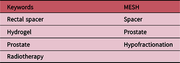
A literature search was carried out in June 2019, using a search of electronic databases including Medline, Cinahl and ScienceDirect using the search terms listed in Table 1.
Search keywords and MESH used as part of the literature review

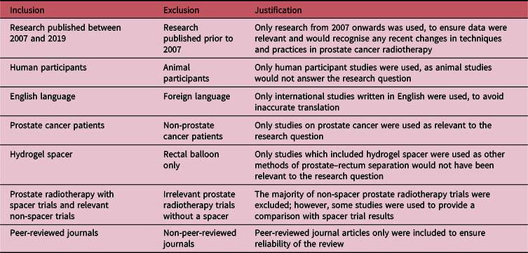
The Boolean operator ‘and’ combined the keywords with Medical Subject Heading (MeSH) to generate articles. Further articles were gained from snowballing the articles generated and using relevant grey literature. Abstracts were reviewed and inclusion/exclusion criteria (Table 2) applied to determine the final articles to be used as part of the literature review.
Inclusion/exclusion criteria to determine relevant research articles

Results
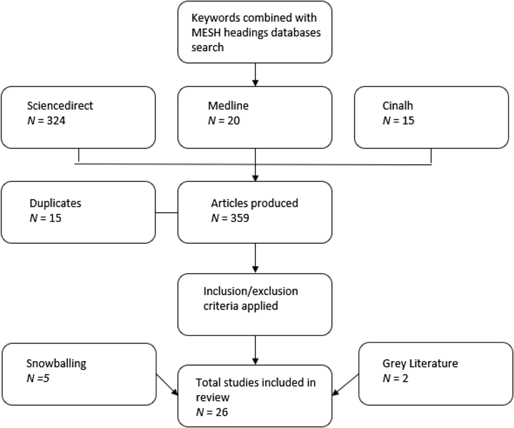
The keyword searches provided 359 articles of which 15 were duplicates. Applying the inclusion/exclusion criteria and focusing on the literature related to the research question led to a total of 26 articles being included as part of the literature review. This included literature from snowballing and grey literature (see Figure 1). The findings from these articles are evaluated in the discussion section under trends related to the research question.
Numbers of articles resulting from database searches.

There were two conflicts of interest declared within the studies: two authors from the PIVATOL trialReference Hamstra, Mariados and Sylvester4 who invested in SpaceOAR™ (Augmenix Inc., Waltham, MA, USA), and others who provided them with consultant services. These are highlighted as this indicates a possible source of bias to their research. However, this would be limited due to the strength of the trial being randomised controlled.
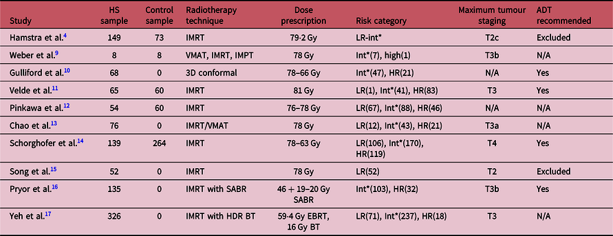
The inclusion criteria included a range of HS trials.Reference Weber, Zilli and Vallee9–Reference Yeh, Lehrich and Tran17 Table 3 provides the details of these trials, showing a range of use of radiotherapy techniques and patient staging. The use of ADT was not always mentioned. However, the theme seemed to be exclusion of LR patients and recommendation for intermediate/HR patients.
Details of HS trials

Int* = intermediate.
Discussion
Spacer impact on rectal dose
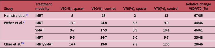
It has been well established that HS reduces the amount of rectum receiving high doses from radiotherapy (Table 4).5,Reference Weber, Zilli and Vallee9,Reference Chao, Ho and Chan13 This is considered a significant factor in late rectal bleedingReference Fiorino, Valdagni and Rancati18 where a systematic review showed rectal planning dose volume, V70 below 25% significantly reduces the incidence of late rectal bleeding. Since this review and with the introduction of inverse-IMRT planning, rectal V70 and V60 constraints have been reduced to below 15–20% and 50–60%, respectively.Reference Dearnaley, Syndikus and Mossop19 Table 4 demonstrates how HS was able to keep rectal volumes significantly within recommended constraints and with a large reduction when compared to no HS insertion.
The percentages of rectal volumes receiving 50–80 Gy (4)

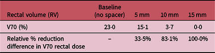
The amount of rectal volumes receiving high dose from prostate radiotherapy was shown to have a significant factor on post-treatment adverse gastrointestinal (GI) toxicity. Table 5 shows results from a previous cadaver study where it was established that a 5-mm spacer gel thickness was sufficient to provide a clinically significant relative reduction of rectal V70 volumes by 33·5%.Reference Susil, McNutt and DeWeese20
The spacer separation effects on the rectal V70 volume and the relative reduction difference in V70 rectal dose compared to the baselineReference Susil, McNutt and DeWeese20

Spacer success rate
Patient safety in the SpaceOAR system pivotal study was examined.Reference Mariados, Sylvester and Shah21 The phase-3 spacer trial found a 99% successful placement rate of SpaceOAR, with no device-related complications, rectal infections, rectal complications or any other adverse events, and with a very high patient satisfaction rate due to the practicality, convenience and comfort provided by the preparation.Reference Hamstra, Mariados and Sylvester4 SpaceOAR insertion is performed as outpatient surgery and, for the phase-3 spacer trial, placed when inserting fiducial markers, a procedure taking less than 5 minutes, on average, from needle-in to needle-out.Reference Fischer-Valuck, Chundury and Gay22 Any hydrodissection training needed to perform these procedures is easy for clinicians to learn, fast and effectiveReference Hatiboglu, Pfitzenmaier and Pahernik23 and 98·7% of radiation oncologists and urologists applying SpaceOAR rated this as easy/very easy.Reference Mariados, Sylvester and Shah21
The peripheral zone located immediately anterior to the rectum is the prostate position most at risk of developing an adenocarcinoma.Reference Susil, McNutt and DeWeese20 However, as this is so close to the rectum, it typically receives the smallest planning target volume (PTV) expansion. From the reviewed literature, the posterior margin used in the spacer trials typically ranged between 4 and 6 mm but reduced to 3 mm for SABR treatment when combined with intra-fraction imaging through exactrac® (Brainlab AG, Munich, Germany).Reference Pryor, Sidhom and Arumugam16
A prospective study by Song et al.Reference Song, Herfarth and Uhl15 assessed if HS can create a mid-gland prostate-inner rectal wall separation of ≥7·5 mm and decrease rectal V70 by ≥25%. The study was conducted on 52 LR patients with a conventional dose prescription of 78, 2 Gy per fraction. Of the initial 52 patients, 4 patients had unsuccessful insertions and were excluded from the trial results. Two more patients were excluded due to missing pre/post-injection treatment plans. Forty-four out of the remaining 46 patients had a ≥25% reduction in rectal V70 resulting in a 95·7% success rate. Of the two unsuccessful patients, one had a 4% increase in rectal V70 and the other had a 39% increase which the authors suggest was due to inaccurate contouring of the prostate including the HS. The latter patient was excluded from the trial results. This suggests the need for T2-weighted magnetic resonance imaging (MRI) has been used in retrospective analysis of spacer rectal wall infiltrations (RWIs)Reference Fischer-Valuck, Chundury and Gay22 but would also guide clinicians in accurately defining the position of HSs. A known value of ≥7·5-mm mid-gland prostate-inner rectal wall separation was achieved in 46/48 patients (95·8%). There are a couple of things to consider with the measurements used in this study: the rectal wall is a sensitive structure and the more relevant measurement used in this study may be the mid-gland prostate–rectal interface, which had a > 5 mm gel thickness in 44/48 patients (91·7%).Reference Song, Herfarth and Uhl15
Pre- and post-spacer average plan conformity index was very similar, suggesting there was no bias in trying to compromise other aspects of the plan to reduce rectal V70. However, there are indications of improved patient compliance in post-HS planning scan with a 27% relative increase in bladder volumes, 299·3 cc mean post-spacer compared to 228·0 cc pre-spacer. This would have eased the planning process achieving bladder dose-constraints post-spacer.Reference Song, Herfarth and Uhl15
Prostate–rectum separation
As HS is inserted through the PRS, it is important to consider how it affects the dose for this structure. A large number of sympathetic, parasympathetic and non-autonomic nerve fibres are found in the PRS, so detrimental effects could be caused by the PRS’s role in facilitating rectal motility, compliance and controlReference Gulliford, Ghose and Ebert10 if fatty atrophy is caused by irradiation.
HS has been shown to create a statistically significant difference of 12 ± 4 mm mean separation compared to a 4 ± 2 mm separation without HS between the prostate and the rectum (p < 0·001).Reference Strom, Wilder and Fernandez24 Since the PIVATOL was published, further work has gone into analysing the quality of spacer placements. A study by Fischer-Valuck et al.Reference Fischer-Valuck, Chundury and Gay22 assessed sub-optimal HS insertions from 149 patients in the phase 3 randomised controlled trial PIVATOL, through spacer placement symmetry and the number of RWI caused from the rectal spacer insertion (RSI). T2-weighted MRI was used to evaluate the post-implants as the HS signal is hyperintense in this modality as it contains approximately 90% water. Symmetry was checked on three slices: mid-gland, 1 cm inferior and 1 cm superior to prostate.
The results showed symmetry in most patients: 92(62%) at the mid-gland and superior slices and 108(72%) in the inferior slice. Uneven HS distribution of ≥2 cm occurred in only 3(2%) patients. However, HS was only present at both the apex and base of the prostate in 48(32%) patients and apex or base in 101(68%) patients.Reference Fischer-Valuck, Chundury and Gay22
Four out of one hundred forty-nine (2·7%) patients did not achieve a ≥25% reduction in rectal V70, of which only 1 patient had RWI and 3 had increased rectal V70. There was a correlation between improved symmetrical HS placements and reduced rectal doses at all volumes between V50 and V80.Reference Fischer-Valuck, Chundury and Gay22
RWI is caused from misplacement into rectal tissue during the hydrodissection procedure which can cause patients’ contra-indications of delays to start radiotherapy until healed and pain in rectum. RWI occurred in 9/149(6·0%) patients (5 level 1, 3 level 2 and 1 level 3). Increase in the chance of acute rectal toxicity occurred in 36/140(26%) of patients without RWI and 5/9(56%) of patients who had RWI. However, there were no cases of late rectal toxicity in patients who had RWI. Following HS insertion, 15(10%) of patients had transient adverse effects from the procedure but only one of these was due to RWI.Reference Fischer-Valuck, Chundury and Gay22
Clinician feedback and success rate was recorded at 99% from the PIVATOL trial.5 However, in terms of optimal spacer placements the rate is significantly lower (62–72%) and HS covering both the apex and base of the prostate in only 32% of patients. Despite a high level of sub-optimal placements,Reference Fischer-Valuck, Chundury and Gay22 HS still made a clinically significant improvement, achieving a ≥25% reduction in rectal V70 in 97% of patients, which has been shown to be an indicator of reduced rectal toxicity from radiotherapy.Reference Fiorino, Valdagni and Rancati18
The safety of HS insertions
Biocompatibility testing on PEG hydrogels shows them to be non-cytotoxic, non-iterating, non-sensitising, non-mutagenic, with no signs of acute or sub-acute systemic toxicity, making them a biochemically safer alternative to hyaluronic spacer insertions.3 No biochemical failures happened to patients in either arm during the follow-up period, although longer follow-up is necessary to confirm these results.Reference Hamstra, Mariados and Sylvester4 PEG hydrogels have several advantages: they are inexpensive to manufacture, show excellent biocompatibility, are easily injected and have polymerised properties for moulding.Reference Patel, Gobin and West25
Another aspect of the safety of HS is to consider whether using HSs could cause under-dosing. A common place for tumour progression in the prostate is the Denonvilliers fascia (located immediately posterior to the prostate), which showed progression in 19% of 243 radical prostatectomy specimens.Reference Susil, McNutt and DeWeese20 However, although spacers were inserted between the Denonvilliers fascia and the anterior rectal wall in this study, tumour invasion beyond this point was never seen, so none of the target volume was displaced and under-dosing did not occur.Reference Hatiboglu, Pinkawa and Vallée26
In a case study of a postoperative prostatectomy patient having a HS insertion for dose-escalated salvage radiotherapy after prostate cancer (PC) recurrence, the disease was located directly on the rectal wall and, after HS placement, it was displaced 1 cm away from the rectal wall. This reduced the amount of rectal wall volume receiving V70 from 2·9 cm3 without spacer to 0 cm3 with a spacer. The HS insertion was successful in allowing for dose-escalation, an effective PSA response from 5·4 ng/mL during treatment to 0·09 ng/mL 5 months post-treatment. The patient had minimal adverse effects noted in post-treatment follow-up.Reference Pinkawa, Schubert, Escobar-Corral and Holy27
Another safety aspect is the insertion process which has been shown to cause RWIs in 6·0% of patientsReference Fischer-Valuck, Chundury and Gay22 which would cause a delay to patients starting treatment, 10·7% of patients being unable to tolerate anaesthesia and failure to hydrodissect in 1% of patients.
There are also conflicting views on using HS on extracapsular PC disease extended into seminal vesicles (SVs). This is demonstrated by MariadosReference Mariados, Sylvester and Shah21 who found limited tumour staging to T2c because of the possibility of a spacer displacing malignant disease away from the planned radiation target. However, Yeh et al.Reference Yeh, Lehrich and Tran17 included HR patients as they did not think it would affect cure rates of the patients due to the very rare incident rate of PC penetrating the Denonvilliers fascia. Several other studies also included extracapsular tumours of ≥T3b as shown in Table 3, Pryor et al. (≤32 patients),Reference Pryor, Sidhom and Arumugam16 Schorghofer et al. (eight patients)Reference Schorghofer, Drerup and Kunit14 and Weber et al. (two patients).Reference Weber, Zilli and Vallee9
Endoscopic GI toxicity scores
Previous results from the PIVATOL trial have shown spacers reduce GI toxicity based on Common Terminology Criteria for Adverse Effects (CTCAEs) scores at 3-year follow-up grade ≥1 and grade ≥2 to 2 and 0%, respectively, compared to grade ≥1 and grade ≥2 to 9·2 and 5·7%, respectively, without spacer.5
A study by Schorghofer et al.Reference Schorghofer, Drerup and Kunit14 compared HS product SpaceOAR insertions with endoballoon product (ProSpace™; BioProtect Inc., Kfar-Saba, Israel) insertions using endoscopy to view rectal damage using a mucosa scoring system (Vienna rectoscopy score, VRS) and CTCAE v4 questionnaires. VRS was used to detect early mucosal defects the day after implantation (TP1), at the end of treatment (TP2) and during follow-ups [6 months (TP3) and 12 months (TP4)]. The dose fractionation used was either conventional 78 at 2 Gy per fraction or moderate hypofractionated 63 at 3 Gy per fraction. One hundred sixteen of the HR patients additionally received 50 Gy in the pelvic nodes.Reference Goldner, Tomicek and Becker28
Spacer insertions were successful in 403/493 (81·6%) of patients. Of the failed insertions, 53 (10·7%) did not receive clearance for anaesthesia. The remaining 38(7·7%) was due to procedural failures, all of which were in the balloon spacer group: value deficiencies resulting in premature balloon deflation (3·2%) and failure to hydrodissect into retroprostatic space (4·5%).Reference Schorghofer, Drerup and Kunit14
VRS showed very low rectal toxicity scores at all stages of assessment, with the highest average score of 0·4 at TP4 which equates to grade 1 congested mucosa and no other adverse GI effects. At TP1, 96·2% of patients presented with VRS score of 0. In contrast, five cases of rectal perforation were recorded in the balloon spacer arm. Although perforations healed without the need for surgical interventions, this caused detrimental effects to patients in the form of delay in starting treatment, rectal pain and shortening treatment prescription. The authors mentioned that the misplacement of spacers was not due to an initial learning curve as the first perforation was not detected until the 59th balloon spacer patient.Reference Schorghofer, Drerup and Kunit14
The CTCAE toxicity scores were also very low with grade 1–2 GI toxicity average of TP1: 2·0% and TP2: 10·9%, TP3: 6·2% and TP4: 6·0%. Acute grade 3 GI toxicity was limited to the five patients who had insertion-related rectal perforations. At TP3/TP4 stage, there was no record of grade 3 GI toxicity. It is important to note that any grade 3 CTCAE score would equate to grade 4 in Radiation Therapy Oncology Group (RTOG) which has been used in some previous trials. Additionally, there was one case of acute grade 3 GU toxicity related to the balloon spacer insertion causing urethral laceration which required surgical intervention.Reference Schorghofer, Drerup and Kunit14
Grade 1–2 GI toxicity scores were likely to be higher if separating to HS-only rectal spacers as the rectal mean dose (24·7 versus 29·9 Gy) and rectal V50 (8 versus 16%) were reduced when using a rectal balloon. The authors mentioned a statistically significant correlation of rectal dose and CTCAE GI toxicity scores (r = 0·201, p < 0·5). However, HS insertions proved to be a safer technique with no grade 3 CTCAE GI toxicity scores, minimal average scores from VRS and a higher success rate. The rectal balloon placements may have caused rectal perforation due to its rigid structure and size.Reference Schorghofer, Drerup and Kunit14
Despite generally low GI toxicity scores from the trial, the results showed rectal spacers cannot be used for all patients with a 81·6% success rate and the possibility of adverse effects from the insertions in particular from rectal balloons where six patients suffered from grade 3 GI or GU toxicity caused from the insertion. The authors recommended restricting the patients who receive rectal spacers to dose-escalated hypofractionated regimes, where the rectal dose reduction benefit is likely to outweigh the risk from insertion.Reference Schorghofer, Drerup and Kunit14
This trial differed from previous spacer trials in that rectal toxicity was measured through VRS. A study by Goldner et al. in Germany–Austria showed there is a correlation between VRS and European Organisation for Research and Treatment of Cancer (EORTC) and RTOG scores as assessments of rectal toxicity from radiotherapy.Reference Goldner, Tomicek and Becker28 However, VRS is a significantly more sensitive technique with a late rectal side effects grade of 1–3 being detected in 68% compared to 43% from EORTC/RTOG (p < 0·05). This was similar to all scores of grading, with grades 0, 1, 2 and 3 scoring 32, 22, 32 and 14%, respectively, using VRS compared with 57, 11, 28 and 3% for EORTC for grades 0, 1, 2 and 3, respectively. The VRS detected radiotherapy-induced effects as in 99% of cases; the pre-radiotherapy VRS score was 0.Reference Goldner, Tomicek and Becker28
Risk factors
When considering the chance of RWI or unsuccessful spacer placements, there are risk factors that can make patients more susceptible to late rectal complications from radiotherapy. Vanneste et al.Reference Vanneste, Hoffman and van Lin6 investigated clinical risk factors associated with increased late GI toxicity from results of Valdagni et al..Reference Valdagni, Kattan and Rancati29 These included diabetes, haemorrhoids, previous abdominal surgery, anticoagulant drugs, ADT and anti-hypertensives. The study investigated if the use of an IRS with IMRT EBRT would improve normal tissue complication probability (NTCP) scores. The results predicted the use of IRS significantly reduced the risk of G2–3 GI toxicity scores in the majority of patients 20/26 (77%). Of these risk factors, the presence of diabetes, haemorrhoids, pelvic node irradiation and previous abdominal surgery had the greatest detrimental effect to NTCP-predicted GI scores.Reference Vanneste, Hoffman and van Lin6
Virtual spacers may be a way to identify which patients will benefit from RSIs. A study by Van Wijk et al.Reference Van Wijk, Vanneste and Walsh30 created a virtual spacer (V-IRS) based on seven CT scans from rectal balloon implant (RBI) patients excluding HS insertions due to the inconsistent placements. The study assessed changes in rectal dose and predicted changes in NTCPs from external models. The predictions were based on the mean rectal dose, the percentage of rectal volume receiving 75 Gy and other suggested clinical risk factors from RTOG/EORTC including AVT, diabetes, pelvic node irradiation, haemorrhoids and the use of anticoagulants. Totally, eight HS and eight RBI patients were used in the trial. CTVs were defined as prostate for RBI and the CTV was extended to additionally include SVs for the HS patients. A known value of 6 mm margins was added in all directions to obtain PTVs.
The two forms of IRS resulted in similar dosimetric improvements in rectal sparing. Discomfort from HS insertions was associated in 6·3% of patients (3·8% moderate and 2·5% severe). This was significantly lower compared to patients who received RBI, where discomfort was associated with 49% of patients (35% moderate and 14% severe), an absolute risk reduction in spacer-related discomfort of 42·7% when patients received HS insertions compared to RBI.
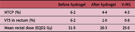
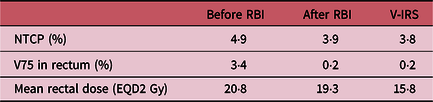
The V-IRS was able to accurately predict changes in rectal doses and NTCP in both techniques of IRS resulting in minimal average differences excluding mean rectal dose using RBI where using V-IRS predicted on average 3·5 Gy less than actual RBI results. The results showed a significantly larger absolute reduction using HS compared to RBI for decreasing mean rectal dose, rectal V75 and NTCP. A known value of 5·6 , 5 Gy and 1·8% absolute reduction from HS compared with 1·5, 3·2 Gy and 1·0% absolute reduction from RBI. The reason for the larger reduction from HS is the significantly higher pre-IRS figures from the HS group. This could be due to the HS group being aimed at HR patients where a larger PTV was used to cover SVs. Therefore, these results suggest a greater benefit for using IRS on advanced staged PC (Tables 6 and 7).Reference Van Wijk, Vanneste and Walsh30
V-IRS predictions compared to actual pre- and post-hydrogel

V-IRS predictions compared to actual pre- and post-rectal balloon insertions

The authors recommend further research into using the V-IRS model to predict the implications of isotoxic dose-escalation and incorporating genetic predictors into the NTCP prediction models.Reference Van Wijk, Vanneste and Walsh30
High-risk patients
Considering poor prognosis of HR-PC compared to LR, extreme hypofractionation trials are being conducted on these patients to try and improve prognosis. However, due to the high dose-escalation, this form of treatment is often associated with high GI toxicity rates.Reference Kothari, Loblaw and Tree31 Pryor et al.Reference Pryor, Sidhom and Arumugam16 investigated IRS using IMRT with a SABR boost for intermediate and HR-PC patients (Table 3). This dose fractionation radiobiologically equated to delivering 110 Gy using conventional 2 Gy per fraction, providing a significantly higher dose equivalent than conventional dose fractionated spacer trials.
This study extended HR patients to include T3b, which has not often been done due to the risk of IRS distorting malignant disease away from the PTV. The CTV included the entire SV with a 3 mm posterior PTV margin. The study aimed to create a benchmark of accurate contours, by verifying initially for each centre involved in the trial by using a peer review system and when any major deviations were required, this was checked by an external reviewer (Table 8).Reference Pryor, Sidhom and Arumugam16
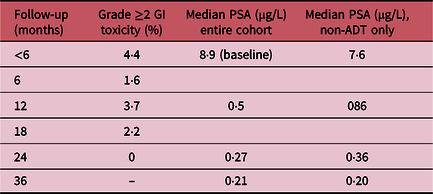
Gastrointestinal toxicity and PSA scores at different stages of follow-upReference Pryor, Sidhom and Arumugam16

Outcomes from the trial were measured using PSA and CTCAE. Despite using extreme hypofractionated SABR treatment, patients experienced low levels of GI toxicity. No patients had an acute grade 3 toxicity score, which accumulatively only occurred in 2% of patients at any stage of follow-up. The treatment enabled 2 years freedom from biochemical recurrence in 98·6% of patients. However, based on PSA nadir scores, there was more benefit when radiotherapy was combined with ADT, which was only used in 54% of patients despite being recommended.Reference Pryor, Sidhom and Arumugam16
This study showed how HS can have a role in increasing dose-escalation for HR patients with minimal grade ≥2 GI toxicity scores and high levels of disease control shown at 3 years post-treatment which appeared to improve when combined with ADT. However, several other factors may have also contributed to the low GI toxicity scores, such as the use of coplanar arc therapy with flattening free filter dose delivery, reduced posterior PTV margins and intra-fraction imaging verification of fiducial markers using exactrac®.Reference Pryor, Sidhom and Arumugam16
Conclusion
HS has been shown to reduce rectal planning doses in radical prostate radiotherapy. There is a correlation between placement symmetry and reduced rectal dose. A gel thickness of 5 mm has been established as a guide to creating a clinically significant reduction in rectal V70 dose. Despite a 99% success rate of spacer placements recorded, the optimal placements results differ with 62–72% <1 cm symmetry at different aspects of the prostate and the apex and base of the prostate being covered by spacer in 32% of patients. However, despite sub-optimal placements, spacers still created a significant reduction of ≥25% of rectal V70 in 95·7% of patients. The use of T2-weighted MRI enhances the visibility of spacer gel placements which helped to assess placement position for symmetry, thickness and possible RWI. The use of VRS endoscopy enabled an accurate way of assessing rectal damage from radiotherapy, which showed HS caused minimal GI toxicity complications from radiotherapy and also insertion, especially compared to RBI.Reference Schorghofer, Drerup and Kunit14 Risk factors have been established for increased risk of rectal complications from radiotherapy which can be used to identify specific patients who will benefit from spacer insertions. Following the results of this study showing reduced rectal planning doses and low levels of RWIs, it is recommended that HS has a role in advance radiotherapy for HR-PC patients, such as extreme hypofractionation through SABR and MRI linacs who have an increased chance of rectal complications compared to conventional radiotherapy dose prescriptions.Reference Kothari, Loblaw and Tree31 However, there are conflicting views on the use of spacers on HR patients.Reference Vanneste, Hoffman and van Lin6,Reference Yeh, Lehrich and Tran17 HS has been used on selected HR patients, depending on the malignancy location, but requires accurate spacer placements.