Introduction
In psychology, insight defines the ability of a person to be aware of his or her own mental state (Markova, Reference Markova2005). In medical terms, insight is used to define awareness of one's medical condition, functioning, or performance. In neuropsychiatric disorders, insight denotes to be consciously aware of one's mental deficits. Patients who fail to realize their mental deficits are considered to have “lack of insight.” Different terms such as “unawareness” and “anosognosia” are also used to define this situation (Sedaghat et al., Reference Sedaghat2010).
Lack of insight (anosognosia) is a common phenomenon in AD patients. Anosognosia may be observed in the early stages of AD, and also in patients with amnestic-type mild cognitive impairment (aMCI), which often represents prodromal stages of AD. In aMCI patients, anosognosia constitutes a risk factor for progression to dementia (Tabert et al., Reference Tabert2002; Ries et al., Reference Ries2007; Edmonds et al., Reference Edmonds2014). It is uncertain whether the severity of anosognosia is related with the severity of dementia. Some studies showed a strong correlation between anosognosia and cognitive impairment and the severity of dementia (Mangone et al., Reference Mangone1991; Verhey et al., Reference Verhey, Rozendaal, Ponds and Jolles1993; Lopez et al., Reference Lopez1994; Vasterling et al., Reference Vasterling, Seltzer, Foss and Vanderbrook1995; Harwood et al., Reference Harwood, Sultzer and Wheatley2000; Vogel et al., Reference Vogel2005; Aalten et al., Reference Aalten2006; Lehrner et al., Reference Lehrner2015), on the other hand others found a weak (Feher et al., Reference Feher1991; Sevush, Reference Sevush1999) or no correlation (Loebel et al., Reference Loebel, Dager, Berg and Hyde1990; Reed et al., Reference Reed, Jagust and Coulter1993; Weinstein et al., Reference Weinstein, Friedland and Wagner1994; Giovannetti et al., Reference Giovannetti, Libon and Hart2002).
In patients with AD, an association between anosognosia and executive functions has been reported (Starkstein, Reference Starkstein2014). Executive dysfunction associated with anosognosia was found to be a predictor of decline in patients with MCI (Scherling et al., Reference Scherling2016). Impairment in episodic memory is the first and most characteristic symptom of AD; memory deficits have also been reported to be associated with anosognosia (Orfei et al., Reference Orfei2010; Clare et al., Reference Clare2013).
Impairment in “directed attention” has been related with neglect syndrome and anosognosia. A critical cortical network including frontal, parietal, and cingulate cortices was proposed as the underlying neural substrate for directed attention (Mesulam, Reference Mesulam1981). In line with these hypothesis, in studies using positron emission tomography (PET) and single positron emission tomography (SPECT), anosognosia was found to be correlated with hypoactivity in frontal, parietal, and cingulate areas in patients with dementia (Salmon et al., Reference Salmon2006; Hanyu et al., Reference Hanyu2008). Both structural and functional magnetic resonance imaging (MRI) studies have also suggested a relationship between ventromedial prefrontal and temporoparietal cortices and anosognosia in patients with dementing disorders (Rosen et al., Reference Rosen2010; Zamboni et al., Reference Zamboni2013).
The objectives of this study were (1) to investigate whether anosognosia is correlated with performance in particular cognitive domains in aMCI versus early-stage AD patients, (2) to explore if there is any correlation between the severity of anosognosia and cognitive functions in both patient groups and total population, and (3) to assess correlations between cortical thickness in frontal–parietal–cingulate areas and anosognosia in these populations.
Patients and methods
Patient recruitment and neuropsychological evaluations
The study included 21 home-living AD patients recently diagnosed according to NINCDS-ADRA (McKhann et al., Reference McKhann1984) criteria and 26 aMCI patients diagnosed according to revised MCI criteria (Winblad et al., Reference Winblad2004). All patients were consecutive and self-referrals. Only early-stage AD patients with Clinical Dementia Rating (CDR) Scores of 0.5 and 1 were included, and patients with CDR scores 2 and 3 and MMSE scores below 20 points were excluded. Further, inclusion criteria were having an age of at least 60 years and a primary caregiver not suffering from a mental illness. Exclusion criteria for all groups were history of a previous major head trauma, stroke, any severe psychiatric disease or alcohol, and drug addiction. Informed consent forms were signed by all patients in aMCI group and by both patients and caregivers in the AD group. After the clinical diagnosis, all participating patients underwent a cranial MRI including high-resolution T1 images in coronal plane. Neuropsychological and anosognosia evaluation were performed in another session within 4 weeks after the MRI acquisition. Neuropsychological test battery included Mini-Mental State Examination (MMSE) test for global cognitive screening; California Verbal Learning Test (CVLT1) for verbal episodic memory; Stroop Test, Wisconsin Card Sorting Test (WCST), and Trail-Making Test (TMT) for executive functions; Digit Span Forward test for simple attention; Digit Span backward test and Digit Ordering test (DOT) for working memory and Clock Drawing Test (CDT) for executive and visuo-spatial functions. CDT was scored using a scoring system ranging between 1 and 10 (Manos and Wu, Reference Manos and Wu1994).
Evaluation of anosognosia
For the evaluation of anosognosia, both Clinician-administered Insight Rating Scale (CIRS) (Ott et al., Reference Ott1996) and Anosognosia Questionnaire-Dementia (AQ-D) (Migliorelli et al., Reference Migliorelli1995) were administered.
clinical insight rating scale (cirs)
This scale assesses anosognosia by evaluating the following aspects: (i) reason for visiting the doctor, (ii) cognitive deficits, (iii) functional deficits, (iv) perception of the progression of the disease. Each of these four domains is scored between 0 and 2. The clinician scores (0) point, if the patient has an insight in the related domain. If the patient's insight is partially preserved, the clinician scores (1) point, whereas (2) points are assigned when there is no insight. Total score of 0 or 1 represent a non-anosognosic state, whereas the scores greater than 1 represent anosognosia. In this scale, a higher total score indicates more severe anosognosia. For the purposes of this study, patients were defined as “anosognosic” if they scored <2 and “non-anosognosic” if they scored ≥2.
anosognosia questionnaire-dementia (aq-d)
AQ-D consists of 30 questions assessing awareness in cognition and behavior. Both patient and caregiver complete two independent forms; the final score is calculated by subtracting caregiver's score from the patient's score. A positive final AQ-D score indicates that the caregiver has rated the patient as more impaired compared to patient's own rating. Scores 13 and below suggest that the patient is aware of his/her symptoms, whereas scores over 31 show that the patient is unaware of his/her own symptoms. The scores between these two scores represent a grayzone (Migliorelli et al., Reference Migliorelli1995).
Patients with CIRS scores of 2 or more were classified as anosognosic and patients with CIRS score of 0 and 1 classified as non-anosognosic in both MCI and AD groups. AQ-D scores were calculated and used as a measure of severity of anosognosia in the analysis.
Measurement of cortical thickness
Two consecutive high-resolution T1-weighted images of each patient were obtained through Philips Achieva 1.5 T MR Scanner. The sequence parameters were as follows: TR\TE ¼ 8.6/4.0 sec, reversing bracket/turning angle ¼ 8, FOV ¼ 240 mm, voxel size ¼ 1.25/1.25/1.2 mm ¼ 1.25/1.25/1.2 mm (structured ¼ 0.94/0.94/1.2 mm), uninterrupted 150 coronal sectioning, scan rate ¼ 7.23 min (for each volume).
In order to calculate cortical thickness, Freesurfer 4.05 running on a Linux operation system installed on a workstation computer was used. Procedure for surface construction was performed as described before (Dale et al., Reference Dale, Fischl and Sereno1999; Fischl and Dale, Reference Fischl and Dale2000). Destrieux atlas (Destrieux et al., Reference Destrieux, Fischl, Dale and Halgren2010) was used for identifying the areas of region-of interest (ROI) including the opercular, orbital, triangular parts of the inferior frontal gyrus, the middle and superior frontal cortex, angular gyrus, supramarginal gyrus, parts of cingulate gyrus (anterior cingulate cortex, anterior and posterior parts of middle cingulate cortex, and posterior and ventral parts of posterior cingulate cortex (PCC)), and superior parietal lobe in both hemispheres. Mean cortical thickness values of these areas were calculated using values from both hemispheres.
Statistical analysis
All statistics were conducted with SAS statis-tical software, version 9.2 (SAS Institute, Cary, NC, USA). Student's t-test and Mann–Whitney U-test were used to compare demographic mea-sures and neuropscyhological test scores between the anosognosic and non-anosognosic groups. Correlations between AQ-D anosognosia scores and neuropsychological test scores as well as correlations between AQ-D scores and mean cortical thickness values were calculated with Spearman's Correlation analysis. A value of p < 0.05 was considered statistically significant for all tests.
Results
Demographics
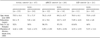
Fourty-seven patients were included in the study, 26 with aMCI and 21 with AD. Demographic values are presented in Table 1. There was a trend toward significance for age (p = 0.058) and education (p = 0.0672) between MCI and AD groups. Although anosognosics and non-anosognosics in the AD group did not show any significant differences in terms of age, gender, and education, anosognosics in the MCI (p = 0.0145) and the total group (p = 0.027) were significantly older than the non-anosognosic patients.
Demographics of the study population

aMCI = Amnestic-type of mild cognitive impairment, AD = Alzheimer's disease.
*Significant difference between anosognosics and non-anosognosics (p < 0.05).
In the total population, mean AQ-D score in the anosognosic group was significantly higher as expected (anosognosics: 20.40 ± 10.70, non-anosognosics: 3.08 ± 7.30, p < 0.0001). In the aMCI subgroup, mean AQ-D score in the anosognosic group was 19.36 ± 9.70 as compared to 1.83 ± 7.17 in the non-anosognosic group (p < 0.0001). In the AD subgroup, anosognosic group had a mean value of 21.22 ± 10.33 as compared to 3.08 ± 7.30 in non-anosognosics (p < 0.0001).
Comparison of cognitive functions between anosognosic and non-anosognosic patients based on CIRS scale
In the total study population, there were 24 anosognosic and 23 non-anosognosic patients based on the CIRS scale. In the aMCI subgroup, 12 patients were anosognosic and 14 were non-anosognosic. In the AD subgroup, 11 were anosognosic and 10 were non-anosognosic.
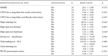
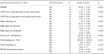
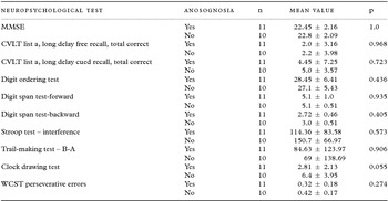
In the total study population, there were significant differences between the two groups in the DOT, Digit Span Test Backward and CDT, non-anosognosics showing better performance than anosognosics (Table 2). In the aMCI subgroup, anosognosic group showed statistically significant worse performance in the same tests, there was also a trend for lower performance in the long delayed cued recall of CVLT as compared to non-anosognsics (Table 3). Although numerical differences were indicating better performance in the non-anosognosic group, there were no statistically significant differences in the neuropsychological test performance of anosognosic and non-anosognosic AD patients (Table 4).
Group comparison of cognitive functions in the total study population including both AD and MCI patients dichotomized as “non-anosognosic” or “anosognosic” based on CIRS score

MMSE = Mini Mental Status Examination, CVLT = California Verbal Learning Test, WCST = Wisconsin Card Sorting Test, AQ-D = Anosognosia Questionnaire-Dementia.
*p < 0.05.
Group comparison of cognitive functions of the aMCI patients dichotomized as “non-anosognosic” or “anosognosic” based on CIRS score

*p < 0.05.
Cognitive functions of the AD group dichotomized as “non-anosognosic” or “anosognosic” based on the CIRS score

Correlation between cognitive deficits and AQ-D anosognosia scores
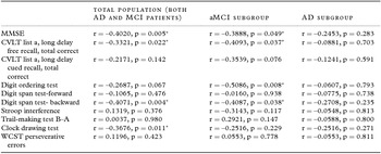
In the total patient population, negative correlations were found between AQ-D scores and MMSE, total score of CVLT Long-Delay Free Recall, Digit Span Backwards and CDT scores. In the aMCI group, a significant negative correlation was found between the AQ-D and MMSE score, total CVLT Long-Delay Free Recall, DOT and Digit Span Backward scores. AQ-D scores in the AD subgroup showed no statistically significant correlations with any of the neuropsychological test scores (Table 5).
Correlation between Anosognosia Questionnaire (AQ-D) and cognitive functions

aMCI = Amnestic type of mild cognitive impairment, AD = Alzheimer's disease, MMSE = Mini-Mental State Examination, CVLT = California Verbal Learning Test, WCST = Wisconsin Card Sorting Test.
*p < 0.05.
The relationship between cortical thickness and anosognosia
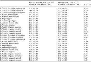
Mean cortical thickness measures of the ROI areas did not significantly differ between the anosognosics and non-anosognosic patients as defined by the CIRS score in the total population (Table 6). Likewise, there were no statistically significant correlations between AD-Q scores and any of the cortical thickness measures of ROI areas in the total population (Table S1, available as supplementary material attached to the electronic version of this paper at www.journals.cambridge.org/jid_IPG).
Cortical thickness measurement of the total population including both AD and MCI patients dichotomized based on the CIRS score

R = Right hemisphere, L = Left hemisphere.
Discussion
Traditionally, three different approaches have been used to assess the presence of anosognosia in patients with AD or MCI. The first is “clinician rating of patients’ awareness” in which the clinician classifies patients into categories including full, partial, or no awareness of deficits after a clinical interview. A major limitation of this method is the non-structured nature of the evaluation. The second strategy designated as “patient–caregiver discrepancy assessment” is based on comparison of the ratings given by patients as opposed to their caregivers on their daily performance. One advantage of this method is the independent information provided by the caregiver. However, factors such as the mood and burden of the caregiver may have an impact on the reliability of caregiver's information. A third method is the “prediction of performance discrepancy” based on patients' report on their level of performance on a given neuropsychological test as opposed to their actual performance. In the current study, both clinician rating of patients’ awareness and patient–caregiver discrepancy assessments were used to assess the presence and severity of anosognosia instead of a single strategy in order to reduce the limitations of both methods.
In this study, we found a correlation between AQ-D scores, which measures severity of anosognosia, and several cognitive functions including the global score of cognitive function, MMSE as well as tests of verbal episodic memory, working memory, and executive functions in the total study population of patients with prodromal phase and early-stage AD. These associations largely remained intact in the aMCI patients were, however, not present in the AD subgroup. The latter finding may be because severity of anosognosia may increase in a non-linear way and may remain stable after the early stages of disease (Clare et al., Reference Clare2012). We found a correlation between the severity of anosognosia and episodic verbal memory deficits, the core and initial symptom of AD. In aMCI but not AD patients, memory was relatively more impaired with more severe anosognosia. Similar to the results of our study, anosognosia was associated with verbal memory impairment in MCI patients, but not in AD (Orfei et al., Reference Orfei2010). Although another study conducted in AD patients could not find any significant correlation between CVLT performances and anosognosia (Green et al., Reference Green, Goldstein, Sirockman and Green1993), other studies found anosognosic AD patients having worse memory impairment and other cognitive and behavioral problems (Reed et al., Reference Reed, Jagust and Coulter1993; Starkstein et al., Reference Starkstein1996). Damage in some specific components of the memory may impair self-perception (Mograbi et al., Reference Mograbi, Brown and Morris2009). It was proposed that there is a “working-self,” modulating the new information to be encoded on the basis of the concept of self/insight (Conway and Pleydell-Pearce, Reference Conway and Pleydell-Pearce2000; Conway, Reference Conway2005). Here, memory allows for the storage of past information as well as providing the sustenance of the ability to animate the future scenarios and “selfhood experience” (Addis and Tippett, Reference Addis and Tippett2004). In other words, memory establishes the relationship that gives meaning to what the “self” was in the past, what it is now, and what it is likely to become in the future (Conway, Reference Conway2005). Binding failure of the past and present due to memory impairment may cause a deficit in awareness deficit.
When patients were dichotomized for presence or absence of anosognosia, main cognitive domains which were found to be more impaired in anosognosic patients were working memory (DOT and Digit Span backwards) and executive functions (CDT). Executive function deficits were described to be associated with anosognosia in AD patients in some studies (Mangone et al., Reference Mangone1991; Lopez et al., Reference Lopez1994; Michon et al., Reference Michon1994; Ott et al., Reference Ott1996; Starkstein et al., Reference Starkstein1997; Kashiwa et al., Reference Kashiwa2005), whereas in others no association was found (Migliorelli et al., Reference Migliorelli1995; Starkstein et al., Reference Starkstein, Jorge, Mizrahi and Robinson2006). In one study, it was proposed that memory or executive dysfunction in AD patients might affect the immediate ability to judge cognitive performance in a domain-specific manner (Hannesdottir and Morris, Reference Hannesdottir and Morris2007). We found poorer performance on the CDT in anosognosic patients, which assesses executive functions such as planning ability, whereas in other tests assessing executive functions, including WCST, Stroop, and TMT, there were no statistically significant differences between the two groups. This may be because each of these tests assesses different aspects of executive function, CDT tapping more insight/foresight along with planning.
Neural basis of anosognosia in AD patients was assessed in several studies. In one study using functional magnetic resonance imaging (fMRI), medial prefrontal and anterior temporal cortices were associated with impaired self-awareness (Zamboni et al., Reference Zamboni2013). In another study using SPECT, right inferior parietal region was found to show hypoperfusion in anosognosic AD patients (Sedaghat et al., Reference Sedaghat2010). A recent study using PET revealed that anosognosia was correlated with hypometabolism in orbitofrontal (OFC) and PCC cortices (Perrotin et al., Reference Perrotin2015). In another PET study, negative correlation was found between hypoperfusion in the right inferior frontal gyrus and anosognosia test scores only in anosognosic patients, but not non-anosognosic patients (Vogel et al., Reference Vogel2005). In addition to functional imaging, there have been also structural studies using voxel-based morphometry (VBM) evaluating anosognosia in patients with AD and other neurodegenerative disorders. In one of them, a correlation was found between anosognosia and ventromedial and frontopolar prefrontal atrophy (Hornberger et al., Reference Hornberger2014). In this study, an insight questionnaire with five subscales evaluating social interaction, emotion, diagnosis/treatment, language, and motivation was used but memory was not a primary target of the questionnaire. Another VBM study including 9 AD and 24 non-AD dementia patients using prediction of performance discrepancy method showed an association between OFC and ventromedial prefrontal cortex atrophy and anosognosia in the whole group (Rosen et al., Reference Rosen2010). In another study using VBM, in MCI patients that converted to dementia in the follow-up period, anosognosia was associated with gray matter volume reduction in anterior cingulate and frontal cortex as well as cerebellum (Spalletta et al., Reference Spalletta2014). The current study is the first one in which cortical thickness analysis was used to evaluate neuroanatomical basis of anosognosia including aMCI and early-stage AD patients. We found no difference between anosognosic and non-anosognosic patients in cortical thickness in any of the analyzed areas. A possible explanation for the difference in the results of this study as opposed to those using VBM may be the use of “region of interest” method in our study as compared to vertex-wise analysis and the use of Destrieux atlas for parceling the cortex. Subregions in Destrieux atlas may be broad enough to detect more subtle changes. Functional deficits rather than structural changes may be another possible explanation for anosognosia in patients with early stages of the disease such as those included in this study. Finally, the relatively small sample size in our study may have been a limitation to detect possible differences.
In terms of functional deficits, anosognosia in early-stage AD and aMCI may be related to impaired connectivity between certain areas. It was proposed that anosognosia in AD may reflect a disconnection syndrome resulting from degeneration of temporofrontal fibers (Ott et al., Reference Ott1996). In a study using resting-state functional magnetic resonance imaging (rsFMRI) including MCI and early-stage AD patients, anosognosics showed decreased functional connectivity between medial prefrontal cortex (also known as anterior node of default mode network (DMN)) and dorsolateral prefrontal cortex, bilateral caudate, and left hippocampus; anosognosia was associated with hypoconnectivity of default mode network (Ries et al., Reference Ries2012). Another study combining PET with rsFMRI revealed a similar pattern of hypoconnectivity between anterior and posterior node of the default mode network and functional changes in these cortical midline structures (Perrotin et al., Reference Perrotin2015). Alteration in DMN is a very early feature of AD and DMN has been suggested as a key node in the network for self-referential processing (Raichle et al., Reference Raichle2001). Thus, anosognosia seen in the early stages of AD may be associated with impairment in DMN connectivity rather than structural changes.
In conclusion, in this study which included aMCI and early-stage AD patients, we found an association between severity of anosognosia and severity of impairment in global cognition, episodic verbal memory, working memory, and executive function in the total patient population and in those with aMCI. In the aMCI group, patients with anosognosia showed more impaired working memory and executive function, no such association was found in patients with early-stage AD. Measures of cortical thickness showed no difference between anosognosic and non-anosognosic aMCI or early-stage AD patients. Anosognosia is a phenomenon that may appear already in early stages of the disease; it may be associated with impairment in certain cognitive areas such as memory and executive functions. Its elucidation needs reliable information from a knowledgeable third-party.
Conflict of interest
None.
Description of author's role
Murat Emre and Başar Bilgiç designed the study and Gulben Senturk, Hakan Gurvit, and Hasmet Hanagasi collected the data. Ali Bilgin Arslan and Ali Bayram supervised the analysis. Gulben Senturk, Basar Bilgic, and Murat Emre wrote the paper.
Acknowledgments
The authors thank Mr. Cagdas Kenar for his help in statistical analysis.
Supplementary material
To view supplementary material for this article, please visit https://doi.org/10.1017/S1041610216001812.