Introduction
Musculoskeletal symptoms are extremely common, with almost one-third of people aged 75 and over having a significant musculoskeletal problem.Reference Martin, Meltzer and Elliot1 Musculoskeletal conditions are of particular concern to elderly people; preservation of mobility may be seen as second only to maintenance of cognitive function in safeguarding an independent existence.Reference Calkins and Isenberg2 Joint pain due to inflammatory arthritis is important to identify, as it is potentially treatable. A number of inflammatory conditions increase in prevalence with age, such as polymyalgia rheumatica; in addition some rheumatological conditions, e.g. gout, are associated with co-morbid conditions or use of drugs that are more common in older patients. With the ageing population, inflammatory arthritis is an increasing problem, and it is imperative that physicians involved in the care of elderly people are up to date with the assessment and management of common rheumatological diseases.Reference Arthur3
Establishing the diagnosis and managing joint problems in elderly patients can be complex. In the patient presenting for the first time with inflammatory joint symptoms, degenerative joint disease often co-exists, making interpretation of symptoms confusing. Indeed, more than one inflammatory arthritis can co-exist. Inflammatory arthritides often present differently in the elderly population than in younger patients. In addition, co-morbid conditions and polypharmacy may limit management options. Finally, psychosocial factors are of extreme importance to the elderly patient who may be suffering from reduced mobility and independence.
In this paper, we seek to update the previous reviewReference Kamath and Hassell4 in this area with new developments in diagnosis and management. We will first discuss the general considerations in the clinical evaluation and management of a patient with inflammatory joint symptoms, before discussing in more detail diagnosis and management of the common causes of inflammatory arthritis. Relevant literature was identified by searching the databases MEDLINE, EMBASE and CINAHL, and the Oxford Textbook of Rheumatology and guidelines found using the National Health Service (NHS) Evidence Health Information Resources website.5
Clinical evaluation of the elderly patient with inflammatory joint symptoms
History
The patient may present primarily with musculoskeletal symptoms or with systemic upset, including malaise, weight loss, fever and decline in general. Thus a brief musculoskeletal screen is essential in the clerking of the unwell elderly patient. If they present with joint symptoms, the hallmarks of inflammation (swelling, warmth, erythema and tenderness) may be present. In addition, morning stiffness is common in rheumatoid arthritis (RA) and crystal arthropathies and is usually present for 30 minutes or longer. Stiffness in peripheral joints is common in osteoarthritis, in the morning or after periods of immobility (‘gelling’), but typically does not last as long.
Differential diagnosis
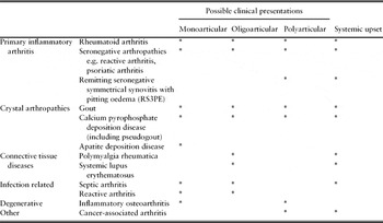
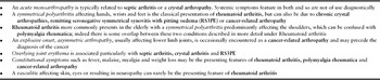
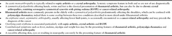
The spectrum of conditions associated with joint inflammation is detailed in Table 1. The pattern and onset of joint involvement is often useful diagnostically; the patient may present with a monoarthritis, oligoarthritis or polyarthritis, and possible presentations are also shown in Table 1. A selection of patterns of presentation are listed in Table 2 and more detail about the clinical presentation of specific conditions is described in the second section of this article under disease headings.
Conditions associated with joint inflammation in elderly patients

Examples of patterns of disease presentation
Other relevant enquiry
A detailed drug history is important in considering disease causality as well as potential treatments. There are a number of drugs that have been implicated in acute arthropathy and a detailed review in this area is beyond the scope of this article. However, a history of recent medication initiation may be relevant and prompt a search of the literature. Two examples are clopidogrel and tamoxifen, which have both been associated with acute arthritis.Reference Creamer6, Reference Garg, Radvan and Hopkinson7 Diuretic use, with both loop and thiazides, predisposes to gout.
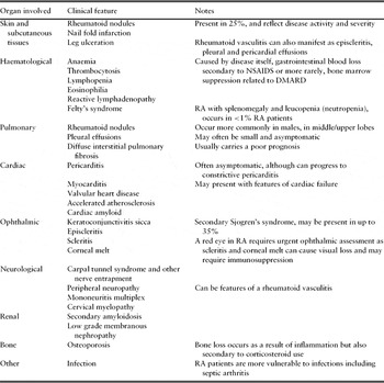
Co-morbid conditions are of relevance to the diagnosis, e.g. cardiovascular disease and gout, and are likely to influence management and medication choice. A full systems review may elicit symptoms related to extra-articular manifestations of rheumatoid arthritis (Table 3) or symptoms of possible underlying malignancy.
Extra-articular manifestations and complications of rheumatoid arthritis

A full socioeconomic history is essential, including screening for depressive symptoms, particularly in those patients with chronic disease or musculoskeletal disability.
Clinical examination
Examination of the joints includes assessment of deformities, joint tenderness and boggy swelling, suggestive of active synovitis. The pattern of joint involvement should be noted, along with erythema and loss of function. The skin should be inspected for rheumatoid nodules, gouty tophi, psoriatic skin and nail changes and the presence of any vasculitic rash. Lymphadenopathy may be indicative of an underlying malignant process, although this may be associated with active rheumatoid arthritis.
Investigations
Initial blood tests should include full blood count including white cell differential, erythrocyte sedimentation rate (ESR) and C-reactive protein (CRP) as measures of inflammation. One would expect elevated ESR and CRP in most, if not all inflammatory arthropathies including crystal arthritis. A low or normal CRP is uncommon in infections and probably is more useful than an elevated CRP. Urea and electrolytes and liver function tests are useful baselines when considering drug treatment. Uric acid levels may be requested in a mono-, oligo- or polyarthritis, although normal levels do not exclude an acute attack of gout. Rheumatoid Factor (RF) may be checked in suspected RA, although is not diagnostic in isolation. The presence of RF is commonly quoted as being more prevalent in elderly people,Reference Ruffatti8 although one study has suggested RF is only increased in prevalence in the chronically unwell.Reference Juby9 Anti-cyclic citrullinated protein (anti-CCP) antibodies provide a more specific test for rheumatoid arthritis (90–95% in published studies) where available, and may be predictive of disease severity.Reference Nishimura10 A septic screen including blood cultures, urine culture and chest X-ray is pertinent in any patient with systemic symptoms and fever, and in an acute ‘flare’ of existing inflammatory arthritis such as RA.
In a mono- or oligoarthritis, joint aspiration is useful diagnostically and is mandatory in the acute hot joint. The aspirate should be sent for microscopy (including polarizing microscopy), culture and sensitivity, and an urgent gram stain requested in the acute hot joint. The number of white cells detected in microscopy can be a useful pointer to diagnosis: a primary inflammatory arthritis typically results in a raised synovial fluid white count, usually >2000/mm3. Counts >50,000/mm3 are more likely to be encountered with a septic joint or with an acute crystal arthropathy.
Plain films are useful in mono- or oligoarthritis and, in the case of a widespread symmetrical polyarthropathy, plain radiographs of hands and feet are indicated to ascertain if any diagnostic erosive changes are present. Chondrocalcinosis is a common incidental finding in older people, particularly in the knee and radiocarpal joint, but in the presence of a suggestive history and/or synovial fluid findings may be a pointer to a diagnosis of calcium pyrophosphate deposition disease. In a suspected monoarthritis that may involve peri-articular tissues e.g. Milwaukee shoulder, ultrasound can be a useful imaging modality. Alternatively magnetic resonance imaging (MRI) would identify adjacent soft tissue pathology and may be indicated in the case of an acute septic joint to identify any adjacent osteomyelitis.
Management
A multidisciplinary team including the treating physician, physiotherapist, occupational therapist, nurses, orthotics, dietician, podiatrist and clinical psychologist may be involved in the management of patients, particularly those with chronic disease. Elderly patients have far more complex needs and may require an in-patient and/or home assessment by therapists. Active physiotherapy is important to maintain muscle strength and function, and physiotherapists can also assist with treatment of pain with acupuncture, heat or ice treatment, TENS machines and local ultrasound. Occupational therapists’ interventions include instruction on joint protection and energy conservation, instruction about assistive devices and provision of splints.
The drugs used to treat rheumatological conditions have special considerations in older people; non-steroidal anti-inflammatories and corticosteroids are discussed below and more specific agents are discussed under disease headings.
Non-steroidal anti-inflammatory drugs (NSAIDs). NSAIDs and selective cyclo-oxygenase-2 (COX-2) inhibitors have been the subject of much attention in the media and medical press since our first review was published. The therapeutic value of these drugs is well known, but their use has dramatically reduced, probably as a result of increasing concerns over their safety. The safety concerns predominantly relate to renal impairment, gastrointestinal side-effects and cardiovascular events.
NSAID use accounts for 15% of causes of acute renal failure and elderly patients are particularly at risk.11 In addition, co-morbidities such as hypovolaemia, congestive heart failure, liver cirrhosis and multiple myeloma, or co-prescription of angiotensin converting enzyme (ACE) inhibitors, angiotensin II receptor antagonists or diuretics all increase the risk. NSAIDs should be avoided in patients with impaired glomerular filtration. Where NSAIDs are prescribed, renal function should be monitored, particularly in the high-risk groups described above.
All patients over the age of 65 years can also be considered at increased risk of gastrointestinal side-effects; the risk is twice as high in men than women, and risk of gastrointestinal bleeding doubles with each increasing decade of life after 55 years.12 Concomitant gastric protection with a proton pump inhibitor should therefore be considered in older patients. Prescribing an NSAID alongside low dose aspirin dramatically increases the gastrointestinal risk and should be avoided if at all possible, the aspirin for cardioprotection taking priority.
With regard to cardiovascular risk, all the selective COX-2 inhibitors have been associated with increased risk of thrombotic events.Reference Kearney13 Diclofenac has been shown to have an equivalent risk to etoricoxib. High-dose naproxen (1000 mg daily) and low-dose ibuprofen (1200 mg daily) do not appear to be associated with increased risk of thrombotic events.Reference Kearney13 The NHS Clinical Knowledge Summaries suggest that all patients over the age of 65 are at increased cardiovascular risk;12 however, recent evidence suggests that thrombotic events are increased regardless of cardiovascular risk factors.14 It seems reasonable to conclude that, if an NSAID is deemed appropriate, low-dose ibuprofen or naproxen is the NSAID of choice in elderly people.
Despite the risks, there will always be patients with residual pain and functional limitation in whom anti-inflammatory drugs provide the best therapeutic option. Taking into account these risks, the general advice regarding NSAIDs in older people is to prescribe alternative analgesia where possible, prescribe topical rather than oral if possible, and to regularly review the need for, and dose of NSAID, when used.
Oral corticosteroids. Corticosteroids are first line treatment in polymyalgia rheumatica (PMR) and relapsing seronegative symmetrical synovitis with pitting oedema (RS3PE) and may be used in RA, seronegative arthritides, crystal arthritis and cancer-associated arthritis. According to UK Royal College of Physicians guidance, all patients treated with corticosteroids for three months or more and aged over 65 years should be prescribed osteoporosis prophylaxis (e.g. bisphosphonates unless contraindicated, in addition to calcium and vitamin D supplementation), without need for bone densitometry monitoring.15 This guidance reflects recent research which demonstrates that bone loss occurs early after commencing steroids. European guidelines state that screening for risk factors for adverse events should take place before prescription.Reference Hoes16 Therefore as patients over 65 years are at increased risk of gastrointestinal haemorrhage, one could argue that gastric protection should also be co-prescribed. These guidelines also suggest that during treatment, patients should be monitored for body weight, blood pressure, peripheral oedema, cardiac insufficiency, lipids, blood and/or urine glucose and ocular pressure depending on individual patient's risk.Reference Hoes16
The clinical presentation and management of specific inflammatory arthritides Rheumatoid arthritis (RA)
Clinical presentation, diagnosis and disease assessment
In considering RA in older people, we shall first discuss RA presenting for the first time and secondly, the challenges of the patient with pre-existing RA.
Late onset RA
RA often presents differently in older patients to the classical presentation seen in younger people. Late onset RA (LO-RA) differs in that:
1 The female to male ratio approaches 1:1
2 There is a higher frequency of systemic features such as weight loss
3 There is more frequent involvement of the shoulder girdle
4 An acute presentation is more common
5 There may be high acute phase reactantsReference Bajocchi17
RA can present abruptly or insidiously, with almost any pattern of joint involvement (polyarticular, oligoarticular or monoarticular) and with systemic features or extra-articular features at onset. The possible extra-articular manifestations are listed in Table 3.
Myalgia is present in 16% of patients with LO-RA and conversely, patients with polymyalgia rheumatica (PMR) may have synovitis (which usually rapidly responds to corticosteroids); this can cause some diagnostic difficulty between LO-RA and PMR.Reference Pease18 In patients with wrist and small joint (metacarpalphalangeal or proximal interphalangeal) synovitis occurring together the diagnosis is likely to be LO-RA.Reference Pease18 Anti-CCP has also been used to try and discriminate between PMR and RA.Reference Lopez-Hoyos19 Seronegative LO-RA has been described as a variant of PMR; however, seronegative RA can be erosive, which calls into question this theory. In practice, the clinical presentation and autoantibody profile are useful pointers but sometimes the distinction is difficult and rheumatologists sometimes use the term polymyalgia/rheumatoid overlap.
The other differential diagnoses to be considered in possible LO-RA include crystal arthritides, psoriatic arthritis, RS3PE syndrome and cancer-associated arthritis. Investigations including RF, anti-CCP, joint aspiration if possible and X-rays should help discriminate. Calcium pyrophosphate deposition arthropathy can be particularly difficult to distinguish from RA, especially in the absence of radiographic erosions. The prognosis of LO-RA is debated in the literature, although it would seem that any increase in mortality in LO-RA is probably due to co-morbidities.Reference Calkins and Isenberg2 The titre of RF does not appear to correlate with disease severity in the over-85s.Reference Calkins and Isenberg2
Pre-existing RA
Clinical assessment of the patient with pre-existing RA includes an evaluation of their current disease activity. This can be measured using objective assessments such as the Disease Activity Score 28 (DAS 28), and functional status can be monitored using assessments such as the Health Assessment Questionnaire (HAQ). The DAS 28 is a validated research measure of disease activity, which is also used in clinical practice and comprises a score of tender and swollen joints, a recent ESR and a patient global score.
In reviewing RA patients, the clinician should be alert to the presence of any complications of the disease. These are listed in Table 3 with extra-articular manifestations. In particular, an evaluation of cardiovascular risk factors such as lipids, hypertension and diabetes is important due to the increased incidence of cardiovascular events in RA patients; RA in isolation is a cardiac risk factor.Reference del Rincón20, Reference Luqmani21 Other important complications include osteoporosis (secondary to the inflammation of RA, in addition to any corticosteroid therapy) and atlanto-axial subluxation resulting in cervical myelopathy. With regard to the latter, X-rays of the cervical spine in flexion and extension are prudent before patients with RA undergo general anaesthetic or upper gastrointestinal endoscopy to screen for neck involvement which could be exacerbated by cervical extension. RA clinical assessment should also pay attention to the patient's social support, home environment and their psychological state. Depression is increased in patients with rheumatoid arthritis and may be correlated with extent of inflammation.Reference Kojima22 However, there is some evidence that depression is more prevalent in younger patients with RA.Reference Wright23
In the RA patient presenting with an apparent ‘flare’ it is important to screen for underlying infection and check thyroid function, as sepsis, hypo- and hyperthyroidism can cause an exacerbation of disease.Reference Calkins and Isenberg2
Management
RA is managed using a multidisciplinary approach, as described above. Patient education is administered by all members of the team, but particularly by specialist nurses, physiotherapists and occupational therapists, and in some areas by dedicated patient education programmes.
The pharmacological management has changed in recent years in light of evidence that joint damage occurs early in the disease; therefore almost all patients are commenced on disease modifying anti-rheumatic drugs as soon as practicable following diagnosis.Reference Nell24 Oral corticosteroid use for RA may have declined as a result. However, steroids play an important role in providing rapid relief of symptoms and reduction in inflammation at diagnosis or during a flare, especially intra-articularly or intramuscularly.
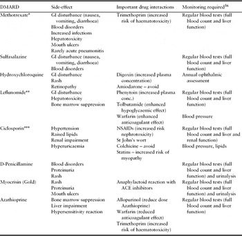
Disease modifying anti-rheumatic drugs (DMARDs). The DMARDs licensed for use in RA are listed in Table 4, along with commonly encountered side-effects and required monitoring. In practice, methotrexate, sulfasalazine, leflunomide and hydroxychloroquine are probably the most commonly used. There is evidence that these drugs are well tolerated in elderly patients with no increase in adverse events;Reference Alivernini25, Reference Dahl26 however, despite this there is evidence that older people are less likely to receive DMARD treatment.Reference Tutuncu27, Reference Schmajuk28 The ongoing management includes regular reviews, aggressive DMARD dosage escalation and the use of concomitant DMARDs (combination therapy) until acceptable disease control is achieved.Reference del Rincón20 The National Institute for Health and Clinical Excellence (NICE) now recommends combination DMARD therapy in all patients with early RA29 although specific studies of such regimens in elderly people are lacking. Cyclosporin, gold and D-penicillamine have a serious potential for renal side-effects in older patients, and the latter two also tend to cause rashes in this age group.Reference Gardner and Furst30 Live vaccines are contraindicated with most of the DMARDs; the seasonal flu vaccine is recommended.
Side-effects and monitoring of DMARDs

*Concomitant folic acid prescription reduces toxic effects;Reference Chakravarty57 reduce dose in renal impairment; **leflunomide has a long half-life and a wash-out procedure is recommended in a serious adverse event;Reference Chakravarty57 ***avoid grapefruit ingestion.
NB. This is not an exhaustive list, but summarizes the most commonly encountered problems in the authors’ experience.
Biological therapy. Biological therapy in regular use for RA consists of the three anti-TNF agents, infliximab, etanercept and adlumimab, and rituximab (anti-CD20). The anti-TNF agents are approved by NICE for use in moderate to severe RA that has not responded to two or more DMARDs.31 Side-effects of these agents include increased incidence of tuberculosis and non-melanoma skin cancer, and injection site reactions in the agents administered subcutaneously (etanercept and adlumimab). There is increasing evidence that the risk of adverse events with these drugs in elderly people is no different than in the younger population.Reference Bathon32–Reference Genevay34 However, as with DMARDs, an observational study indicates that elderly patients may be less likely to receive anti-TNF compared with younger patients.Reference Radovits35
Surgical therapy. Surgical intervention is useful for patients with longstanding rheumatoid arthritis who have developed secondary osteoarthritis in addition to patients with co-existing primary osteoarthritis. Surgery is recommended mainly for pain relief and, in the case of hip and knee replacements, improving mobility.
Seronegative spondyloarthropathies
The seronegative spondyloarthropathies include ankylosing spondylitis (AS), psoriatic arthropathy (PsA), reactive arthropathy (ReA), enteropathic arthropathy and undifferentiated spondyloarthropathy. Of these, ReA and PsA are the most likely to present in elderly people.
Clinical presentation
ReA and PsA are classified with the spondyloarthopathies; however, these conditions usually present as peripheral arthritis and can mimic other inflammatory arthritides. PsA can present with monoarticular, oligoarticular, polyarticular or spinal symptoms. Nail changes of psoriasis, including onycholysis and nail ridging, are seen in about 70% of patients with psoriatic joint disease. ReA is usually mono- or oligo-articular; extra-articular features may be helpful in diagnosis and include conjunctivitis, preceding urethritis or diarrhoea.
Diagnosis and management
The drug management of ReA tends to be symptomatic with intra-articular steroids, NSAIDs, analgesics and physiotherapy, as most cases are self-limiting. Sulfasalazine or methotrexate may be used if symptoms become chronic. In PsA, the spectrum of severity ranges from mild pain without dysfunction, through to an aggressive erosive arthritis. Patients with more severe disease are treated similarly to those with RA, particularly with methotrexate as this can be effective at treating the skin as well as the joints.Reference Salvarani, Cantini and Oliveri36 Anti-TNF therapy is approved by NICE for moderate to severe PsA that has not responded to two or more DMARDs.37, 38
Gout
Gout is becoming more common worldwide and has a relatively equal sex distribution in old age.Reference Wise39 Gout first presenting in elderly people is usually secondary to another cause of hyperuricaemia and important risk factors include diuretic use, renal impairment and hypertension.Reference Wise39, Reference Roddy, Zhang and Doherty40 Pre-existing osteoarthritis is also an important risk factor; monosodium urate crystals are thought to deposit more readily in osteoarthritic joints.Reference Roddy, Zhang and Doherty40 Hyperuricaemia and primary gout are associated with obesity, the metabolic syndrome, and alcohol use and there is some evidence that the incidence of primary gout is increasing in elderly men.Reference Roddy, Zhang and Doherty40
Clinical presentation
In the elderly patient, gout presents less commonly with classic podagra or an acute monoarthritis, and more commonly with a chronic polyarthropathy with gouty tophi, which is more prevalent in females. Tophi develop early, and may occur at sites of Heberden's nodes, which may result in diagnostic difficulty.Reference Wise39 They usually appear yellowish in colour, may exude chalky, pus-like material and are not usually radio opaque. Systemic upset including fever is relatively common in contrast to septic arthritis, when up to 50% of patients may be apyrexial. Therefore timely joint aspiration is essential in the case of an acute monoarthritis.
Diagnosis and management
In acute attacks, the history of joint swelling, overlying erythema and pain that reaches a peak in 6–12 hours is highly suggestive of gout, although demonstration of monosodium urate crystals in synovial fluid is considered the gold standard for confirmatory diagnosis.Reference Zhang41 Uric acid levels may be normal during an acute attack and hyperuricaemia may be asymptomatic; hence serum levels do not confirm or exclude gout.Reference Zhang41 X-rays in chronic gout may demonstrate well-defined erosive changes, typically with an overhanging edge.Reference Wise39
Recent comprehensive guidelines have been published concerning the diagnosis and management of gout in the UK and Europe.Reference Zhang41–Reference Zhang43 In the patient presenting with an acute monoarthritis, septic arthritis should first be excluded by means of joint aspiration, unless the first metatarsalphalangeal joint is affected when gout can be diagnosed clinically.Reference Coakley44 For acute attacks, control of pain and inflammation can be achieved with either low dose colchicine (0.5 mg once to three times daily) or an oral NSAID with proton pump inhibitor cover. In practice, NSAIDs are often inappropriate for the elderly patient who may be at increased risk of cardiovascular, renal and gastrointestinal side-effects. Colchicine can be used with caution in impaired renal function and, if used at low dose, is usually effective and tolerated; high dose regimes (0.5 mg four times daily or more frequent) almost always result in significant gastrointestinal upset and should be avoided. Glucocorticoids are useful alternatives, particularly in elderly patients where NSAIDs or colchicine are contraindicated or for refractory cases, and can be administered orally, intra-articularly or intramuscularly when septic arthritis has been excluded;Reference Jordan42, Reference Zhang43 intra-articular steroids are the most effective treatment for gout and achieve relief by rapidly reducing intra-articular hypertension in addition to anti-inflammatory effects. Diuretic therapy for cardiac impairment should not be stopped, but if prescribed for hypertension, an alternative drug should be used if possible.Reference Jordan42 The addition of non-pharmacological measures such as ice treatment and rest are appropriate and evidence-based.Reference Jordan42
Lifestyle advice is important, especially for elderly patients in whom drug treatment may be complicated due to polypharmacy, renal impairment and increased side-effects. Advice on weight loss, alcohol avoidance and dietary advice, including avoidance of high-protein diets and purine-rich foods have been shown to be beneficial.Reference Jordan42, Reference Zhang43 In patients with recurrent acute attacks, gouty tophi, chronic arthropathy or radiological changes, long-term urate lowering therapy should be considered, which may be started 2 weeks after an acute attack has settled.Reference Zhang43 The first line drug is allopurinol, a xanthine oxidase inhibitor, which may be started at 50–100 mg daily, and subsequently increased at 50–100 mg increments, four-weekly, to achieve a uric acid level below 360 μmol/l.Reference Zhang43 Prophylaxis against acute attacks with low dose colchicine or NSAID should be co-prescribed in the first months of starting urate-lowering therapy. Allopurinol can cause a life-threatening acute hypersensitivity reaction characterized by fever, leucocytosis, hepatitis and renal failure, which is more common in patients with renal impairment, and the dose should therefore be reduced in this patient group. Allopurinol also has an important drug interaction with warfarin. In patients who have had mild cutaneous reactions to allopurinol, desensitization may be possible. Clear explanation regarding the treatment plan is mandatory and may influence compliance in elderly patients, which has been reported as poor.Reference Solomon45 Febuxostat is an alternative xanthine oxidase inhibitor that has been endorsed by NICE as an alternative to allopurinol, although there are currently supply issues with this drug.46 Alternative uricosuric agents available include probenecid, sulfinpyrazone and benzbromarone. Only benzbromarone is suitable for patients with impaired renal function; however, in the UK and other European countries its use has been restricted due to rare cases of hepatotoxicity.Reference Jordan42, Reference Zhang43, Reference Perez-Ruiz47 Finally, recognition and treatment of co-existing co-morbidities that comprise the metabolic syndrome should be considered as part of the treatment of gout. Some of these treatments may be beneficial for gout and there is some evidence to suggest that losartan and fenofibrate are mildly uricosuric,Reference Puig48, Reference Bastow, Durrington and Ishola49 although only in patients with normal renal function.
Calcium pyrophosphate dihydrate(CPPD) arthropathy
Pseudogout is the clinical syndrome of acute inflammatory arthritis associated with intra-articular deposition of CPPD and is a common cause of an acute inflammatory monoarthritis in elderly people. However, pseudogout is part of a wider spectrum of CPPD deposition arthropathy that includes a chronic inflammatory arthritis.
Clinical presentation
Acute pseudogout may be difficult to distinguish from septic arthritis and can cause marked synovitis, overlying erythema, systemic upset with fever, malaise and may be associated with marked elevation in acute phase reactant and leucocytosis; common joints affected include the knees and wrists. Chronic CPPD deposition disease can mimic RA, although may be asymmetrical and often affects the wrists.
Diagnosis and management
Diagnosis is made by joint aspiration which reveals positively bifringent crystals, and is also important to rule out septic arthritis. Demonstration of chondrocalcinosis on X-ray is helpful when associated with the symptoms of inflammation, but one must remember that chondrocalcinosis is present in up to 40% of individuals over the age of 80 years, and is associated with osteoarthritis.Reference Wise39
The management of acute pseudogout is very similar to acute gout; intra-articular steroids are useful when colchicine or NSAIDs are contraindicated and when symptoms are severe. However, attacks may be short-lived and rest and aspiration may sometimes be sufficient relief.Reference Singh and Torralba50 Low-dose colchicine may be useful in preventing recurrent attacks.Reference Singh and Torralba50 Chronic CPPD arthropathy can be difficult to treat and antimalarials and methotrexate have been used.Reference Announ and Guerne51
Apatite deposition disease (ADD)
Apatite crystals may be present in 30–60% of synovial fluid in patients who have osteoarthritis, and seem to be associated with more severe osteoarthritis.Reference Wise39 In practice, apatite crystals cause disease when deposited intra-articularly or peri-articularly and the spectrum of ADD includes calcific periarthritis, acute or chronic monoarthritis, destructive arthropathy, tendinitis and bursitis.
Clinical presentation
ADD in elderly patients presents typically as a chronic destructive arthropathy and is associated with aggregates of apatite crystals in synovial fluid of the affected joint. The shoulder is the most commonly affected joint, commonly known as ‘Milwaukee shoulder’, and this is often bilateral, associated with rotator cuff defects, joint instability and massive joint effusions. Women are more commonly affected and present with pain with reduced active shoulder movement. The acute presentation also typically affects the shoulder and can mimic gout.
Diagnosis and management
Apatite crystals may be difficult to detect with light microscopy and are not bifringent.Reference Wise39 Plain X-rays may demonstrate peri-articular calcification and/or glenohumeral degeneration or displacement of the glenohumeral head.Reference Wise39 Ultrasound and MRI may be useful diagnostically. Treatment is difficult and frequently unsatisfactory. Joint aspiration and corticosteroid injection may provide relief. Simple analgesics and physiotherapy should be considered and suprascapular nerve blocks and transcutaneous nerve stimulation may be of use in refractory cases.
Polymyalgia rheumatica (PMR)
Clinical presentation
PMR is characterized by proximal pain and stiffness, which is usually symmetrical and associated with elevated inflammatory markers. Patients may have systemic symptoms of fever and weight loss and peripheral synovitis, and the possible overlap and diagnostic distinction between PMR and RA is discussed above. In addition, 10–20% of patients with PMR may have symptoms of giant cell arteritis (GCA)Reference Dasgupta, Matteson and Maradit-Kremers52 including scalp tenderness, visual disturbance, temporal headache and jaw claudication; these patients should be referred to rheumatology and will need higher corticosteroid doses (40–60 mg).
The differential diagnosis of PMR is broad, and it is suggested that referral to rheumatology be sought if the patient has normal inflammatory markers, a prominent systemic response, lack of shoulder involvement or inflammatory stiffness, a protracted onset, or lack of response to corticosteroids.Reference Dasgupta, Matteson and Maradit-Kremers52 In addition, the diagnosis may not be clear cut if the patient has a pre-existing condition such as an inflammatory rheumatological condition, cancer, infection, hip or shoulder disease, or has been using statins.Reference Dasgupta, Matteson and Maradit-Kremers52
Diagnosis and management
Prednisolone should be commenced at 15 mg daily,Reference Dasgupta, Matteson and Maradit-Kremers52 along with appropriate gastroprotection and osteoporosis prophylaxis. A response should be evident in 1 week, although typically patients feel better within 48 hours of starting treatment. The dose should be gradually tapered over a period of approximately 18 months. PMR is monitored both clinically, by the patient's reported stiffness, and by the ESR and CRP. Relapses while reducing the dose are common and should be managed by returning to the last effective dose. Difficulty in reducing the steroid dose may be an indication for introducing a steroid sparing agent such as azathioprine or methotrexate, and this would be a further indication for referral to rheumatology. Some patients remain on a small dose of prednisolone indefinitely.
Relapsing seronegative symmetrical synovitis with pitting oedma (RS3PE)
Clinical presentation
RS3PE was first described in 1985 as a new syndrome consisting of a symmetrical synovitis affecting predominantly the upper limbs (flexor tendon sheaths, wrists and hand joints particularly) associated with pitting oedema, usually of the hands; nine of the first ten cases described were over the age of 65, and it is a condition of old age.Reference McCarty53 In addition to ‘pure’ type which is suggested to be part of the clinical spectrum of PMR, inflammation with pitting oedema of the dorsum of the hands and/or feet has also been described in conjunction with other inflammatory rheumatic diseases (RA, sarcoidosis, connective tissue disease such as systemic lupus erythematous, Whipple's disease and seronegative arthritis). In addition, RS3PE can be paraneoplastic and has been associated with haematological and solid malignancies.Reference Olivieri, Salvarani and Cantini54, Reference Cantini55
Diagnosis and management
Patients with RS3PE are seronegative and may have raised inflammatory markers. X-rays will be non-erosive, which is of use when trying to distinguish between RS3PE and RA. There may be some characteristic findings on MRI,Reference Cantini55 although the diagnosis is largely clinical. RS3PE is readily treated with low-dose corticosteroids and usually has a good prognosis. Concomitant osteoporosis prophylaxis should be prescribed. Poor response to steroid, systemic symptoms or an asymmetrical presentation may be indicators of an underlying neoplastic process.
Septic arthritis
Clinical presentation
Septic arthritis presents as an acute monoarthritis with a hot, swollen, painful restricted joint. The patient may be systemically unwell, confused and febrile, but fever can be absent in up to 50% of cases. Risk factors include an underlying prosthetic joint, recent joint surgery or aspiration, diabetes and immunocompromise. Septic arthritis is rare in the absence of risk factors; the differential diagnosis being a crystal arthritis or a mono-articular presentation of a polyarthritis. Septic arthritis can co-exist with crystal and other inflammatory arthritis. If untreated, septic arthritis carries a mortality of 11%.Reference Coakley44
Diagnosis and management
If septic arthritis is suspected then urgent joint aspiration for gram stain and culture should be performed prior to starting antibiotics, and blood cultures taken. Suspected infected prosthetic joints should always be referred to orthopaedics for aspiration. The absence of organisms on gram stain or culture does not exclude the diagnosis of septic arthritis.Reference Zhang43 Antibiotic choice may be influenced by local guidelines; British guidelines suggest high-dose intravenous Flucloxacillin in the absence of risk factors for gram-negative or atypical organisms.Reference Coakley44 Subsequent management may include recurrent aspiration to dryness or recurrent arthroscopic wash-out depending on local policy.
Cancer-associated arthritis
Cancer-related arthropathy forms part of a spectrum of musculoskeletal manifestations of malignant disease. It is a paraneoplastic syndrome that affects men and women equally; in women the most common primary tumour is breast, and in men it is most often associated with underlying lung cancer.Reference Symmons and Isenberg56
Clinical presentation
The most common pattern of presentation is an explosive onset arthritis usually affecting the lower limbs, although carcinomatous arthropathy can also mimic RA.
Diagnosis and management
Investigations usually reveal mild elevation of inflammatory markers and X-rays are non-erosive; rheumatoid factor is usually negative. The arthritis often responds to NSAIDs and steroids and resolves rapidly if the underlying tumour is removed.Reference Symmons and Isenberg56
Conclusion
Inflammatory arthritides are a common problem in the elderly and significantly impact on patients' well-being and functional ability. Thorough clinical evaluation supported by appropriate investigations is paramount in arriving at a diagnosis. A multidisciplinary approach to management is essential. Multiple co-morbidities and polypharmacy present challenges to pharmacological management, highlighting the need for treatment to be tailored to the individual's needs.
Conflicts of interest
The authors have no conflicts of interest to report.
Acknowledgements
The authors would like to acknowledge the assistance of Dr Edward Roddy with the crystal arthritides section of the manuscript.