Introduction
The human nervous system is arguably the ultimate control system that governs all human functions—from the “automatic” impulse for us to breathe, to our ability to sense the world outside, to our ability to manipulate the world outside through actuation or movement. In fact, the human nervous system determines our emotional well-being as well as our sense of self. The substrate that enables this amazing range of tasks includes the cells of the nervous system and their connections to each other. Essentially, our nervous system’s architecture allows precise connections between electrically active cells (neurons), supported by a range of glial cells that maintain an environment around the neural cells that modulates their level of activity and supports neural cell function.
Given the complexity of this system, it is understandable that pathologies of the nervous system exist that essentially involve three kinds of functional deficits: imbalances due to specific neurons being unable to secrete neurochemicals such as dopamine at specific locations in the brain; traumatic deficits where connections within the network of neurons are broken, as is the case in spinal cord injury; or some combination of the two, as seen in traumatic brain injury or Alzheimer’s disease, where both neuronal loss and neural connectivity are adversely affected.
Such cases represent a strong motivation for materials scientists and engineers to develop technologies to “modulate” neural function. The basis of neural function is its ability to code information through electrical signals, therefore using electronic systems that can modulate, in a spatially distinct manner, the activity of specific neurons that can have a profound impact on neural function. Such modulation, to achieve specificity of action, essentially hinges on a successful interface between external electronics and the neuronal cells at specific locations in the nervous system as each region specializes in different activities. The quality of this interface ultimately rests on the specifics of the material design that, in turn, enables a long-lasting functional interface. The challenge for materials science is to safely design, with minimal inflammation or cell death, neural electrodes or probes that can modulate the function of individual or sets of neurons in a manner that can be controlled in space and time.
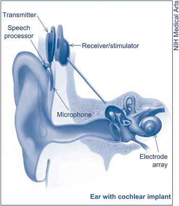
Neural interfaces are devices that are implanted into the nervous system for bidirectional communication (i.e., to both stimulate and receive recordings from the neural tissue). The seed for the concept of interfacing technology was sown approximately a century and a half ago when Fritz and Hitzig showed that the brain motor cortex, cerebral cortex regions that are involved in formulating and executing voluntary movement, could be electrically stimulated, and subsequently there have been extensive scientific efforts to develop “devices” to electrically stimulate the nervous system.Reference Mussa-Ivaldi and Miller1 This potential, however, could only be put to scientific experimentation with recent advances in microelectrodes and computer technology. Initial validation of this concept came from experiments with rats with microwire implants in the motor cortex and their ability to perform certain functions by controlling a robotic arm; this strategy was reproduced by numerous other researchers.Reference Chapin, Moxon, Markowitz and Nicolelis2 Further research in this area led to one of the earliest successful neuroprosthetics—cochlear implants for hearing (Figure 1),Reference Loeb3, 4 followed by interfacing/stimulation efforts in the visual cortexReference Bak, Girvin, Hambrecht, Kufta, Loeb and Schmidt5, Reference Normann, Maynard, Rousche and Warren6 and retina.Reference Zrenner7
A cochlear implant allows deaf patients to experience sound by stimulating the auditory nerve in the inner ear (cochlea). Sound picked up by the microphone is organized by the speech processor, which in turn is sent to the transmitter and receiver/stimulator for conversion to electric impulses. The electric impulses are gathered by the electrode array (group of electrodes) and passed on to the auditory nerve. Reproduced with permission from NIDCD.

Currently, a number of electrodes are available for applications in the central and peripheral nervous systems (for details, see References Reference Mussa-Ivaldi and Miller1, Reference Green, Lovell, Wallace and Poole-Warren8, and Reference Grill, Norman and Bellamkonda9). From a materials perspective, current electrode interfaces are made of conductive materials such as gold, platinum, iridium oxide, and glassy carbon; however, they fail to conform to the biological tissue properties, resulting in an inability to produce complete integration. Several properties of these interfaces such as the degree of inflammatory reaction at the interface and the distance between the interface and the tissue to be stimulated/recorded determine the success of neuroprosthetics.
Successful interfacing of external electronics to the human nervous system has profound potential to increase our understanding of neural function, to modulate neural function electrically to address pathologies, and to enable neuroprosthetics. The clinical implications of neural interfacing include the potential to modulate function after spinal cord injuries, neurodegenerative diseases such as amyotrophic lateral sclerosis, and other debilitating neural diseases/injuries.Reference Chapin, Moxon, Markowitz and Nicolelis2, Reference Carmena, Lebedev, Crist, O’Doherty, Santucci, Dimitrov, Patil, Henriquez and Nicolelis10–Reference Wessberg, Stambaugh, Kralik, Beck, Laubach, Chapin, Kim, Bigg, Srinivasan and Nicolelis16 In cases of severe injuries (i.e., the loss of limbs or traumatic accidents), the ability to efficiently interface prosthetic devices to the nervous system directly affects the quality of life of individuals. In principle, neuroprosthetics allow an individual to operate them just by thinking about a certain task, which is then successfully executed.
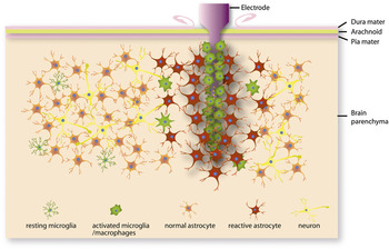
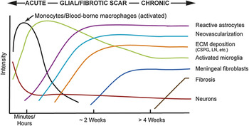
However, the widespread use of implantable neural electrodes is currently hindered by their inability to reliably record neural signals (especially in the chronic phase) due to degradation of electrode performance. The dominant hypotheses imply that electrode implantation–induced local inflammation, a breached blood brain barrier, or scar formation may contribute to the failure of neural interfaces (Figure 2).Reference He, Bellamkonda and Reichert17 Neuronal function around the electrodes generally degrades over time either due to (1) insertion-associated injury to neurons, (2) the chronic presence of a foreign material, or (3) mechanical mismatch between the stiffness of the electrode and the brain, causing chronic strain at the interface.Reference Block and Hong18–Reference Turner, Shain, Szarowski, Andersen, Martins, Isaacson and Craighead26 A close examination of these hypotheses suggests that the challenge and the solution might lie in the materials realm. In the case of chronic implantation of electrodes, the challenge lies in minimizing the plethora of cellular and biochemical events that progressively develop in the electrode/tissue interphase and contribute toward electrode failure (Figure 3).Reference He, Bellamkonda and Reichert17
Depiction of cellular changes induced in brain tissue on electrode implantation. As a response to the foreign material, several cellular events have been documented in the electrode-implanted tissue. Various non-neuronal cell types such as microglia and astrocytes are involved in this process. The change in cell shape of microglia to a macrophage-like morphology indicates that they are activated. Similarly, the enlarged size of astrocytes and their greater level of glial fibrillary acidic protein expression (darker staining) indicates the activation state of these cells. Reproduced with permission from Reference Reference He, Bellamkonda and Reichert17. ©2008, CRC Press.

Postulated sequence of the wound healing response to implanted materials in the central nervous system. The time and magnitude of the events are determined by the severity of the implantation injury as well as a number of electrode parameters, including material properties of the electrode. In addition to the induction of the activation of astrocytes and microglia, several other changes take place. Formation of new blood vessels (neovascularization) and production and deposition of a number of surface materials (i.e., extracellular matrix [ECM]) is evident. The predominant matrix materials found are chondroitin sulfate proteoglycans (CSPG) and laminin (LN). The sequence of events culminates in scar formation (fibrosis). Reproduced with permission from Reference Reference He, Bellamkonda and Reichert17. ©2008, CRC Press.

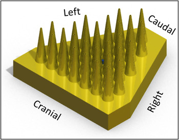
Successful interfacing technologies can have an impact on several aspects of both healthy and diseased states. Breakthroughs in neural interfacing are critical for realizing the potential of neuromodulation technologies. Neuromodulation, in turn, has the potential to impact the debilitating challenges of autism, Alzheimer’s disease, Parkinson’s disease, loss of limbs or control of limbs through traumatic brain or spinal cord injuries, depression, and severe mental illness. The technological challenge is to design interfaces such as multi-electrode arrays that can integrate well with living tissue(s);Reference Hochberg, Serruya, Friehs, Mukand, Saleh, Caplan, Branner, Chen, Penn and Donoghue11, Reference Velliste, Perel, Spalding, Whitford and Schwartz27 a schematic of one such array is depicted in Figure 4. In this context, the convergence of efforts in materials science and neuroscience are likely to lead to success. The articles in this issue of MRS Bulletin explore how materials considerations, from synthetic to natural, from stiff to soft, from micro- to nanoscale, directly impact neural interfaces—arguably the last link between sophisticated prosthetics and neural control of such prosthetics. We explore the evolution of neural interfacing technologies from a materials perspective and future challenges governing the design of neural interfaces.
A schematic of a multi-electrode array for achieving high spatial resolution for neural interfacing.

In vitro experimentation is ground zero for the field of neural interfacing and for developing a deeper understanding of the interaction of cultured neuronal cells and their networks. Arguably, a significant portion of our current understanding of neural behavior comes from cell culture studies. This is the staple of neuroscience, wherein cells are deconstructed from their networks and are probed in a culture dish. Nam discusses nano- and microscale technologies that enhance our ability to interact and probe neural cultures. In vitro studies provide valuable information on the utility of specific materials for developing interfaces that would be potentially functional in vivo. Some key areas where laboratory experimentation could shed light on the appropriateness of materials to be incorporated into interfaces are in the sensing and stimulation aspects of neurons, as well as identifying substrates that favor neuronal growth. Nam delves into these areas and discusses the utility of microelectrode arrays as a platform for electrophysiological research. Furthermore, development of microfluidic interfaces and micropatterning with proteins has enabled better maintenance of neuron networks in vitro. Ultimately, functionalization of “inert” interface materials such as gold and platinum, by conjugating proteins using appropriate chemistries, has provided a wealth of information with respect to utility of these materials for in vivo application. Recent developments in nanotechnology have also informed this line of investigation and have revealed that nanoscale patterns provide topographical cues to favor neuronal growth.
Several researchers have pursued research in the area of implantation of electrodes/devices in the peripheral nervous system (i.e., peripheral nerve interfacing [PNI]) in the last few decades. PNI presents special challenges on several fronts. These range from the design of the device to the ability of the nerve to grow through the electrode material provided. Extracting information from the peripheral nerves adds yet another tier of complexity. The general consensus in this context is that there is no simple electrode design, given that the signal-to-noise ratios can be very small. Kim and Romero-Ortega, in this issue, address the various constraints of metal electrodes that limit the use of these interfaces. A number of events that contribute to the failure of these devices include epineurial fibrosis, nerve fiber loss, and inflammatory response culminating in lack of reproducibility of recordings and variability of stimulation.
Yet another impediment to the success of these electrodes is the electrode-tissue stiffness mismatch and the resultant strain-induced scar formation at the neural electrode-brain interface. Kim and Romero-Ortega discuss various approaches taken in the incorporation of biological entities into electrode design, including neural cell adhesion molecules and growth factors such as axonin-1, NgCAM, and NGF-1. While it is challenging to obtain recordings selectively from sensory and motor neurons, the authors highlight some of the recent work in this area where compartmentalized delivery of certain neurotrophic factors have allowed specific outgrowth of different types of sensory neuron. Challenges still exist in determining whether these advances can translate to achieving sensory specific stimulation and selective recording from motor neurons.
While PNI is challenging, brain-electrode interfacing has unique materials challenges that thwart progress. The soft nature of brain tissue along with the fact that electrodes need to penetrate the protective membrane of the dura mater provide a challenging venue in which the conducting electrodes need to be within 100 microns of a cell to pick up signals with single neuron acuity. How does one design materials whose mechanical constraints vary over the course of their implantation and use? Capadona et al. describe “bioinspired” nanocomposites that offer a possible solution. The choice of a bionanocomposite is based on their observation that certain invertebrates can change the stiffness of their dermis. Specifically, materials based on the architecture of the sea cucumber dermis can provide a rigid structure initially, which can transform into soft matter via a chemical switching mechanism that disrupts the intermolecular interactions between collagen fibrils.Reference Capadona, Shanmuganathan, Tyler, Rowan and Weder28 Capadona et al. mimic this and are able to design an electrode that is rigid initially for insertion but later softens in an aqueous environment to conform to match the elasticity of the brain tissue. Initial characterization portends their superior biocompatibility over rigid electrodes.
One potential design solution for implantable electrodes is to minimize their physical and mechanical footprints in the brain by designing thin-film-based electrodes. However, the design of thin films presents significant challenges in the physiological environment when design requirements mandate functional lifetimes over 20 years. Ordonez et al. discuss the key features that could contribute to the success of thin-film technology for neuroprosthetics. Adhesion of different material layers constituting thin-film electrodes is a critical parameter in need of optimization. Several adhesion promoting materials, such as nanoscale layers of silicon carbide, are discussed. The authors also highlight other interfacing approaches and applications of lithography based on micromachining.
While neural interfacing is generally synonymous with neural-electrode interfaces, the increasing interest in optogenetics, an area combining genetics and optical tools to control biological events, and related technologies might lead to a day where the interface is optical rather than electrical in nature. Chernov et al. address the material considerations for using optical neural interfacing. One of the strong arguments in favor of optical interfacing is that it overcomes some of the deficiencies of electrode-based measurements. These include difficulty in pairing stimulation with recording, lack of spatial selectivity, and the ability to measure only membrane potential. The authors also draw parallels between design of the electrode and fiber-optic interfaces. The need for biocompatible materials for long-term optical neural interfacing is also stressed.
All of the materials currently used for electrode design are synthetic, hence it is an intriguing possibility to explore the use of extracellular matrix (ECM)-based materials for the design of electrodes to decrease “foreign body reaction” in the brain and lead to more biocompatible electrodes. This leads to the concept that long-term compatibility depends on biology to engineer the interface with minimal or no foreign material footprint. If the ECM serves to “organize” cells into tissues, could the ECM be the basis of designing implants that are more compatible and less “foreign”? Chen and Allen discuss the advantages of adapting techniques developed for Si-based materials to soft hydrogels and ECM polymers. The advantages of using ECM coatings on electrodes to circumvent adverse events such as inflammation are discussed. In addition to potentially minimizing inflammation at the electrode/tissue interface, ECM incorporation into the electrode design should provide additional benefits such as providing natural biochemical cues for neuronal growth and function. The biocompatible and biodegradable nature of ECM materials is ideal for interfacing, as it eliminates micro-injuries and minimizes elasticity mismatch. Scaffolds made using ECM materials should also find applications in several areas, especially in directional neuronal guidance. Further, ECM scaffolds “loaded” with biological molecules can be used to deliver molecules such as neurotrophic factors or cytokines.
Outlook
The current state of neural interfacing technology clearly indicates the significant advances that have taken place over the last several years. This, in large part, is due to a better understanding of physical, chemical (material), and biological aspects associated with the electrodes. Rapid advances that are occurring in methodologies in various disciplines will continue to propel the field further. With material considerations as the central theme, researchers are trying to decipher the critical elements needed for developing a viable interface. Microscale determination of physical interactions at the electrode/tissue interface is currently possible. In addition, a vast array of entities, synthetic to natural, is being explored for generating successful interfaces.
Recent advances in neuroscience are providing greater impetus toward the development of interfaces that can overcome the deficiencies of earlier technologies. Specifically, advances made through cell and molecular biological studies are allowing for an understanding of the sequence of events that takes place in the neuronal tissue subsequent to incorporation of an electrode. Knowledge gained from this line of investigation on biological changes could profoundly influence our approaches to all aspects of designing neuroprosthetics, especially materials. Synergistic interactions between the physical, chemical, and neuroscience disciplines should culminate in the development of neuroprosthetics with long-term functionality in the near future. This should fill the void that currently exists for clinical management of a number of neuronal diseases and provide much needed care for individuals suffering from these ailments.
Acknowledgments
We would like to thank Jessica Falcone for providing the image of the multi-array electrode for use in the article, and Dr. Lohitash Karumbiah, Gaurangkumar Patel, and Kristin Loomis for help with the preparation of the concept for the cover page. We would also like to acknowledge funding by the Defense Advanced Research Projects Agency MTO under the auspices of Dr. Jack Judy through the Space and Naval Warfare Systems Center, Pacific Grant/Contract No. N66001-11-1-4014.