Introduction
The collared peccary (Pecari tajacu) is a type of ‘wild pig’ whose population is globally classified as one of least concern; however, the species is already extinct in eastern and southern Argentina and is declining in some biomes (Gongora et al., Reference Gongora, Reyna-Hurtado, Beck, Taber, Altrichter and Keuroghlian2011). For Latin American communities, peccaries are economically important because of the consumption of its meat and use in leather production by the international market (Santos et al., Reference Santos, Mendes, Nogueira and Nogueira-Filho2009). In addition, the species is currently being used for the development of assisted reproductive techniques that have the potential to be applied for the conservation of closely related endangered species (Silva et al., Reference Silva, Campos, Maia and Borges2017), such as the Chacoan peccary (Catagonus wagneri).
To establish strategies for conservation of the germplasm of female peccaries, their preantral follicle (PF) population was first estimated to be 33,273.45 follicles per ovary (Lima et al., Reference Lima, Santos, Luz, Rodrigues and Silva2013). Subsequently, some efforts for the cryopreservation of peccaries’ ovarian tissue were conducted and more than 70% of the morphologically normal and viable PFs could be preserved (Lima et al., Reference Lima, Luz, Alves, Lunardi, Souza, Peixoto, Rodrigues, Oliveira and Silva2012; Moreira et al., Reference Moreira, Campos, Silva, Praxedes, Bezerra, Apolinário and Silva2017). Recently, the efficiency of an in vitro culture system for peccary ovarian tissue was demonstrated (Lima et al., Reference Lima, Luz, Lima, Rocha, Castro, Castelo, Rodrigues, Figueiredo and Silva2018). However, studies conducted on swine, the domestic species more closely related to peccaries (Cavalcante-Filho et al., Reference Cavalcante-Filho, Miglino, Machado, Bevilacqua and Neves1998), have highlighted the need for culturing isolated PFs so that they can reach subsequent developmental stages (Lima et al., Reference Lima, Rubessa, Rocha, Winters, Milner, Campello, Figueiredo and Wheeler2017; Sirotkin et al., Reference Sirotkin, Florkovičová (Koničková), Schaeffer, Laurincik and Harrath2017). Despite the phylogenetic proximity of collared peccary to the domestic swine (Gongora et al., Reference Gongora, Reyna-Hurtado, Beck, Taber, Altrichter and Keuroghlian2011), there are marked differences between the species with relation to its reproductive physiology (Mayor et al., Reference Mayor, Fenech, Bodmer and Lopez-Bejar2006; Lima et al., Reference Lima, Santos, Luz, Rodrigues and Silva2013) and application of assisted reproductive technologies (Lima et al., Reference Lima, Santos, Luz, Rodrigues and Silva2013; Silva et al., Reference Silva, Campos, Maia and Borges2017), which justifies the investigation of protocols established for the peccaries.
The current methods for the isolation of PFs can be classified as enzymatic and/or mechanical. The enzymatic method consists of exposing the ovarian tissue to the action of enzymes, such as collagenase, as reported for swine (Choi et al., Reference Choi, Seung and Jeong2008). However, using this method, the basal membrane or theca cells are damaged in many of the isolated PFs, especially when the incubation time is not strictly controlled (Rossetto et al., Reference Rossetto, Lima, Saraiva, Lima-Verde, Sales and Figueiredo2011). Conversely, the mechanical method has the advantage of maintaining the integrity of the follicular and basal membrane structure, as well as the interactions among the oocytes, granulosa cells, and theca cells. Despite being slower and more laborious than the enzymatic method and allowing the recovery of a low number of follicles, it has been recommended for the isolation of porcine PFs (Ahn et al., Reference Ahn, Lee, Park, Kim, Park, Choi and Lim2012). To optimize the isolation of PFs, a combination of enzymatic and mechanical methods was successfully applied for cattle (Figueiredo et al., Reference Figueiredo, Hulshof and Van Den Hurk1993), human (Dong et al., Reference Dong, Ma, Shi, Dai, Liu, Su, Guo, Wang and Sun2014), and sheep (Sadeghnia et al., Reference Sadeghnia, Akhondi, Hossein, Mobini, Hosseini, Naderi and Shirazi2016). After isolation, PFs are usually evaluated for viability and development through in vitro culturing followed by evaluation with fluorescent probes, but electron microscopy has now emerged as an effective method to provide accurate details of the ultrastructure of PFs (Klein et al., Reference Klein, Buhr, Frase and Hawkes2012).
To establish a protocol for the isolation of collared peccary PFs, the purpose of the present study was to compare the efficiency of mechanical and enzymatic methods, alone or in combination, in conserving the morphology and viability of peccary PFs. Moreover, we confirmed the viability of the PFs obtained using the most effective method through scanning electron microscopy (SEM) and epifluorescence microscopy after a short in vitro culture.
Materials and methods
Source of ovaries
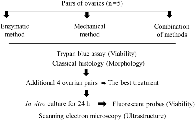
The ovaries (n = 12 ovaries) from six adult (∼3.5 years old) collared peccaries were collected. The animals belonged to the Center of Wild Animal Multiplication (IBAMA Register No. 1478912), UFERSA, Mossoró, RN, Brazil (5°10′S, 37°10′W). This centre presently shelters 200 collared peccaries that are used in studies that focus on the development of productive management practices. A programmed slaughter is conducted every year for population control, and the carcasses are used in various studies in the fields of morphology, histology, physiology, pathology, etc. For our study, immediately post mortem, the ovaries were washed in 70% alcohol and TCM-HEPES and then transported to the laboratory, in which they were weighed and the length, width, and thickness were measured using a pachymeter. Then, ovaries were destined to an experimental design (Fig. 1) in which an initial screening trial was conducted by comparing different protocols for PFs isolation, followed by a validation trial at using the best protocol.
Experimental design for the evaluation of different methods for isolation of collared peccary ovarian preantral follicles.

Comparison of protocols for the isolation of PFs
For all the ovaries, the corpora lutea and antral follicles, if present, were removed. The ovarian cortexes from the same individual were randomly assigned to the mechanical or enzymatic methods for isolation of PFs.
For the enzymatic procedure (Figueiredo et al., Reference Figueiredo, Hulshof and Van Den Hurk1993; with slight modifications), the ovarian cortex was divided into small fragments (1 × 1 × 1 mm), which were then suspended in TCM-HEPES after the addition of 0.5 mg/ml type IV collagenase for 20 min at 37°C in a water bath. Every 5 min, the suspensions were gently homogenized 40 times with a Pasteur pipette. After 20 min, the collagenase activity was blocked through the addition of 10% fetal bovine serum (FBS) to the suspensions that were gently homogenized using a Pasteur pipette; the suspensions were then added to TCM-HEPES with 1% BSA and incubated for 10 min. The suspensions were filtered first through 500-μm and then through 200-μm nylon mesh filters, and the filtrates were centrifuged at 1600 g for 5 min and evaluated.
For mechanical isolation, the ovarian cortex was placed in a Petri dish and subjected to dissociation by using a scalpel blade. The suspension was then transferred to a 50 ml Falcon tube containing 5 ml of TCM-HEPES and gently homogenized 40 times with a Pasteur pipette. The suspensions were then filtered first through 500-μm and then through 200-μm nylon mesh filters. The filtrates were then centrifuged at 1600 g for 5 min and evaluated (Figueiredo et al., Reference Figueiredo, Hulshof and Van Den Hurk1993).
For the combination of the enzymatic and mechanic isolations, the remaining filtrate from the nylon mesh filters used during mechanical isolation was submitted to the enzymatic procedure as previously described.
Initial screening trial
After each method, the number of isolated PFs in the suspension was quantified and classified according to the method of Hulshof et al. (Reference Hulshof, Figueiredo, Beckers, Bevers and Van Den Hurk1994) using an inverted microscope (Leica, Episcopic Fluorescent Attachment EFA Halogen Lamp Set, Leica Microsystems Inc., Bannockburn, USA). In summary, the primordial follicles were considered to be those that had an immature oocyte in the centre of the follicle surrounded by a layer of pre-granulosa cells in a pavement format; primary follicles, those with an immature oocyte in the center of the follicle surrounded by a layer of granulosa cells arranged cubically; and secondary follicles, those with an immature oocyte surrounded by two or more layers of cubic granulosa cells in the presence of theca cells (Hulshof et al., Reference Hulshof, Figueiredo, Beckers, Bevers and Van Den Hurk1994). Furthermore, the denuded oocytes (follicles without granulosa cells or partially surrounded by them) were also counted (Lopes et al., Reference Lopes, Santos, Celestino, Melo, Chaves, Campello, Silva, Báo, Jewgenow and Figueiredo2009).
For viability analysis, the suspension obtained after isolation procedures was centrifuged for 10 min, and 10 μl trypan blue (0.4%) (Sigma Chemical Co., St. Louis, MO, USA) was added to the precipitate at 25°C. After 5 min, the samples were evaluated under an inverted microscope (Eclipse TS-100F, Nikon Corporation, Tokyo, Japan), and PFs were classified as viable, if unstained, and not viable, if stained blue (Lopes et al., Reference Lopes, Santos, Celestino, Melo, Chaves, Campello, Silva, Báo, Jewgenow and Figueiredo2009).
Scanning electron microscopy analysis
To evaluate details related to the ultrastructural integrity of the isolated PFs, ovaries derived from four additional adult females were subjected to the most effective isolation method, and the PFs were fixed in 2% glutaraldehyde. After fixation, the PFs were recovered using a pipette and mounted onto stubs with the aid of a carbon tape. For metallization, the stubs were placed on a metallizer (Q150T ES; Quorum Technologies, Guelph, Ontario, Canada) and metalized with a 20-nm layer of gold. SEM (Quanta 450-FEG; FEI, Hillsboro, OR, USA) observations of at least 10 different PFs were conducted according to the methodology described by Bustos-Obregon and Fléchon (Reference Bustos-Obregon and Fléchon1975).
In vitro culture of PFs
To obtain more reliable results than those observed through the use of vital staining during the initial screening trial, ovaries derived from four additional females were subjected to the most effective isolation method, subjected to a 24 h in vitro culture and then evaluated for viability by using fluorescent probes (Santos et al., Reference Santos, Biondi, Cordeiro, Miranda, Dantas, Figueiredo and Ohashi2006). Here, 20 intact follicles were selected and individually cultured in 20 μl of the culture medium with mineral oil for 24 h at 38.5°C in a humidified atmosphere of 5% CO2 in air (adapted from Demeestere et al., Reference Demeestere, Delbaere, Gervy, Van Den Bergh, Devreker and Englert2002). The basic culture medium consisted of TCM199 (pH 7.2–7.4) supplemented with ITS (insulin 10 μg/ml, transferrin 5.5 μg/ml, and selenium 5.0 ng/ml), 0.23 mM pyruvate, 2 mM glutamine, 2 mM hypoxanthine, and 1.25 mg/ml BSA.
After culture, the isolated PFs were incubated in a solution constituted by a mixture of propidium iodide (2 µM in salt solution) and Hoechst 33342 (40 µM in dimethyl sulfoxide) for 10 min at 37°C to detect follicular viability and to enable the counting of nuclei, respectively. After labelling, the stained follicles were placed on a glass microscope slide and examined under an epifluorescence microscope equipped with a digital camera. Oocytes and granulosa cells were classified as degenerating if the chromatin was stained positively with propidium iodide and as viable if the chromatin was unlabelled with propidium iodide. The percentages of viable granulosa cells were calculated in relation to the total number of Hoechst-positive nuclei. Follicles with a viable oocyte surrounded by ≥90% viable granulosa cells were considered viable (Santos et al., Reference Santos, Biondi, Cordeiro, Miranda, Dantas, Figueiredo and Ohashi2006).
Statistical analysis
Data were expressed as mean ± standard error and were initially subjected to Smirnov–Kolmogorov and Bartlett tests to confirm the normal distribution and homogeneity of variance, respectively. Analysis of variance (ANOVA) was then carried out using the Statview (5.0) software, and Fisher’s protected least significant difference (PLSD) test was applied for comparison of the isolation methods (enzymatic, mechanical, or combined). Comparisons among the treatments in relation to the percentage of viable PFs were conducted using the chi-squared test. Values were considered statistically significant at P-value < 0.05.
Results
The mean weight of the ovaries used in the present study was 1.85 ± 0.32 g. The length, width, and thickness were 1.68 ± 0.13 cm, 1.29 ± 0.09 cm, and 1.11 ± 0.06 cm, respectively.
Compared with the mechanical method (434.3 ± 88.9 PFs), the enzymatic method (961.7 ± 132.9 PFs) provided a higher number of isolated PFs (P < 0.05), but the number of isolated PFs did not differ from the combined method (743.2 ± 92.8 PFs) (Table 1). The highest number of isolated follicles was classified as primary, regardless of the method used (Table 1). It was possible to verify the presence of denuded oocytes in all the methods, but there was no statistical difference among them (mechanical: 10.3 ± 6.1; enzymatic: 3.2 ± 3.2; combined: 10.3 ± 6.1 oocyte). From this initial analysis, the highest percentage of viable PFs (P < 0.05), as determined by the trypan blue assay, was obtained by the enzymatic method (98.7 ± 0.6%) in comparison with the mechanical (89.2 ± 1.6%) or the combined (90.2 ± 1.9%) method.
Mean values (mean ± SEM) for collared peccaries’ ovarian preantral follicles isolated by the mechanical or enzymatic methods, or the association of both (n = 6 ovarian pairs)

a,bSuperscript lowercase letters compare follicular categories into each treatment.
A,BSuperscript capital letters compare methods regarding the total number of isolated follicles (P < 0.05).
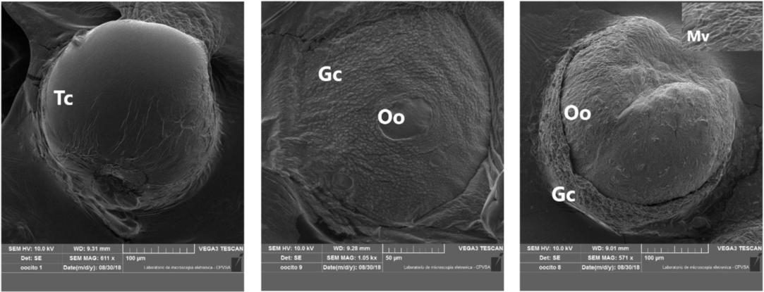
We confirmed the integrity of PFs obtained by the enzymatic method by using SEM that provided images of the follicular surface (Fig. 2). From these images, we were able to observe the structural elements to be the theca cells and granulosa cells, in which the oocytes remained intact in the PFs evaluated. The theca layer was distributed as a continuous thin layer with a smooth surface appearance. The granulosa cells formed a compact layer around the oocyte, whose surface was covered by numerous microvilli.
Scanning electron microscopy images showing intact structures on the surface of collared peccaries’ ovarian preantral follicles isolated by the enzymatic method. Note that it is possible to observe the theca cells (Tc) layer surface, the compacted granulosa cells (Gc) and the oocyte (Oo) presenting numerous superficial microvilli (Mv, highlighted in the inset).

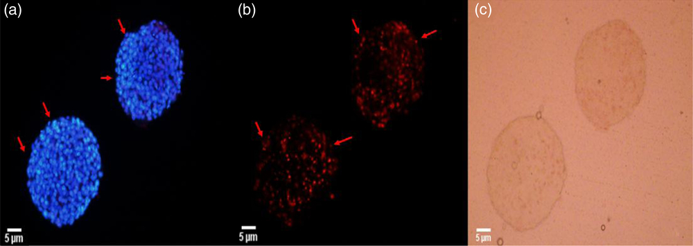
Visualization through epifluorescence microscopy following a short in vitro culture indicated that enzymatic procedure is capable of providing an amount of 86% viable PFs (Fig. 3).
Representative images of collared peccaries’ ovarian preantral follicles submitted to a short in vitro culture for 24 h after enzymatic isolation. Note the presence of granulosa cells of marked in blue by the Hoechst probe indicating viable follicles (a) with the absence of the propidium iodide impregnation (b), and also the follicles evaluated under the clear field (c).

Discussion
To improve the technologies for the recovery and storage of the germplasm of female collared peccaries, we demonstrate, for the first time, the application of protocols for the isolation of PFs from this species. Although viable PFs can be obtained by any of the methods tested here, the enzymatic procedure proved to be the most efficient. This fact highlights the effective action of the enzyme collagenase that promotes efficient dissociation of PFs from peccaries, in which ovaries are surrounded by a layer of fibrous connective tissue (Mayor et al., Reference Mayor, Fenech, Bodmer and Lopez-Bejar2006).
Even if the enzymatic method had provided the recovery of the highest number of ovarian follicles from the peccary ovaries, this number was many times fewer than those observed for domestic swine (∼185,000 PFs per ovary; Greenwald and Moor, Reference Greenwald and Moor1989). This can be explained as the domestic swine presents a follicular population (>400,000 PFs per ovarian pair; Gosden and Telfer, Reference Gosden and Telfer1987) many times higher than that of peccaries (∼60,000 PFs per ovarian pair; Lima et al., Reference Lima, Santos, Luz, Rodrigues and Silva2013). It is necessary to highlight that in addition providing the highest number of isolated PFs from peccaries, the enzymatic method also provided a higher percentage of viable follicles (98.7 ± 0.6%) than that by the mechanical (89.2 ± 1.6%) or the combined (90.2 ± 1.9%) methods, as evaluated using the trypan blue assay. Such values were even higher than those reported for domestic swine in which 74% viable follicles were recovered after enzymatic isolation (Ahn et al., Reference Ahn, Lee, Park, Kim, Park, Choi and Lim2012).
The collagenase used in the present study was a partially purified bacterial enzyme (Clostridium histolyticum) that could attack native collagen without affecting related proteins and damaging the epithelial tissue (Mandl et al., Reference Mandl, Zipper and Ferguson1958). The efficiency of type IV collagenase enzyme has previously been proven in bovine (Figueiredo et al., Reference Figueiredo, Hulshof and Van Den Hurk1993), caprine (Machado et al., Reference Machado, Rodrigues, Brasil, Amorim, Matos, Santos and Figueiredo2002), porcine (Choi et al., Reference Choi, Seung and Jeong2008), human (Lierman et al., Reference Lierman, Tilleman, Cornelissen, De Vos, Weyers, T’Sjoen, Cuvelier and De Sutter2014), and murine (Young et al., Reference Young, Drummond, Akers, Bartle, Kennedy and Asaduzzaman2017) tissues. In addition, the crucial factors of this enzyme are its concentration and duration of digestion (Shuttleworth et al., Reference Shuttleworth, Pipkin and Hunter2002). In the present study, the enzyme incubation time was 20 min, which was sufficient to isolate a large number of viable peccary PFs, as has been previously reported for rabbits (Nicosia et al., Reference Nicosia, Evangelista and Batta1975), bovines (Figueiredo et al., Reference Figueiredo, Hulshof and Van Den Hurk1993), equines (Telfer and Watson, Reference Telfer and Watson2000), porcines (Choi et al., Reference Choi, Seung and Jeong2008), and humans (Lierman et al., Reference Lierman, Tilleman, Cornelissen, De Vos, Weyers, T’Sjoen, Cuvelier and De Sutter2014), in all of which, the incubation time should not exceed 30 min. Moreover, the addition of up to 5% FBS in the collagenase solution seemed to ensure better preservation of the follicle cytostructure without interfering with the enzymatic activity and final yield (Nicosia et al., Reference Nicosia, Evangelista and Batta1975). In the present experiment, 10% FBS was used, which was enough to prevent the basal membrane digestion in some follicles.
Compared with the enzymatic method, we highlighted that it is possible to isolate viable peccary PFs by the mechanical method, even if a lower amount was provided. Probably, the scalpel blade used for this purpose was not efficient enough to dissociate the connective tissue and, therefore, released less follicles (434.3 ± 88.9) than those obtained for prepubertal gilts (599.160 ± 74.089; Alves et al., Reference Alves, Alves, Araújo, Beletti, Gambarini and Jacomini2012). Furthermore, it is worth noting that the mechanical method is a cheap and easily accessible procedure that can be applied at any laboratory. In addition, it is evident that the combination of mechanical and enzymatic methods can improve the efficiency of the mechanical method and, therefore, increase the number of viable follicles isolated from peccaries as previously reported for buffalos (Santos et al., Reference Santos, Biondi, Cordeiro, Miranda, Dantas, Figueiredo and Ohashi2006), humans (Dong et al., Reference Dong, Ma, Shi, Dai, Liu, Su, Guo, Wang and Sun2014), and sheep (Sadeghnia et al., Reference Sadeghnia, Akhondi, Hossein, Mobini, Hosseini, Naderi and Shirazi2016).
By comparing the follicular categories, we verified that most of the isolated follicles were primary, regardless of the method used. This result is interesting because it has been established previously that most of the peccary follicle population is composed of primordial (91.56%) follicles, followed by primary (6.29%) and secondary (2.15%) follicles (Lima et al., Reference Lima, Santos, Luz, Rodrigues and Silva2013). This discrepancy is possibly because of the primordial follicles being more intimately embedded in the tunica albuginea, which may make it difficult for the follicles to dissociate, as previously reported for other species, including caprine (Machado et al., Reference Machado, Rodrigues, Brasil, Amorim, Matos, Santos and Figueiredo2002), swine (Kerong et al., Reference Kerong, Xuefeng, Liying, Dengke, Yunhai, Yonghui, Xuemei, Xiaoxiang, Changxin and Ning2007), and humans (Dong et al., Reference Dong, Ma, Shi, Dai, Liu, Su, Guo, Wang and Sun2014).
We also highlighted the presence of denuded oocytes after the isolation of follicles in all the tested methods. This can be attributed to the action of collagenase or mechanical instruments that can destroy the membranes of many follicles during the isolation, therefore, denuding them (Dong et al., Reference Dong, Ma, Shi, Dai, Liu, Su, Guo, Wang and Sun2014). In the present experiment, an average of 3.2 ± 3.2 oocytes was observed in the enzymatic method with no differences observed between methods (P > 0.05). In bovines, however, some studies have shown that the presence of denuded oocytes is the most evident in the enzymatic method, suggesting that the enzyme can rupture the follicular membrane (Figueiredo et al., Reference Figueiredo, Hulshof and Van Den Hurk1993), which did not occur in the present study.
As an additional attempt to confirm the integrity of PFs isolated by the enzymatic method, which was the most efficient method that we observed here, we conducted SEM analysis that provided unprecedented detailed ultrastructural images of the surface of peccary PFs. Transmission electron microscopy, associated with other in vitro analytical studies, has a well recognized diagnostic–prognostic role in the assessment of ovarian follicle and oocyte viability (Nottola et al., Reference Nottola, Cecconi, Bianchi, Motta, Rossi, Continenza and Macchiarelli2011). However, SEM is not commonly used to verify the PF viability. In the present study, SEM provided important information regarding the maintenance of the PF surface architecture after enzymatic isolation. The ultrastructural view of peccary PF surface structures presented general similarities with those that have been described previously for mice, although the double theca layer usually described for this species (Nottola et al., Reference Nottola, Cecconi, Bianchi, Motta, Rossi, Continenza and Macchiarelli2011) was not observed in the peccary PFs evaluated here. In addition, we highlighted the presence of numerous microvilli on the peccary oocyte surface that are probably involved in apposition and fusion of the sperm and oocyte membranes during fertilization (Runge et al., Reference Runge, Evans, He, Gupta, McDonald, Stahlberg, Primakoff and Myles2007).
To support the interpretations of the SEM results, the follicles isolated by the enzymatic method were cultured for 24 h. This short-culture has been shown to be an effective procedure in the evaluation of follicular viability as the follicular damage appears only after a few hours, when the follicle returns to its physiological conditions in which case follicular growth does not occur anymore (Vanhoutte et al., Reference Vanhoutte, Cortvrindt, Nogueira and Smitz2004). In the present study, peccary PF viability was confirmed using fluorescent probes that confirmed the presence of an intact basal membrane around isolated follicles, similar to that for domestic swine (Ahn et al., Reference Ahn, Lee, Park, Kim, Park, Choi and Lim2012), sheep (Lakshminarayana et al., Reference Lakshminarayana, Praveen Chakravarthi, Brahmaiahb and Rao2014), and mice (Zhang et al., Reference Zhang, Wu, Weng, Xu, Chen, Zheng, Lin, Liu and Zhou2017).
In conclusion, we clearly demonstrated that the enzymatic method is an efficient procedure for the isolation of peccary ovarian PFs. However, we highlighted that it is alternatively possible to obtain viable follicles by the mechanical method or by combining both methods. The information generated here presents ample evidence for the positive effect on the availability of competent oocytes in terms of future applicability for in vitro development and fertilization. Such techniques, in association with cryopreservation, will contribute to the conservation and multiplication of germplasm derived from valuable endangered tayassuids.
Acknowledgements
The authors thank CEMAS/UFERSA for providing the animals used in the experiment.
Ethical standards
The ethics committee of the UFERSA approved the experimental protocols, as well as the animal care procedures used (no. 23091.006525/2016-82). The study was authorized by the Chico Mendes Institute for Biodiversity (SISBio no. 37329). All chemicals used in this study were purchased from Sigma Chemical Co. (St. Louis, MO, USA) unless indicated otherwise.
Financial support
This study was financed in part by the Coordenação de Aperfeiçoamento de Pessoal de Nível Superior – Brasil (CAPES, Financial Code 001) and the National Council for the Scientific Development (CNPq, Process No. 407302/2013–1).
Statement of interest
The authors have no conflicts of interest to declare