Introduction
In the current era, extracorporeal membrane oxygenation (ECMO) has become an integral component in the management of paediatric patients undergoing surgery for complex congenital cardiac malformations. Outcomes of congenital cardiac surgery have improved significantly over the past decades; however, a small proportion of patients (3.2–8.4%) still require ECMO support in the perioperative period due to cardiorespiratory failure. Reference Khorsandi, Davidson and Bouamra1,Reference Baslaim and Jamjoom2 In selected cases, ECMO may be initiated before surgery as a bridge to cardiac repair. Reference Khorsandi, Davidson and Bouamra1 The primary indication for veno-arterial extracorporeal membrane oxygenation (VA-ECMO) in the operating room is failure to wean from cardiopulmonary bypass (CPB) due to severe low cardiac output syndrome, borderline haemodynamics requiring high inotropic support, or respiratory failure caused by pulmonary hypertension or pulmonary oedema. Reference Perry, Brown, Misfeldt, Lehenbauer and Cooper3–Reference Brown, Moynihan and Deatrick5 In such situations, ECMO acts as a bridge to recovery by providing myocardial rest following cardiopulmonary bypass and cardioplegic arrest while maintaining adequate systemic perfusion. Reference Perry, Brown, Misfeldt, Lehenbauer and Cooper3–Reference Brown, Moynihan and Deatrick5 It also allows time to identify and correct residual lesions through transcatheter or surgical interventions. In cases where cardiac function does not recover, ECMO may serve as a bridge to transplantation or facilitate end-of-life decision-making. Reference Khorsandi, Davidson and Bouamra1,Reference Alsoufi, Al-Radi and Gruenwald6 In the post-operative period, sudden cardiac arrest requiring emergency ECMO initiation, also known as extracorporeal cardiopulmonary resuscitation (ECPR ECMO), remains an important indication. Reference Perry, Brown, Misfeldt, Lehenbauer and Cooper3,Reference Guerguerian, Sano, Todd, Honjo, Alexander and Raman7 Persistent low cardiac output syndrome despite maximal medical therapy, respiratory failure, septic shock, and organ dysfunction are additional indications. Reference Yuerek, Rossano, Mascio and Shaddy8 Despite growing experience, data on long-term outcomes following ECMO remain limited. This study aims to evaluate incidence, short-and long-term outcomes, and factors affecting ECMO outcomes after congenital cardiac surgery.
Materials & methods
All neonatal and paediatric patients (age <16 years) who required VA-ECMO before or after congenital cardiac surgery between January 2013 and December 2022 were included in this retrospective study. Patients who underwent VA-ECMO in the perioperative period were identified from the institutional ECMO database, and detailed clinical information was collected from hospital medical records.
Patients were classified according to the timing of ECMO initiation in the perioperative period: (A) pre-operative ECMO, including patients requiring ECMO before surgery, with some successfully weaned after surgery and others continued on ECMO; (B) intra-operative ECMO, including patients requiring ECMO in the operating room at the end of surgery due to failure to wean from cardiopulmonary bypass; (C) post-operative ECMO, including patients requiring ECMO in the immediate post-operative period (<24 h after surgery) or early post-operative period (>24 h after surgery) while in the ICU; and (D) extracorporeal cardiopulmonary resuscitation ECMO (ECPR ECMO), including patients requiring emergency ECMO following sudden cardiac arrest in the perioperative period.
The decision to initiate ECMO was made by a multidisciplinary team. The full surgical team, including surgeons, anaesthesiologists, perfusionists, nurse specialists, and ECMO specialists (where applicable), was present during every ECMO initiation. For ECMO maintenance in the PICU, one nurse specialist and one perfusionist were assigned to continuously monitor the ECMO circuit under the supervision of an ECMO specialist. In addition, a dedicated ECMO coordinator supervised and coordinated the overall ECMO programme, including record maintenance and quality oversight.
ECMO cannulation strategy was individualised based on patient size, anatomy, timing of ECMO initiation, and clinical status. Central cannulation through the great vessels was the preferred approach for most intra-operative, immediate post-operative, and ECPR cases. Cervical cannulation was selectively utilised in pre-operative or late post-operative patients. In patients requiring prolonged ECMO support, conversion from central to peripheral cannulation was occasionally performed to facilitate delayed sternal closure and reduce complications associated with prolonged open chest management.
Patients were risk stratified according to the Risk Adjustment for Congenital Heart Surgery (RACHS) score based on the type of structural heart disease and surgical procedure performed. Follow-up data for survival were obtained from hospital records and outpatient follow-up documentation.
Statistical analysis
The presentation of the categorical variables was done in the form of numbers and percentages (%). Quantitative data were presented as the means ± SD and as median with 25th and 75th percentiles (interquartile range). Data normality was checked by using Kolmogorov–Smirnov test. In cases in which data was not normal, we used nonparametric tests. The following statistical tests were applied for the results:
-
1. Association of the variables which were quantitative and not normally distributed in nature was analysed using the Mann–Whitney test and variables which were quantitative and normally distributed in nature were analysed using an independent t-test. Kruskal Wallis test was used for the association of weight (kg) with age.
-
2. Association of the variables which were qualitative in nature was analysed using the Chi-square test. If any cell had an expected value of less than 5 then Fisher’s exact test was used.
-
3. A receiver operating characteristic curve was used to assess the cut-off point, sensitivity, specificity, positive predictive value, and negative predictive value of CPB and cross-clamp time (minutes) and duration of ECMO for predicting mortality.
-
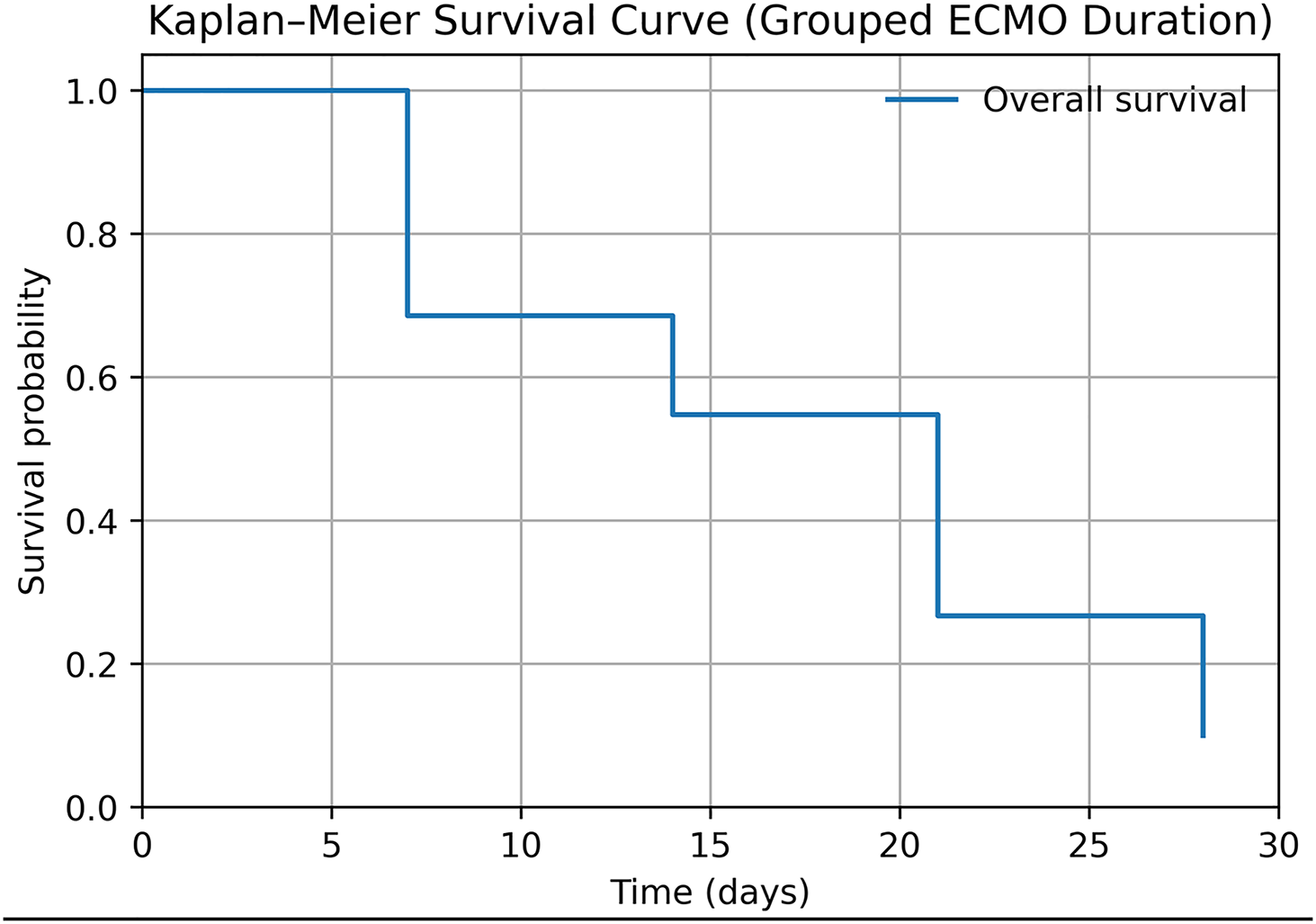
4. Kaplan–Meier survival curves were constructed using grouped survival data derived from ECMO duration categories. Survival probabilities were approximated at defined time intervals due to the absence of individual time-to-event data.
-
5. Multivariate logistic regression analysis was performed to identify independent predictors of mortality. Variables with p < 0.1 on univariate analysis and clinically relevant variables were included in the model. Adjusted odds ratios with 95% confidence intervals were calculated. Model fit was assessed using standard goodness-of-fit measures.
Data entry was done in the Microsoft EXCEL spreadsheet, and the final analysis was done with the use of the Statistical Package for Social Sciences (SPSS) software, IBM manufacturer, Chicago, USA, ver. 25.0. For statistical significance, p value of less than 0.05 was considered statistically significant.
Results
Over the last 10 years, 150 patients required ECMO during the perioperative period of congenital cardiac surgery. Among them, 123 patients (82%) were successfully separated from ECMO, 91 patients (60.7%) were weaned from mechanical ventilation, and 81 patients (54%) survived to hospital discharge. A total of 173 ECMO runs were performed, with 132 patients requiring a single ECMO run and 18 requiring multiple runs.
Demographic characteristics
There were 83 male and 67 female patients, with no significant association between sex and mortality (p = 0.295). The age ranged from 1 day to 6067 days (mean 514.5 days, median 59.5 days), and the mean body weight was 8.01 kg (median 3.9 kg). Most patients were younger than 1 year. Neonates showed higher mortality compared to infants and older children; however, the association was not statistically significant (Supplementary Table 1).
Pre-operative risk factors
Pre-operative mechanical ventilation and cardiac arrest were not associated with poor outcomes. Premature birth, chromosomal abnormalities, non-cardiac systemic abnormalities, pre-existing sepsis, pulmonary complications, and gastrointestinal complications were associated with an increased risk of mortality. However, the risk is statistically significant only for the patients with associated non-cardiac systemic abnormalities (Supplementary Table 2).
Operative factors
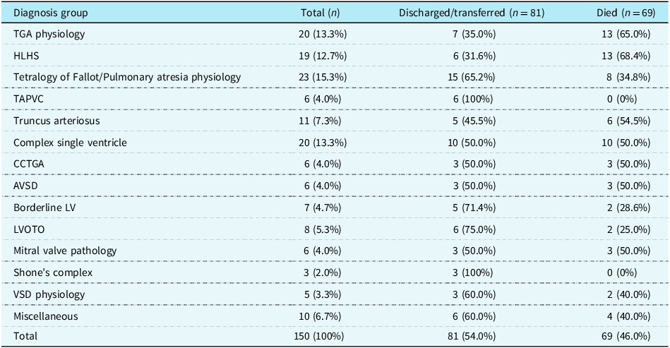
The diagnoses and surgical procedures varied widely, with tetralogy of Fallot with pulmonary atresia, transposition of the great arteries, and hypoplastic left heart syndrome being the most common. Complex single-ventricle physiology also constituted a significant proportion of cases, although diagnosis was not significantly associated with survival (Table 1 and Supplementary Table 6).
Diagnosis distribution and association with survival

Table 1. Long description
A table with 150 total cases, 81 discharged or transferred, and 69 deaths. The table has 15 rows and 3 columns. Column headers are Diagnosis group, Total (n), Discharged/transferred (n = 81), and Died (n = 69). Row labels include various diagnosis groups such as TGA physiology, HLHS, Tetralogy of Fallot/Pulmonary atresia physiology, TAPVC, Truncus arteriosus, Complex single ventricle, CCTGA, AVSD, Borderline LV, LVOTO, Mitral valve pathology, Shone’s complex, VSD physiology, Miscellaneous, and Total. Each row lists the total number of cases, the number of discharged or transferred cases, and the number of deaths for each diagnosis group. Notable trends include high mortality rates for TGA physiology, HLHS, and Complex single ventricle, while TAPVC and Shone’s complex have zero deaths.
Data are presented as number (percentage). Percentages for diagnosis groups are calculated from total cohort (N = 150). p value derived using Fisher’s exact test (p = 0.125); not statistically significant.
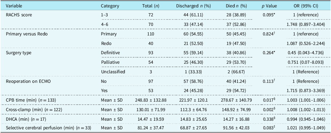
Cardiopulmonary bypass (CPB) and aortic cross-clamp duration were significant predictors of mortality. Receiver operating characteristic analysis demonstrated that cardiopulmonary bypass duration >221 minutes and cross-clamp time >125 minutes were associated with increased mortality after ECMO. Deep hypothermic circulatory arrest (DHCA) and selective cerebral perfusion were not associated with increased mortality. Surgical complexity based on the Risk Adjustment for Congenital Heart Surgery (RACHS) score and redo surgery did not significantly affect survival (Table 2 and Supplementary Table 3).
Association of operation-related factors with survival

Table 2. Long description
A table comparing operation-related factors with survival rates. The table has 11 rows and 7 columns. Column headers are Variable, Category, Total (n), Discharged n (%), Died n (%), p Value, OR (95% CI). Row labels include RACHS score, Primary versus Redo, Surgery type, Reoperation on ECMO, CPB time (min), Cross-clamp (min), DHCA (min), Selective cerebral perfusion (min). Each row provides data on the number of patients discharged and died, p values, and odds ratios with confidence intervals. Notable trends include significant p values for CPB time and Cross-clamp time, indicating their association with increased mortality.
CI = Confidence interval.
* Fisher’s exact test.
†Chi-square test.
‡Independent t-test.
§Mann–Whitney U test.
Type and timing of ECMO
Most ECMO runs occurred in the post-operative period, including 54 in the immediate post-operative period and 57 in the early post-operative period, while 51 were instituted intraoperatively and 11 preoperatively. ECPR ECMO were required in 63 cases, mostly in the post-operative period. ECMO initiated after 24 h of surgery was associated with significantly higher mortality compared with other groups (p = 0.026) (Table 3 and Figure 1).
Association of different types of ECMO runs with ECMO survival, PICU survival, and Hospital Survival.

Association of different types of ECMO run with survival

Table 3. Long description
The table compares survival rates and mortality for different types of ECMO runs. It has five rows and six columns. The columns are labeled Type of ECMO run, Total, Survival to discharge, Expired, and p Value. The row labels are Pre-operative, Intra-operative, Immediate post-operative (<24 h), Early post-operative (>24 h), and ECPR. The values in the table are as follows: Pre-operative: Total 11, Survival to discharge 8 (72.73 percent), Expired 3 (27.27 percent), p Value 0.117. Intra-operative: Total 51, Survival to discharge 22 (43.14 percent), Expired 29 (56.86 percent), p Value 0.5. Immediate post-operative (<24 h): Total 54, Survival to discharge 31 (57.41 percent), Expired 23 (42.59 percent), p Value 0.067. Early post-operative (>24 h): Total 57, Survival to discharge 20 (35.09 percent), Expired 37 (64.91 percent), p Value 0.026. ECPR: Total 63, Survival to discharge 29 (46.03 percent), Expired 34 (53.97 percent), p Value 0.832.
* Fisher’s exact test.
†Chi-square test.
Out of the 173 ECMO runs, 146 (84.9%) utilised central cannulation, 18 (10.5%) utilised cervical cannulation, and 9 (5.2%) underwent conversion from central to peripheral cannulation. Exploratory analysis did not demonstrate a statistically significant association between cannulation strategy and ECMO outcomes.
ECPR ECMO
Among the 63 ECPR ECMO runs, 6 were initiated preoperatively, 33 during the immediate post-operative period, and 24 during the early post-operative period. Comparative analysis between ECPR and non-ECPR patients demonstrated no significant difference in short-or long-term survival outcomes. However, the ECPR group had significantly lower body weight compared with the non-ECPR cohort (median 3.6 versus 4.6 kg, p = 0.016). Within the ECPR cohort, multivariable logistic regression analysis was performed to identify predictors of hospital mortality. No independent predictors reached statistical significance, likely reflecting the relatively small sample size and limited number of mortality events. However, a higher Risk Adjustment for Congenital Heart Surgery (RACHS) score demonstrated a trend toward association with mortality (adjusted OR 1.83, 95% CI 0.92–3.65, p = 0.085).
ECMO duration and number of ECMO runs
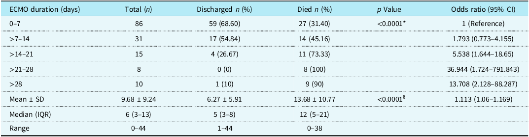
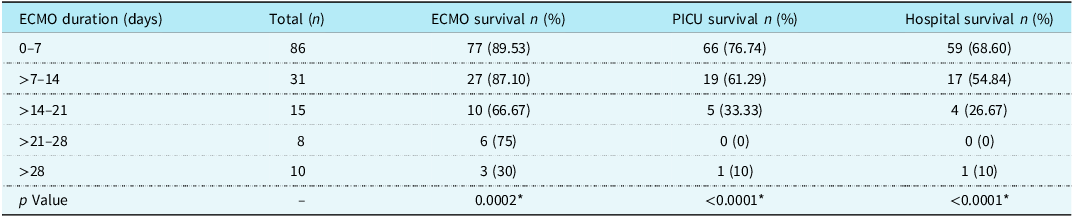
The mean ECMO duration was 9.68 days (median 6 days). Survival decreased significantly with increasing ECMO duration, with survival highest for duration <7 days and less than 10% when ECMO exceeded 21 days (p < 0.001). Receiver operating characteristic analysis showed ECMO duration >10 days predicted higher mortality (Tables 4 and 5). Kaplan–Meier analysis demonstrated a progressive decline in survival with increasing ECMO duration (Figure 2).
Association of ECMO duration with survival

Table 4. Long description
The table has six rows and six columns. The columns are labeled ECMO duration (days), Total (n), Discharged n (%), Died n (%), p Value, and Odds ratio (95% CI). The rows are grouped by ECMO duration ranges: 0-7 days, >7-14 days, >14-21 days, >21-28 days, and >28 days. Additionally, there are summary rows for Mean ± SD, Median (IQR), and Range. Each row provides the total number of cases, the number and percentage of patients discharged, the number and percentage of patients who died, the p value, and the odds ratio with a 95% confidence interval. Notable trends include a decrease in the percentage of patients discharged and an increase in the percentage of patients who died as ECMO duration increases. The odds ratio also increases with longer ECMO durations, indicating higher mortality risk.
CI = Confidence Interval.
* Fisher’s exact test.
§Mann–Whitney U test.
Association of ECMO duration with ECMO survival, PICU survival, and hospital survival

Table 5. Long description
The table presents data on the association of ECMO duration with survival rates. It has five columns: ECMO duration (days), Total (n), ECMO survival n (%), PICU survival n (%), and Hospital survival n (%). The table is divided into five rows based on different ECMO duration ranges: 0-7 days, >7-14 days, >14-21 days, >21-28 days, and >28 days. Each row provides the total number of cases and the survival rates for ECMO, PICU, and hospital. Notable trends include higher survival rates for shorter ECMO durations, with a significant drop in survival rates as the duration increases beyond 7 days.
* Statistically significant (p < 0.05).
Kaplan–Meier survival curves stratified by ECMO duration. Patients with shorter ECMO duration demonstrated significantly improved survival compared with those requiring prolonged support. Survival differences between groups were assessed using the log-rank test.

Multiple ECMO runs were associated with higher mortality compared to single runs (61.1% versus 43.9%), although this was not statistically significant (p = 0.17) (Table 6).
Association of number of ECMO runs with survival

Table 6. Long description
A table comparing the association of the number of ECMO runs with survival. The table has four rows and six columns. The columns are labeled Number of ECMO runs, Total (n), Discharged (n %), Died (%), p Value, and Odds ratio (95% CI). The row labels are Single, Multiple, and Total. Row 1: Single, 132, 74 (56.06), 58 (43.94), 0.17, 1 (Reference). Row 2: Multiple, 18, 7 (38.89), 11 (61.11),, 1.95 (0.713-5.329). Row 3: Total, 150, 81 (54.0), 69 (46.0),, . Notable trends include a higher mortality rate for multiple ECMO runs compared to single runs, although this difference is not statistically significant.
CI = Confidence Interval.
†Chi-square test.
ECMO complications
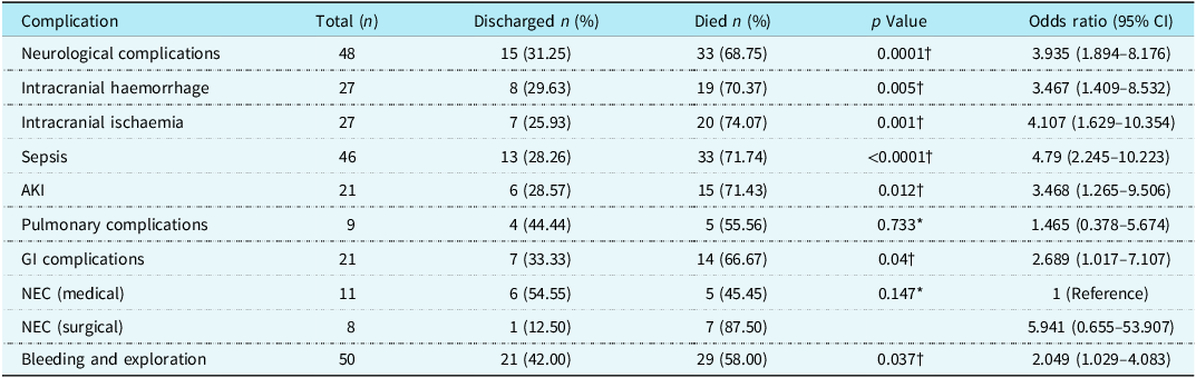
Major complications included mediastinal bleeding, neurological injury, sepsis, acute kidney injury, gastrointestinal complications, and pulmonary complications. Neurological complications and sepsis were strongly associated with mortality, with intracranial haemorrhage and intracranial ischaemia showing mortality rates exceeding 70% (Table 7). Prolonged ECMO duration was significantly associated with intracranial haemorrhage, sepsis, and acute kidney injury (Supplementary Table 4).
Association of ECMO-related complications with survival

Table 7. Long description
A table with seven rows and eight columns comparing different complications with survival rates and statistical values. The columns are labeled Complication, Total (n), Discharged n (%), Died n (%), p Value, and Odds ratio (95% CI). The rows list different complications: Neurological complications, Intracranial haemorrhage, Intracranial ischaemia, Sepsis, AKI, Pulmonary complications, GI complications, NEC (medical), NEC (surgical), and Bleeding and exploration. Each row provides the total number of cases, the number and percentage of discharged patients, the number and percentage of patients who died, the p value, and the odds ratio with a 95% confidence interval. Notable trends include high mortality rates for neurological complications and sepsis, with intracranial haemorrhage and intracranial ischaemia showing mortality rates exceeding 70%.
CI = Confidence Interval.
* Fisher’s exact test.
†Chi-square test.
Short- and long-term outcomes
One-year survival after ECMO was 50.36%, and five-year survival was 41.56% (Supplementary Table 5). The distribution of immediate, short-term, and long-term survival across different age groups is shown in Figure 3.
Association of age of ECMO institution with survival.

Mortality predictors
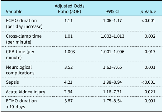
On multivariate logistic regression analysis, prolonged ECMO duration, increased cardiopulmonary bypass (CPB) time, and longer aortic cross-clamp duration were independently associated with mortality. In addition, major complications including neurological injury, sepsis, and acute kidney injury (AKI) were significant predictors of poor outcome. ECMO duration greater than 10 days was strongly associated with increased mortality risk. These findings highlight both procedural complexity and post-ECMO complications as key determinants of survival (Table 8).
Multivariate logistic regression analysis of predictors of mortality

Table 8. Long description
A table with eight rows and three columns showing the results of a multivariate logistic regression analysis of predictors of mortality. The columns are labeled Variable, Adjusted Odds Ratio (aOR), 95% CI, and p Value. The rows list different variables and their corresponding adjusted odds ratios, confidence intervals, and p values. The variables include ECMO duration (per day increase), Cross-clamp time (per minute), CPB time (per minute), Neurological complications, Sepsis, Acute kidney injury, and ECMO duration greater than 10 days. Each row provides specific values for the adjusted odds ratio, the 95% confidence interval, and the p value for each variable.
Multivariate logistic regression model including variables significant on univariate analysis. Odds ratios are adjusted for confounding.
CI = Confidence interval; CPB = cardiopulmonary bypass.
Discussion
Over the past two decades, survival rates for paediatric patients requiring perioperative ECMO have gradually improved due to advances in surgical techniques, perioperative intensive care, and ECMO technology. Early multicentre data from the United States reported survival to hospital discharge of 48.1% in children undergoing ECMO after cardiac surgery. Reference Gupta, Robertson and Beam9 Subsequent studies have demonstrated comparable outcomes, with decannulation success of 70% and discharge survival of 44% in a Scottish cohort, Reference Khorsandi, Davidson and Bouamra1 41% survival in an Indian series, Reference Agarwal, Hardison and Saville10 and 37% survival in patients requiring mechanical circulatory support for refractory cardiorespiratory failure. Reference Alsoufi, Al-Radi and Gruenwald6 Meta-analyses have further confirmed these findings, reporting successful decannulation rates of approximately 60% and survival to discharge around 43%, while highlighting limited long-term outcome data and persistent late mortality among survivors. Reference Wu, Zhao, Li, Wu, Wu and Wei11–Reference Zhou, Wang and Zhao13,Reference Yu, Yang and Guo16
In the present study, hospital survival of 54% is consistent with contemporary international registry data, where paediatric post-cardiotomy ECMO survival ranges between 40% and 55%. Importantly, the one-and five-year survival rates of 50.4% and 41.6% provide valuable insight into long-term outcomes, demonstrating sustained survival in a substantial proportion of patients while also reflecting late attrition due to complications such as neurological injury and recurrent cardiac dysfunction. Reference Weeda, Van der Palen and Bunker-Wiersma14 These findings emphasise the importance of long-term follow-up in this high-risk population.
Survival varied according to the timing of ECMO initiation. Immediate post-operative ECMO showed the highest survival, whereas delayed ECMO (>24 h after surgery) was associated with significantly higher mortality, consistent with published data linking delayed ECMO initiation to multiorgan dysfunction and poorer outcomes. Reference Deng, Haller and Moss15 Pre-operative ECMO, although infrequently used (11 patients), showed encouraging survival (72.7%) in our series, likely reflecting early stabilisation and optimised end-organ perfusion prior to surgery. The underlying diagnoses included obstructed total anomalous pulmonary venous return (TAPVR), severe mitral valve pathology, recurrent left ventricular outflow tract obstruction (LVOTO), pulmonary atresia associated with other complex congenital lesions, ruptured sinus of Valsalva and hypoplastic left heart syndrome (HLHS) with abnormal pulmonary venous drainage requiring emergent stabilisation. Postoperative ECMO remained the most common indication, particularly for failure to wean from cardiopulmonary bypass and low cardiac output syndrome, as reported in major registries such as the Society of Thoracic Surgeons database. Reference Faheem, Khan, Cheema, Akhtar and Ashraf12,Reference Deng, Haller and Moss15
Risk factors for mortality in our cohort were broadly consistent with existing literature. Neonatal age, prematurity, and comorbid conditions were associated with higher mortality, although not always statistically significant. Procedural complexity, prolonged cardiopulmonary bypass and cross-clamp time, and longer ECMO duration were important predictors of poor outcome. ECMO duration exceeding 10 days was strongly associated with mortality, which aligns with previous studies identifying prolonged ECMO support as a key determinant of adverse outcomes. Reference Zhou, Wang and Zhao13 These findings reinforce the importance of early decision-making, timely weaning strategies, and careful patient selection.
Complications during ECMO were major contributors to mortality. Neurologic complications included seizures, neurodeficits, abnormal pupillary responses, and brain death. Renal dysfunction was defined according to KDIGO criteria as an increase in serum creatinine by ≥0.3 mg/dL within 48 h or an increase to ≥1.5 times the pre-operative baseline within 7 days. Gastrointestinal complications included gastrointestinal bleeding and necrotising enterocolitis. These complications were associated with poor outcomes, highlighting the complex and resource-intensive nature of ECMO management. The high mortality observed in patients with intracranial haemorrhage and ischaemic brain injury underscores the importance of meticulous anticoagulation management, early neurologic surveillance, and rapid intervention. Similarly, the association between prolonged ECMO support and organ dysfunction emphasises the importance of infection control, end-organ protection, and multidisciplinary critical care management.
This study has several limitations. Its retrospective single-centre design limits generalisability and prevents causal inference. The heterogeneous patient population and relatively small subgroup sizes reduce statistical power for certain comparisons, particularly in patients requiring multiple ECMO runs. Precise CPR duration data were not consistently available in our retrospective dataset, and therefore, we were unable to reliably evaluate the relationship between CPR duration and outcomes in the extracorporeal cardiopulmonary resuscitation (ECPR) cohort. In addition, cost-effectiveness, quality-adjusted life years, and detailed neurodevelopmental outcomes were not evaluated. The absence of year-wise trend analysis also limits assessment of the impact of evolving ECMO technology and surgical practice.
Future research should focus on multicentre collaboration to improve statistical power and enhance generalisability, particularly by including centres from low-and middle-income countries where ECMO access and outcomes may differ. Prospective studies incorporating predictive modelling, biochemical markers, and standardised ECMO protocols could improve patient selection and timing of intervention. Long-term neurodevelopmental follow-up and cost–benefit analysis will be essential to determine the broader impact and sustainability of ECMO programmes.
In conclusion, veno-arterial extracorporeal membrane oxygenation (VA-ECMO) remains a critical life-saving strategy in complex congenital cardiac surgery, with hospital survival of 54% in our cohort. Early initiation, shorter ECMO duration, and prevention of complications are key determinants of improved outcomes. Continuous multidisciplinary collaboration, systematic auditing, and long-term follow-up are essential to optimise ECMO utilisation and improve survival in this vulnerable population.
Supplementary material
The supplementary material for this article can be found at https://doi.org/10.1017/S1047951126123658.
Acknowledgements
We are thankful to our colleagues from departments of paediatric cardiology, paediatric intensive care, and paediatric perfusion for their help and support.
Financial support
This research received no specific grant from any funding agency, commercial or not-for-profit sectors.
Competing interests
None.