INTRODUCTION
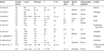
Non-parasitic associations between nematodes and aquatic multicellular animals have been known since as early as 1834, when what is now thought to be one of the first known marine non-parasitic nematodes, Odontobius ceti Roussel de Vauzème, Reference Roussel de Vauzème1834, was described from the glutinous substance covering baleen plates of a southern right whale (Roussel de Vauzème, Reference Roussel de Vauzème1834; Baylis, Reference Baylis1923). But it was not until the beginning of the 20th Century, when the first descriptions of commensalism between nematodes and aquatic (or semi-aquatic) invertebrates first appeared in the scientific literature. The first records were of members of the order Monhysterida (to which Odontobius also belongs) occurring in gill chambers of land crabs from the genera Gecarcinus and Cardisoma from the supralittoral of the Caribbean islands (Baylis, Reference Baylis1915; Cobb, Reference Cobb1920). Later on numerous associations between free-living nematodes and aquatic organisms were described in the literature. Nematodes from different taxonomic groups were found both in sessile organisms like sponges (Schuurmans Stekhoven, Reference Schuurmans Stekhoven1942a; Tsalolikhin, Reference Tsalolikhin1974; Bongers, Reference Bongers1983), bryozoans (Schuurmans Stekhoven, Reference Schuurmans Stekhoven1933) and bivalve molluscs (Schuurmans Stekhoven, Reference Schuurmans Stekhoven1942b), as well as in actively moving animals like annelids (Hopper, Reference Hopper1966; Platonova & Potin, Reference Platonova and Potin1972) and crustaceans (Sudhaus, Reference Sudhaus1974; Lorenzen, Reference Lorenzen1986a). Marine Monhysterida are by far the most species rich, common and widespread commensals of aquatic and semi-aquatic animals. The genera Odontobius, Gammarinema Kinne & Gerlach, Reference Kinne and Gerlach1953, Monhystrium Cobb, Reference Cobb1920 and Tripylium Cobb, Reference Cobb1920 are exclusively epibiotic (Lorenzen, Reference Lorenzen1986b; Tchesunov, Reference Tchesunov2006) and numerous groups of crustaceans are the most common hosts for free-living nematodes (Tchesunov, Reference Tchesunov and Sveshnikov1989). Here we describe the first record of close epibiotic (inquilinistic) association between the non-parasitic marine nematode Endeolophos skeneae sp. nov. and the marine deep-sea gastropod Skenea profunda (Friele, Reference Friele1879).
MATERIALS AND METHODS
During an expedition with RV ‘G.O. Sars’ in the north-east East Atlantic in June 2007 a large wood fall was discovered at 2830 m depth using the ROV ‘Bathysaurus’ at 73°33.19′ N 08°16.900′ E. The wood fall was too large to recover whole, so a block approximately 30 × 40 cm was broken off and brought to the surface. Immediately the block was divided into four pieces, one of each was either frozen in −80°C, fixed in 7% formaldehyde or fixed in 96% alcohol, respectively. The fourth piece was split and sorted under the dissecting microscope on board. The wood was heavily penetrated by the bivalve Xylophaga sp. Additional fauna were picked out under dissecting microscope and consisted mainly of rissoid gastropods, amphipods and Skenea profunda. The fauna collected were fixed in ethanol or formaldehyde. Additional samples were later sorted in the laboratory in a similar way.
Skenea profunda was originally described by Friele (Reference Friele1879) from sunken wood from 2440 m depth west of Svalbard. A second shallower record (229 m) was disregarded by Bouchet & Warén (Reference Bouchet and Warén1979) who argued that the second position was likely a contamination from a previous haul and this opinion is shared by Høisæter (Reference Høisæter2009). Friele (Reference Friele1886) also reported the species from 1187 m depth west of Senja, but this record has never been re-examined. Additional records are listed in Bouchet & Warén (Reference Bouchet and Warén1979) from between 2900 and 3300 m depth in the Norwegian Sea. Little is known of the biology of S. profunda but the species is obviously closely associated with sunken wood since Friele (Reference Friele1886) reported that the species was abundant in wood perforated by ‘Teredo’ at the type locality, which is also confirmed by our own observations.
Specimens of the epibiotic nematode Endeolophos skeneae sp. nov. were picked from the shells of the gastropods for studies by light (LM) and scanning electron microscope (SEM). For LM, specimens were transferred to pure glycerine by a slow evaporation method and mounted on permanent slides in glycerine with paraffin wax as support for the coverslip. Measurements are given in the format: mean ± standard deviation (range). For SEM studies, several specimens were rehydrated and preserved in 1% formaldehyde solution. They were postfixed in 1% osmium tetroxide (OsO4) and transferred to pure acetone through an acetone/distilled water series. Specimens were critical point dried in liquid CO2, mounted on a stub, gold-plated under vacuum to a thickness of 200 Å using a sputter coater, and examined in a Hitachi S-4300 SEM at an accelerating voltage of 10 kV.
Measurements are in µm. Abbreviations are as follows: a, body length/maximum body diameter; b, body length/pharynx length; c, body length/tail length; c', tail length/anal body diameter; V-A/Tail, distance from vulva to anus/tail length; V, distance from the anterior end of body to vulva/body length as %; T, distance from the anterior end of body to testis/body length as %.
SYSTEMATICS
DIAGNOSIS
Body cuticle with homogeneous ornamentation along the body: each annule with very fine and numerous longitudinal ridges. Lateral differentiation in shape of a subcuticular discontinuity in cuticle pattern along the lateral sectors of the body. The six outer labial and four cephalic sensilla setiform are arranged in a single circlet. Amphidial fovea a transverse slit. Buccal cavity has a small dorsal tooth and tiny ventrosublateral denticles. Pharynx has no defined terminal bulb. Female reproductive system is didelphic, amphidelphic. Vagina has no sclerotizations. Male reproductive system is monodelphic, prodelphic. Spicules well developed. Gubernaculum is slightly arcuate, plate-like, with or without apophyses. Telamons absent. Precloacal and postcloacal supplementary organs are absent. Marine.
TYPE SPECIES
Endeolophos minutus (Gerlach, Reference Gerlach1967) Boucher, Reference Boucher1976
1967 Nygmatonchus minutus—Gerlach, ‘Meteor’ Forschungsergebnisse D 2: 34–35, figure 19.
1985 Endeolophos minutus—Blome, Microfauna Marina 2: 291–293, figures 24–26, 28, 37.
1976 Endeolophos minutus—Boucher, Bulletin du Muséum National d'Histoire Naturelle 352, Zoologie 245: 35.
OTHER SPECIES
Endeolophos fossiferus (Wieser, Reference Wieser1954) Boucher, Reference Boucher1976
1954 Nygmatonchus fossiferus—Wieser, Lunds Universitets Årsskrift 50: 80, figure 141.
1976 Endeolophos fossiferus—Boucher, Bulletin du Muséum National d'Histoire Naturelle 352, Zoologie 245: 35.
Endeolophos spinosus (Gerlach, Reference Gerlach1957) Boucher, Reference Boucher1976
1957 Prochromadorella spinosa—Gerlach, Mitteilungen aus dem Zoologischen Museum in Berlin 33: 439–440, figure 9g–l.
1959 Neochromadora bicoronata—Wieser, University of Washington Publications in Biology 18: 58–59, figure 59.
Reference Inglis1969Nygmatonchus bicoronata—Inglis, Bulletin of the British Museum (Natural History) Zoology, London 17: 200.
1970 Nygmatonchus bicoronatus—Riemann, Veröffentlichungen des Instituts für Meeresforschung in Bremerhaven 12: 390–392, figures 30–35.
1976 Endeolophos spinosus—Boucher, Bulletin du Muséum National d'Histoire Naturelle 352, Zoologie 245: 35.
1985 Endeolophos spinosus galapagensis—Blome, Microfauna Marina 2: 293–295, figures 29–31, 37.
Endeolophos subterraneus Blome, 1982
1982 Endeolophos subterraneus—Blome, Mikrofauna des Meeresbodens 86: 126–128, figure 25A–D.
Endeolophos skeneae sp. nov.
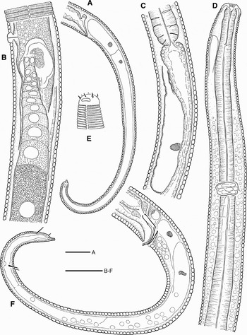
Endeolophos skeneae sp. nov. (A–E) female paratype; (F) male paratype. (A) Female caudal region; (B) posterior part of female reproductive system; (C) pharyngeal–intestinal junction and renette cell; (D) pharyngeal region; (E) anterior end, surface view; (F) male caudal region. Scale bars: 20 µm.

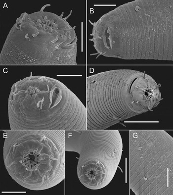
Endeolophos skeneae sp. nov., scanning electron microscope. (A) Anterior end, oblique view; (B) anterior end, lateral view; (C, D) anterior end, oblique view; (E) anterior end, apical view; (F) pharyngeal region showing setae, apical view; (G) cuticle, showing annules, longitudinal ridges, one body seta and body pore. Scale bars: A–C, E, G, 5 µm; D, F, 10 µm.

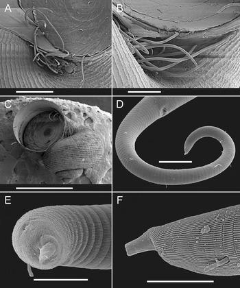
Endeolophos skeneae sp. nov., scanning electron microscope. (A–C) Nematodes on the shell of Skenea profunda; (D) male caudal region; (E) male tail end, apical view; (F) male tail end, lateral view. Scale bars: A, 500 µm; B, 250 µm; C, 2 mm; D, 20 µm; E, F, 5 µm.

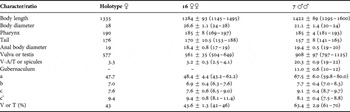
Measurements (in µm) of Endeolophos skeneae sp. nov. from Skenea profunda (Gastropoda: Skeneidae) presented as mean ± SD (range).

TYPE MATERIAL
Holotype is female (collection No. 8017), there are five female and four male paratypes (collection Nos. 8017–8019) deposited in the type collection of the Department of Invertebrate Zoology, Swedish Museum of Natural History, Stockholm, Sweden; eleven female and three male paratypes deposited in the University of California–Riverside Nematode Collection, Riverside, USA.
TYPE LOCALITY AND HABITAT
North-east East Atlantic (73°33.19′ N 08°16.900′ E), 2830 m depth, wood fall, on the gastropod Skenea profunda; collected by C. Schander in June 2007. Nematodes were found attached to the shells of Skenea profunda especially in the umbilicus and also scattered around the edges of the aperture, sometimes partly enclosed by the operculum. Forty-seven out of 50 examined specimens of S. profunda had nematodes attached.
ETYMOLOGY
Species name derived from the genus name of the associated gastropod Skenea.
DESCRIPTION
Adults: body long and thin, tapering anteriorly in pharyngeal region and posteriorly on tail; ventrally curved upon fixation, more strongly curved ventrad in posterior part in males. Cuticle is annulated; annuli 2.1–2.8 µm wide at midbody. Each annule has very fine and numerous longitudinal ridges, equally distributed around the body. External lateral alae are absent, but there is a subcuticular discontinuity in the cuticle pattern along the lateral sectors of the body. Epidermal glands absent; small body pores present, scattered throughout the body. Somatic setae present, not connected to epidermal glands, evenly distributed throughout body. Lip region is rounded, lips low and basally fused. First annulus appears posterior to the cephalic sensilla and amphidial fovea level. Inner labial sensilla are small and setiform with an apical pore, arranged immediately around the oral opening. Outer labial sensilla and cephalic sensilla setiform are equal in length, arranged in two close circles on the outer surface of lips. Subcephalic and cervical sensilla and ocelli are absent. Amphidial fovea a transverse slit, surrounded by expanded cuticle, located 3–4 µm from anterior end. Nerve ring surrounds the pharynx at about its midpoint. Secretory–excretory system is present; renette cell ovoid, located ventrally, opposite to the anterior part of intestine. Excretory pore located just posterior to labial region, on the 11th annule. Buccal cavity is funnel-shaped, 6.7–8.7 µm long. Cheilostom is wide, with twelve longitudinal ridges in a propeller-like pattern visible through the oral opening. Gymnostom very short if present at all. Stegostom narrow, with small dorsal tooth and tiny ventrosublateral denticles/ridges/thickenings; with thin cuticular lining; surrounded by muscular lining continuous with that of corpus. Pharynx cylindrical anteriorly, gradually widening posteriorly; heavily muscularized with uniformly thickened lumen throughout, lacking valves and bulbs. Pharyngeal lumen penetrated by the dorsal gland orifice somewhat posterior to buccal cavity and by two ventrosublateral gland orifices just posterior to nerve ring level. Tail is elongate–conoid to subcylindrical, arcuate ventrad. Three caudal glands present, their nuclei incaudal. Spinneret is functional.
Females: reproductive system didelphic, amphidelphic; ovary branches reflexed antidromously. Anterior ovary located on right-hand side of intestine (dextral), posterior ovary located on left-hand side (sinistral). Gonoduct broad and filled with granular content in most specimens, and is not divided into oviduct and uterus. Spermatheca as a separate structure is absent. Small and thin spermatozoa enclosed in an ovoid spermatophore-like structure with hyaline outer shell. Vagina is straight and short, without sclerotizations. Intrauterine egg(s) is not seen. Rectum relatively short, about one anal body diameter long; surrounded by three gland-like cells at junction with intestine: one flattened dorsal cell and two more rounded ventrosublateral cells. Caudal setae present, one subventral pair just posterior to anal opening, and two–three pairs in the posterior part of the tail.
Males: reproductive system monodelphic, prodelphic. Spicules paired, well developed, L-shaped, with ovoid manubrium, narrow blade and thin velum. Tip of the spicules are rounded but only visible when they protrude beyond body contour. Gubernaculum is slightly arcuate, plate-like, without apophyses. Telamons are absent. Precloacal and postcloacal supplementary organs are absent. Caudal setae present, one subventral pair just posterior to anal opening, few scattered along the tail and one subventral pair in posterior part of the tail and one subdorsal pair near tail terminus.
Juveniles: these were not found.
DIAGNOSIS
Endeolophos skeneae sp. nov. is particularly characterized by homogeneous ornamentation of the cuticle with annules with very fine and numerous longitudinal ridges, equally distributed around the body, subcuticular discontinuity in cuticle pattern along the lateral sectors of the body, relatively long body (1.1–1.6 mm) and relatively long tail (141–188 µm, c' = 7.5–11.4), short (19–22 µm) L-shaped spicules with ventrally bent blade and simple plate-like gubernaculum without apophysis.
RELATIONSHIPS
Recent specimens are most similar to the genus Endeolophos Boucher, Reference Boucher1976 in having ‘normally’ developed spicules (spicules are reduced or modified in some closely related genera, like Actinonema Cobb, Reference Cobb1920 and Rhips Cobb, Reference Cobb1920) and gubernaculum and relatively simple morphology of the cuticle with homogeneous ornamentation across most of the body and simple lateral differentiation of the cuticle. The genus Endeolophos so far includes only five valid species and one subspecies. Major distinguishing characters for species identification are body measurements and ratios, as well as shape of the spicule tip and presence and shape of gubernaculum apophysis (see Table 2). Endeolophos skeneae sp. nov. most closely resemble Endeolophos subterraneus Blome, Reference Blome1982 in general body size, body measurements and cuticle structure, but differs from it by a somewhat longer body (1.1–1.6 mm versus 1.1–1.3 mm) and longer tail (141–188 µm versus 128–135 µm), differently shaped spicules (L-shaped with ventrally bent blade versus arcuate with straight blade) and simple plate-like gubernaculum (versus with arcuate dorsal apophysis). The new species can be easily separated from the type species E. minutus (Gerlach, Reference Gerlach1967) Boucher, Reference Boucher1976 (based on Gerlach's (1967) and Blome's (1985) descriptions) by having longer body (1.1–1.6 mm versus 0.7–1.0 mm), longer tail (c' = 7.5–11.4 versus c' = 5.2–5.6) and different shape of spicula (spicules with rounded versus bilobed tip). It differs from E. fossiferus (Wieser, Reference Wieser1954) Boucher, Reference Boucher1976 by having much shorter body (1.1–1.6 mm versus 2.0–2.4 mm), more anterior vulva position (V = 42–46 versus V = 50–52), shorter spicules (19–22 µm versus 30 µm) and different spicule shape (angular with straight manubrium versus curved with ventrally inclined manubrium). Finally, the new species is distinct from E. spinosus (Gerlach, Reference Gerlach1957) Boucher, Reference Boucher1976 (based on Gerlach's (1957), Wieser's (1959), Riemann's (1970) and Blome's (1985) descriptions) in the longer body (1.1–1.6 mm versus 0.8–1.2 mm), longer tail (141–188 µm versus 94–126 µm), more anterior vulva position (V = 42–46 versus V = 49–50), and differently shaped gubernaculum (without versus with caudal apophysis).
Comparisons of diagnostic characters (measurements in µm) in different species and populations of the genus Endeolophos Boucher, Reference Boucher1976.
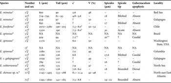
1, after Gerlach (Reference Gerlach1967) and calculations from there; 2, after Blome (Reference Blome1985) and calculations from there; 3, after Wieser (Reference Wieser1954) and calculations from there; 4, after Gerlach (Reference Gerlach1957) and calculations from there; 5, after Wieser (Reference Wieser1959) and calculations from there; 6, after Riemann (Reference Riemann1970) and calculations from there; 7, after Blome (Reference Blome1985) and calculations from there; 8, after Blome (Reference Blome1982); 9, recent material; *, only one range given for adults; **, recalculated based on figures and/or measurements.
DISCUSSION
Nematodes have a vast range of community associations with molluscs from symbiotic to the most prevalent parasitism. The most commonly observed parasitic relationships are nematodes using terrestrial molluscs as paratenic or intermediate hosts and definite hosts (Grewal et al., Reference Grewal, Grewal, Tan and Adams2003). In the first case, the host serves as a transitional stage in the nematode's life-cycle, but generally no death of the host occurs. In the second case on the contrary, the host is killed after the nematode completed its life-cycle. The majority of the current literature reports nematode parasitism in terrestrial molluscs, with very few examples in the existing literature discussing symbiotic relationships between non-parasitic nematodes and aquatic molluscs, particularly bivalves.
The first studies of nematode commensals in marine bivalves date back to 1942, when Schuurmans Stekhoven (Reference Schuurmans Stekhoven1942b) listed 16 nematode species that he found associated with oyster Ostrea edulis L., 1758 from the North Sea. These nematodes were, however, collected from the exterior surface of the oyster shells (Korringa, Reference Korringa1954). In the Pacific oysters, Crassostrea gigas (Thunberg, 1793), the majority of nematodes (species was not identified) were located on the gill surfaces near the food grooves and within the mantle fold, and are thought to seek shelter and avoid desiccation or exposure during periods of low tide (Dinamani, Reference Dinamani1986). Similarly, Pontonema vacillatum Leidy, 1855, was found within the mantle and in the folds of the siphon in the soft-shelled clam, Mya arenaria L., 1758 (Anderson & Bourne, Reference Anderson and Bourne1960). Another nematode species, Monhystera cameroni Steiner, 1958, was most abundant in the necrotic mantle or gill tissues, but also present in blood sinuses and in vessels of the vesicular connective tissue of the captive American oyster, Crassostrea virginica (Gmelin, 1971) (Meyers et al., Reference Meyers, Elston and Georgi1985).
In freshwater, symbiotic relationships between nematodes and bivalves were described only for zebra mussels, Dreissena polymorpha (Pallas, 1771), where free-living nematodes were commonly observed in the mantle cavity. As far as it is known, however, there is no obligate association between nematodes and D. polymorpha and the nematodes enter the mantle cavity with the water current or by active crawling among byssal threads (Molloy et al., Reference Molloy, Karatayev, Burlakova, Kurandina and Laruelle1997). Chromadorina bioculata (Schultze in Carus, 1857) was the most common species in samples of zebra mussels from Lake Erken in Sweden and in different freshwater habitats in Belarus (Karatayev et al., Reference Karatayev, Mastitsky, Burlakova, Molloy and Vezhnovets2003; Mastitsky et al., Reference Mastitsky, Lucy and Gagarin2008). Several other nematode species were found in lower numbers. Chromadorina leuckarti (de Man, 1876), Punctodora ratzemburgensis (Linstow, 1876), Eumonhystera vulgaris (de Man, 1880), Tridentulus floreanae (Abebe, 1995), Tobrilus tenuicaudatus Gagarin, 1989 and Monhystrella sp. were found in zebra mussels from several water bodies in Belarus (Karatayev et al., Reference Karatayev, Mastitsky, Burlakova, Molloy and Vezhnovets2003; Mastitsky & Gagarin, Reference Mastitsky and Gagarin2004). Dorylaimus stagnalis Bastian, 1865 was found in zebra mussels from the Volga Basin (Kuperman et al., Reference Kuperman, Zhochov and Popova1994) and Laimydorus sp. in zebra mussels from Lake Erken (Mastitsky et al., Reference Mastitsky, Lucy and Gagarin2008). Studies of zebra mussels in North America, in the St Lawrence River and in Lake Erie, revealed five not identified nematode species, including members of Enoplida, Mononchida and Dorylaimida (Toews et al., Reference Toews, Beverly-Burton and Lawrimore1993; Conn et al., Reference Conn, Babapulle, Klein and Rosen1994). The high proportion of literature on Dreissena polymorpha and their symbionts is not surprising in view of the problems caused globally by zebra mussels, since there is a need to understand how they alter community structures.
The case described in the present paper seems to be the first known symbiotic relationship between a nematode and an aquatic gastropod. However, the important differences between bivalves and gastropods are that: (1) bivalves are generally sessile suspension feeders attached to the substrate with a microhabitat created by the fluid-filled space between the shells and by the current of water over the gills, supporting a community of symbiotic organisms like ciliates, nematodes and oligochaetes; while (2) gastropods are most often actively moving on the substrate and generally lack the suspension feeding mode of the bivalves.
Currently, the exact mechanisms of symbiotic association of nematodes with aquatic bivalves or gastropods are unknown. It seems possible that nematodes find food and shelter in the mantle cavity of the bivalve molluscs, and in the umbilicus and aperture of the gastropods, but further studies are needed to address these questions.
ACKNOWLEDGEMENTS
We want to thank the crew of RV ‘G.O. Sars’ for excellent working conditions, and the pilots of the ROV ‘Bathysaurus’ for skilfully recovering the wood. This is a publication from the Centre of Excellence in Geobiology, University of Bergen.