INTRODUCTION
Dictyocaulus viviparus is an important pathogenic lungworm of cattle in temperate climatic areas. We have been studying the acetylcholinesterases (AChE, EC 3.1.1.7) of this parasitic nematode, with a view to identifying their potential as novel drug targets or vaccine candidates. The classical role of AChE is to regulate synaptic transmission at neuromuscular junctions via the hydrolysis of acetylcholine (ACh). However, several parasitic nematodes also secrete this enzyme into their environment during the course of infection (Lee, 1996). D. viviparus is no exception to this; during its migration through the lungs, this nematode secretes a number of AChE isoforms which are the targets of circulating antibody responses in infected cattle (McKeand et al. 1994). Previously, we isolated 2 genes (Dv-sace-1 and Dv-sace-2) from D. viviparus which encode secreted AChEs (Lazari et al. 2003). The products of these genes were classified as secreted forms of the enzyme on the basis of their truncated C-termini, which indicated that the proteins would form soluble monomers. These observations were substantiated after we characterized the corresponding recombinant proteins (Lazari et al. 2003). Subsequently, we established that the genes encoding these secreted AChEs were distinct from those genes which encode neuromuscular forms of AChE in D. viviparus (Lazari et al. 2004). In the latter studies, we isolated a gene which encoded a form of AChE analogous to the glycosylphosphatidylinositol (GPI)-anchored forms identified previously in mammals and invertebrates. This gene, Dv-ace-2 (formerly designated Dv-ace-3), is a homologue of the Caenorhabditis elegans AChE gene, Ce-ace-2 (Combes et al. 2003), and was the first neuromuscular AChE gene to be isolated from any parasitic nematode of animals. In the current study, we describe the isolation and characterization of a cDNA encoding a second class of neuromuscular AChE from D. viviparus. In this case, the enzyme exhibits sequence properties distinct from the GPI-anchored form of the enzyme and has the sequence characteristics of an AChE analogous to the ‘tailed’ (T) subunit AChE of vertebrates (Massoulié et al. 1998) and to ACE-1 from C. elegans (Arpagaus et al. 1994).
MATERIALS AND METHODS
Parasite material
Third-stage larvae (L3) were donated by Intervet (Milton Keynes, UK) and stored in 50 mM phosphate-buffered saline, pH 7·4, at 4 °C prior to infection. To obtain adult parasites, cattle were infected at a maximum dose rate of 10 L3 kg−1 and the parasites were obtained from the lungs between 28 and 35 days post-infection (p.i.), as described previously (McKeand et al. 1994).
Gene cloning and sequence alignment
Total RNA was extracted from ~30 adult D. viviparus and reverse-transcribed using the Superscript First Strand cDNA Synthesis Kit (Invitrogen). An AChE-encoding cDNA fragment was amplified from adult parasite RNA by reverse transcriptase-polymerase chain reaction (RT-PCR) using primers (forward: 5′-GAAATCAATTTCATTTTTGGAGAACC-3′, reverse: 5′-TGGAAGCAAC-AAATGGATAAATGG-3′) designed to conserved regions of the C. elegans ace-1 gene (Arpagaus et al. 1994, Accession no. X75331), but with low identity to the nucleotide sequences of the D. viviparus AChE genes isolated previously (Lazari et al. 2003, 2004, Accession nos. AY546079 [Dv-sace-1], AY218788 [Dv-sace-2], AY218789 [Dv-ace-2]). The PCR was performed under the following cycling conditions: 5 min denaturation at 94 °C; 40 cycles of 94 °C for 1 min, 50 °C for 1 min and 72 °C for 2 min, with a final extension of 72 °C for 10 min. The PCR product obtained was gel purified and cloned into the TA vector, pGem-T-Easy (Promega). Plasmid DNA was isolated using the alkaline lysis method (Qiagen) and the insert sequenced from both strands using vector-specific primers. Following the confirmation that this fragment showed similarity to other AChEs, gene-specific primers were designed to facilitate the isolation of the 5′ (primer: 5′-CCATGACCTGTACGTCGAGCATTAGGGATATG-3′) and 3′ (primer: 5′-AGTAGCAGATTTATGCGATACTGGGCAAA-3′) termini using the rapid amplification of cDNA ends technique (RACE, SMART Kit, Clontech). Subsequently, a PCR fragment spanning the coding region of the Dv-ace-1 cDNA was amplified using primers designed to the start and stop codons (forward: 5′-ATGCTGTTTGCATTGTTTTTAAGCGC-3′, reverse: 5′-TCAGGATCCACTGCA-TGAATCGGA-3′) and cloned and sequenced. Nucleotide sequences were manipulated and aligned using the DNAStar programme (http://www.dnastar.com/web/r3.php). Alignments of AChE protein sequences were performed via CLUSTAL W 1.8 using default settings (http://www.ebi.ac.uk/clustalw/; Thompson et al. 1994) and the numbering of amino acid residues was assigned based on comparison with the Torpedo californica AChE sequence (Accession no. X03439), according to the previously agreed recommendations (Sussman et al. 1991). Sequence homology searches were performed using the FASTA programme (http://www.ebi.ac.uk/fasta33) using default settings and percentage identities of the sequences to Dv-ACE-1 and are quoted in the Results section.
Temporal analysis of transcription
To examine the time-points of infection at which Dv-ace-1 is transcribed, RT-PCR analysis was performed using RNA isolated from parasites harvested at 14, 22, 29 and 35 days p.i. Parasites collected at 14 days p.i. were kindly provided by Dr Harm Ploeger (Utrecht University) and those at 22, 29 and 35 days p.i. were obtained as described previously (McKeand et al. 1994; Johnson et al. 2005). RNA extraction and reverse transcription were performed as described previously (Lazari et al. 2003). The PCR was performed using the Dv-ace-1-specific primers used in the RACE. To confirm integrity of input template cDNA, positive control reactions were included for each sample using primers (forward: 5′-CCTGCTTTTGGTATTATTAGACA-3′, reverse: 5′-CTTACTACATAAGTATCATGCAAC-3′) designed to amplify a fragment of the D. viviparus cytochrome c oxidase subunit 1 (cox1) gene (Accession no. AF263474). The Dv-ace-1 PCR was carried out in the following manner: 1 cycle of 10 min at 94 °C; 30 cycles of 94 °C for 1 min, 60 °C for 1 min and 72 °C for 1 min, with a final extension at 72 °C for 10 min. The cox1 PCR was performed using the following cycling protocol: 1 cycle of 10 min at 94 °C; 35 cycles for 1 min, 50 °C for 1 min and 72 °C for 1 min, with a final extension at 72 °C for 10 min. Amplification products were separated on 1% (w/v) agarose gels stained in ethidium bromide and images captured upon transillumination.
Phylogenetic analysis
A phylogenetic tree of the evolutionary relationship of Dv-ACE-1 with other AChE proteins was constructed using AChE amino acid sequences retrieved from NCBI (http://www.ncbi.nlm.nih.gov/). These protein sequences were selected either on the basis of their high levels of identity to the AChE protein under study or to provide a wide range of AChE T-tailed enzymes from a range of vertebrate and non-vertebrate species with which to compare to Dv-ACE-1. The 3 previously characterized D. viviparus AChE sequences (Dv-ACE-2, Dv-sACE-1 and Dv-sACE-2) were also included in the phylogenetic analysis for comparative purposes. The peptide sequences were aligned using the default settings in the CLUSTAL W 1.8 software programme (as above) and an unrooted Neighbour-Joining phylogenetic tree of these sequences was generated using the MEGA programme, version 3 (http://www.megasoftware.net; Kumar et al. 2001), with 250 bootstrap replicates.
RESULTS
Sequence analysis of Dv-ace-1
The full-length cDNA (designated Dv-ace-1) was 2724 nucleotides (nt), covering 1857 nt of coding sequence, 153 nt of 5′-UTR and 714 nt of 3′-UTR, including the polyA tail. This sequence has been deposited in GenBank under Accession no. DQ375489. By FASTA analysis, the predicted protein sequence (Dv-ACE-1) of 619 amino acid residues showed the highest percentage identity (79%) to the T-tailed AChEs, C. elegans and Caenorhabditis briggsae ACE-1 (Accession nos. P38433 and Q27459, respectively), and 64% identity to Meloidogyne incognita and Meloidogyne javanica AChE (Accession nos. AAD02835 and AAD25921, respectively). Lower percentage identities were observed with the previously characterized D. viviparus AChE proteins: the GPI-anchored Dv-ACE-2 (33%) and the secreted AChEs, Dv-sACE-1 (30%) and sACE-2 (29%).
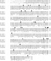
Fig. 1 depicts an alignment of Dv-ACE-1 with its closest homologue, C. elegans ACE-1 (Ce-ACE-1). The peptide sequence of the AChE (TCALACHE) from the electric ray, Torpedo californica, is included to identify residues with critically defined roles in enzyme structure and activity. Like the other D. viviparus AChEs, the Dv-ACE-1 protein contains areas of relative conservation which are associated with known properties of this group of enzymes. The residues which constitute the catalytic triad (S199, E329 and H451; equivalent to S200, E324, H440 in T. californica AChE) are all present in Dv-ACE-1. These residues are located at the base of a deep gorge which is known to penetrate approximately halfway into the structure of the enzyme (Sussman et al. 1991). Fourteen aromatic residues, thought to be involved in substrate guidance, are present in this gorge and these amino acids are conserved among a wide range of species. In Dv-ACE-1, 12 of these 14 residues are conserved or exhibit conservative substitutions when compared with T. californica AChE (Fig. 1). The 2 remaining aromatic residues not conserved between Dv-ACE-1 and T. californica AChE are S68 (also a serine in Ce-ACE-1 and a threonine in Dv-sACE-1, sACE-2 and ACE-2) and A290 (a glycine in Ce-ACE-1 and a methionine in Dv-sACE-1, sACE-2 and ACE-2). Six cysteine residues which contribute to intra-molecular disulphide bonds in T. californica AChE (Sussman et al. 1991) are present in Dv-ACE-1 and, indeed, these have been identified in all D. viviparus AChEs characterized thus far. There are 4 potential sites for N-linked glycosylation in Dv-ACE-1, all of which are conserved in Ce-ACE-1 and 2 of these are conserved in T. californica AChE. Unlike the secreted AChEs of D. viviparus, Dv-ACE-1 does not contain a truncated C-terminus (Lazari et al. 2003). Furthermore, Dv-ACE-1 does not possess a predicted consensus site for GPI addition as described for Dv-ACE-2 (Lazari et al. 2004). The derived C-termini of the sequences of both Dv-ACE-1 and Ce-ACE-1 show features of the tryptophan amphiphilic tetramerization (WAT) domain, identified previously in the C-terminus of vertebrate T AChE type (AChET) subunits (Massoulié, 2002). In vertebrates, this C-terminus is 40 amino acids in length and contains 7 conserved aromatic residues and a cysteine at position -4 from the C-terminus that is involved in dimer formation via disulphide bonding (Massoulié, 2002). AChE T peptides form elongated α helices with the aromatic side-chains clustered into a hydrophobic patch (Bon et al. 2004). This hydrophobic patch allows AChE tetramerization and also enables the enzyme portion to associate with a proline-rich attachment domain (PRAD) on a non-catalytic subunit which anchors the entire protein complex into basal laminae (Belbeoc'h et al. 2004). The C-termini of both Dv-ACE-1 and Ce-ACE-1 contain all 7 conserved aromatic residues. Of these aromatic residues, 3 evenly spaced tryptophans, which are 7 residues apart in vertebrates and 7 or 8 residues apart in Ce-ACE-1 and Dv-ACE-1, are critical for the assembly of AChE tetramers (Belbeoc'h et al. 2004). The nematode AChET enzymes also contain the conserved cysteine residue at position -3 or -4 from the C-terminus.

Fig. 1. Alignments of the sequences of Dv-ACE-1, Caenorhabditis elegans ACE-1 and Torpedo californica AChE. Numbering is from the first residue of the mature proteins. The symbols are: [utri ]-conserved cysteines involved in internal disulphide bonds; [bull ]-substitution in aromatic gorge-lining residues between Dv-ACE-1, Ce-ACE-1 and T. californica AChE; -conserved residues in active site gorge; [squf ]-conserved active site residues; [utrif ]-N-linked glycosylation consensus signals conserved in Dv-ACE-1 and Ce-ACE-1; [starf ]-conserved terminal cysteine; bold – aromatic residues in C-terminal WAT domain conserved between Dv-ACE-1 and Ce-ACE-1.
Expression pattern of Dv-ace-1
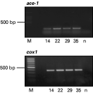
As would be expected for a neuromuscular AChE, and similar to Dv-ace-2, Dv-ace-1 appears to be constitutively expressed with RT-PCR products obtained from parasites isolated at 14, 22, 29 and 35 days p.i. (Fig. 2).

Fig. 2. Transcription pattern of Dv-ace-1. RNA isolated from parasites harvested at 14, 22, 29 and 35 days p.i. was used for RT-PCR employing gene-specific primers (ace-1 panel). The cDNA samples were also amplified with primers specific to the Dictyocaulus viviparus cytochrome oxidase subunit 1 gene (cox1). The negative control sample (‘n’), in which template DNA was not included, is also shown in each panel. Amplicons were resolved on agarose gels.
Phylogenetic analysis
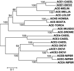
To examine the evolutionary relationship of the 4 types of Dictyocaulus enzymes with other AChEs, phylogenetic analysis was performed (Fig. 3). This analysis indicated that Dv-ACE-1 is most closely related to C. elegans ACE-1 and to the ACE-1 homologues from the root-knot nematodes, M. incognita and M. javanica, and from the mollusc, Loligo opalescens. The ACE-1 AChE sequences from nematodes clustered with the AChET peptide sequences from vertebrates and flies rather than with the other types of AChE proteins (i.e. GPI-anchored and soluble monomeric forms) previously identified in parasitic nematodes (Lazari et al. 2003, 2004; Hussein et al. 2002). As shown previously, Dv-ACE-2 clustered with C. elegans ACE-2 and with the secreted AChE sequences from D. viviparus and Nippostrongylus brasiliensis (Lazari et al. 2004). The results illustrated were derived by the Neighbour-Joining method. Analysis by Maximum Parsimony and Unweighted Pair Group Method with Arithmetic Mean (data not shown) resulted in almost identical tree topologies, with very similar bootstrap values for the nodes. Thus, the tree presented in Fig. 3 would appear to provide a realistic representation of the phylogenetic relationship of the AChEs illustrated.

Fig. 3. Neighbour-Joining tree of the phylogenetic relationship of Dv-ACE-1 with other cholinesterases. The scale bar represents 0.1 nucleotide substitutions/site, and numbers on the tree represent bootstrap values. Protein sequences used were: ACE1-DICVI (boxed): Dictyocaulus viviparus AChE 1 (Accession no. DQ375489 [peptide translation]), ACE2-DICVI: D. viviparus AChE 2 (Accession no. AAO62949), sACE1-DICVI: D. viviparus secreted AChE 1 (Accession no. AAG34705), sACE2-DICVI: D. viviparus secreted AChE 2 (Accession no. AAO62366), ACE1-CAEEL: Caenorhabditis elegans AChE 1 (Accession no. P38433), ACE-MELIN: Meloidogyne incognita AChE (Accession no. AAD02835), ACE-MELJA: Meloidogyne javanica AChE (Accession no. AAD25921), ACE-LOLOP: Loligo opalescens AChE (Accession no. AAD15886), ACHE-HOMSA: Homo sapiens AChE (Accession no. AAA68151), ACE-BOSTA: Bos taurus AChE (Accession no. P23795), ACE-TORCA: Torpedo californica AChE (Accession no. CAA27169), ACE-MUSDO: Musca domestica AChE (Accession no. CAC39209), ACE-DROME: Drosophila melanogaster AChE (Accession no. CAA29324), ACE4-CAEEL: C. elegans AChE 4 (Accession no. AAC14017), ACE3-CAEEL: C. elegans AChE 3 (Accession no. AAC14022), ACE2-CAEEL: C. elegans AChE 2 (Accession no. AAC14016), ACEB-NIPBR: Nippostrongylus brasiliensis AChE B (Accession no. AAC05785), ACEC-NIPBR: N. brasiliensis AChE C (Accession no. AAF04599), ACEA-NIPBR: N. brasiliensis AChE A (Accession no. AAK44221).
DISCUSSION
Invertebrates and vertebrates differ in their genomic organization of AChE genes (Massoulié, 2002, Selkirk et al. 2005). Vertebrates characteristically have a single gene for AChE, with C-terminal-variable catalytic subunits generated by alternative splicing of the 3′-end of the transcript (Massoulié, 2002). In mammals, Type R (‘readthrough’) AChE subunits form soluble monomers and are expressed exclusively during development or in the brain under stress, whereas Type H (‘hydrophobic’) AChE subunits form GPI-anchored dimers and secreted molecules which are largely expressed in haematopoietic cells. T (‘tailed’) subunits, similar to Dv-ACE-1, exist for AChE and BuChE and represent the enzyme forms expressed in mammalian brain and muscle (Massoulié, 2002). These subunits generate a variety of quaternary structures, including homomeric oligomers and hetero-oligomeric assemblies which associate with specific anchoring proteins, collagen Q (ColQ) or the proline-rich membrane anchor (PRiMA). These latter proteins enable the AChE subunits to be anchored to the basal laminae of neuromuscular junctions (Massoulié, 2002). It is not yet known whether Dv-ACE-1 forms tetramers which associate with a non-catalytic subunit, but the information available for AChET homologues in C. elegans (Combes et al. 2000) and Meloidogyne (Chang and Opperman, 1991) support this. The nature of non-catalytic subunit partners remains to be elucidated for the AChEs of nematodes. Homo-logy sequence searches of the relevant databases (e.g. GenBank and Nembase) for homologues of either ColQ or PRiMA have yielded no molecules of significant identity to either of these proteins (Matthews, unpublished observations).
Invertebrates have several AChE genes, each of which encodes a specific enzyme type. C. elegans and C. briggsae each have 4 neuromuscular AChE genes, and 3 of these (ace-2, ace-3 and ace-4) encode GPI-anchored or Class B AChEs, which are hydrophobic and detergent-soluble (Combes et al. 2000). Previously, Dv-ace-2, a homologue of Ce-ace-2, was identified by our group (Lazari et al. 2004). C. elegans ace-1 encodes a Class A AChE homologous to the common WAT-domain-containing AChET enzymes of vertebrates (Arpagaus et al. 1994) and, as indicated by sequence and phylogenetic analyses, this gene is the closest homologue of Dv-ace-1 identified here. AChE sequences homologous to Ce-ace-1 and Ce-ace-2 have been isolated from Meloidogyne species (Piotte et al. 1999; Laffaire et al. 2003) and an amphiphilic, collagen-tailed tetrameric AChE has been defined in N. brasiliensis (Hussein et al. 1999), demonstrating that these types of neuromuscular AChE exist in a variety of parasitic nematode species. No AChE genes similar to C. elegans ace-3 and ace-4 have been isolated from Meloidogyne, although a native enzyme with properties similar to the Class C AChE has been characterized (Chang and Opperman, 1991). Although it remains to be elucidated whether D. viviparus has further homologues of the remaining Caenorhabditis ace genes (i.e. ace-3 and -4), preliminary Southern blot analysis has indicated that there may be a homologue of Ce-ace-3 or -4 in the D. viviparus genome (Lazari, unpublished data). It would seem, however, that expression of the full repertoire of C. elegans ace genes is not essential for survival as only 3 of the 4 AChE classes appear to be enzymatically active, and the ace genes have overlapping phenotypes (Combes et al. 2000). Deletion of any single class results in slight phenotypic changes and ace-1: ace-2 mutants survive, albeit severely uncoordinated (Combes et al. 2000). These latter observations have been made, despite the fact that the C. elegans ace genes appear to be expressed in distinct tissue locations in wild-type worms (Combes et al. 2003).
In addition to the variation at the C-terminus, additional distinct differences were identified between Dv-ACE-1 and the other D. viviparus AChEs, Dv-ACE-2, Dv-sACE-1 and Dv-sACE-2. For example, 12 of the 14 aromatic residues which line the active site gorge are conserved between T. californica AChE and Dv-ACE-1, but in the other D. viviparus AChE sequences, only 11 of these residues exhibited conserved or conservative substitutions when compared with T. californica AChE (Lazari et al. 2003, 2004). The tryptophan at position 297 in T. californica lies at the top of the active site gorge and is a constituent of a loop which contributes to what has been termed the ‘peripheral anionic site’ (PAS; Harel et al. 1993). This residue is conserved in Dv-ACE-1 (W283) and in Ce-ACE-1 (W283), but is substituted with aspartic acid in Dv-ACE-2, tyrosine in Dv-sACE-1 and serine in Dv-sACE-2. Despite variation in this residue amongst the other D. viviparus AChEs, the two enzymes which have been expressed in their recombinant form (Dv-sACE-1 and -2) are susceptible to PAS inhibitors (Lazari et al. 2003, 2004). Another difference between Dv-ACE-1 and the other D. viviparus AChEs is in the number and location of potential sites for N-linked glycosylation. Dv-ACE-1 has the most sites (4) compared with Dv-sACE-1 (3) and Dv-sACE-2 (2) and Dv-ACE-2 (2). The unique hydrophilic sequence reported previously in the secreted AChEs of D. viviparus (Lazari et al. 2003), and which molecular modelling located to the enzyme surface, is not present in Dv-ACE-1. This sequence is also absent from Dv-ACE-2, suggesting that the insertion may have a particular function associated with enzyme secretion in vivo, a hypothesis supported by the presence of unique hydrophilic insertions in the secreted AChEs of N. brasiliensis (Hussein et al. 2002).
As would be expected of neuromuscular AChEs, both genes, Dv-ace-1 and -2, are transcribed at all stages of the parasite life-cycle examined. This is in contrast to the observations made for the secreted AChEs of D. viviparus, transcripts of which are detectable from 14 (Dv-sace-1) and 22 (Dv-sace-2) days p.i. onwards (Lazari et al. 2003). These expression patterns confirm the biochemical excretory-secretory profiles of these enzymes (McKeand et al. 1994) and emphasize the relevance of these secreted AChE variants in this host/parasite relationship. The present findings support the proposal that animal-parasitic nematodes appear to have evolved a separate cohort of genes that encode secreted AChEs important in the relationship of the pathogen with its host. These genes exist in addition to those which encode homologues of the various AChE protein types which are expressed primarily in the neuromuscular systems of vertebrates and other invertebrates. Unlike soluble, monomeric forms of AChE identified in vertebrates (Massoulié, 2002), the nematode secreted AChEs are not merely splice variants of the neuromuscular forms of the enzyme.
This project was funded by the BBSRC. We would like to thank Intervet for supply of D. viviparus L3, Dr Harm Ploeger (Division of Parasitology and Tropical Veterinary Medicine, Utrecht University) for donation of lung stage parasites and Ms Lynne Meikle (Moredun Research Institute) for her expert technical assistance.