INTRODUCTION
Numerous microbes colonize the tissues and systems of humans such as the skin, gut, mouth and genitals. Most of these microbes are bacterial (Rajilić-Stojanović et al. Reference Rajilić-Stojanović, Smidt and de Vos2007) with smaller numbers of Archaea, fungi, viruses, protozoa and arguably helminths during asymptomatic infections. Within the human gut alone there are around 1013–1014 commensal bacterial cells (Gill et al. Reference Gill, Pop, DeBoy, Eckburg, Turnbaugh, Samuel, Gordon, Relman, Fraser-Liggett and Nelson2006; Ley et al. Reference Ley, Peterson and Gordon2006a ; Sekirov et al. Reference Sekirov, Russell, Antunes and Finlay2010) with diverse populations and quantities occupying the jejunum, ileum, caecum and rectum (Hayashi et al. Reference Hayashi, Takahashi, Nishi, Sakamoto and Benno2005). A significant proportion of these species belong to the Gram-positive Firmicutes and Gram-negative Bacteroidetes phyla, with a relatively greater amount of cell turnover and metabolic activity within the Firmicutes (Maurice et al. Reference Maurice, Haiser and Turnbaugh2013). There are also smaller numbers of the Actinobacteria, Proteobacteria, Fusobacteria and Verrucomicrobia groups (Yatsunenko et al. Reference Yatsunenko, Rey, Manary, Trehan, Dominguez-Bello, Contreras, Magris, Hidalgo, Baldassano, Anokhin, Heath, Warner, Reeder, Kuczynski, Caporaso, Lozupone, Lauber, Clemente, Knights, Knight and Gordon2012).
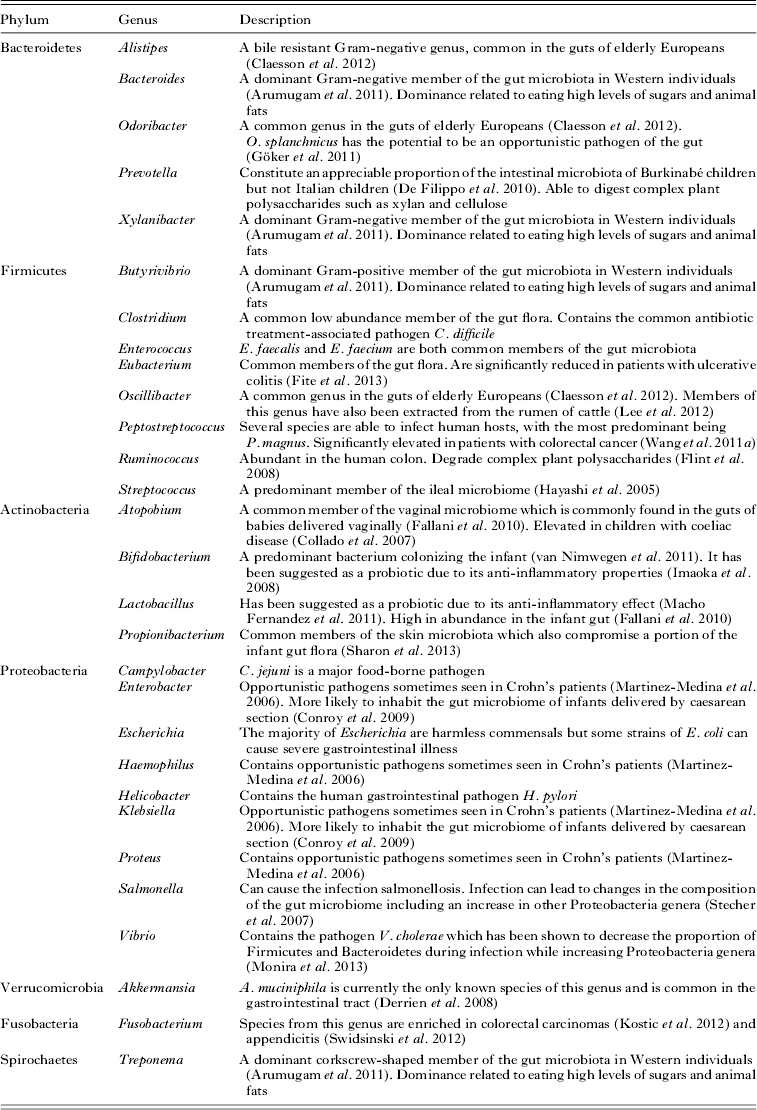
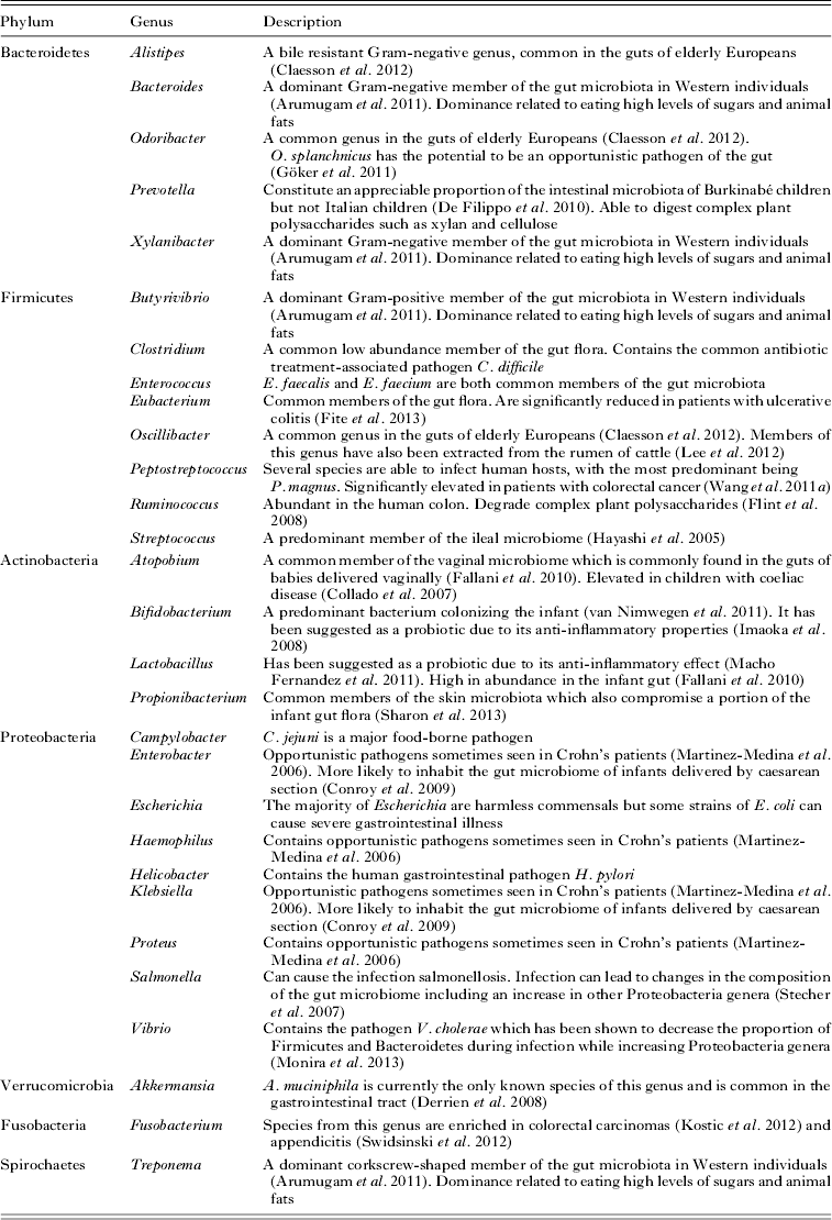
There is now a concerted global effort to characterize the gut microbiome of different human populations e.g. The Human Microbiome Consortium, The European Commission's Metagenomics of Human Intestinal Tract Project, The US National Institutes of Health Microbiome Project and The Canadian Microbiome Initiative. Already the predominant and/or medically important bacteria occupying the human gastrointestinal tract and their major function have been characterized (see Table 1). Now the major focus of research is to understand how these different bacterial groups function as a community in the human gut and how changes in the composition of this community impact on the host's health.
The predominant and/or medically significant bacterial genera represented in the human gut
The gut flora plays a significant role in human health including the development and maturation of the immune system (Chung et al. Reference Chung, Pamp, Hill, Surana, Edelman, Troy, Reading, Villablanca, Wang, Mora, Umesaki, Mathis, Benoist, Relman and Kasper2012), the repair of damaged epithelial tissues (Scales and Huffnagle, Reference Scales and Huffnagle2013), the production of new blood vessels (Reinhardt et al. Reference Reinhardt, Bergentall, Greiner, Schaffner, Östergren-Lundén, Petersen, Ruf and Bäckhed2012) and protection against pathogens including viruses, bacteria and eukaryotes, termed colonization resistance (Lawley and Walker, Reference Lawley and Walker2013). It also plays a role in nutrition by increasing the potential of the host to harvest energy from its diet (Tremaroli and Bäckhed, Reference Tremaroli and Bäckhed2012). For these reasons, it is advantageous for the host to cultivate a large population of commensal gut bacteria. However, correlation studies suggest that not all commensal populations may be advantageous. Different microbial colonizations are associated with disease states in humans including metabolic disorders, autoimmune diseases and allergies, as summarized by Bäckhed et al. (Reference Bäckhed, Fraser, Ringel, Sanders, Sartor, Sherman, Versalovic, Young and Finlay2012), although the causal mechanisms/pathways of these relationships have yet to be elucidated. Thus, the ‘healthy’ host maintains a homoeostatic relationship with the gut microbiota, where infection by pathogens can be prevented while allowing for a diverse community of microorganisms to be present within the gut lumen.
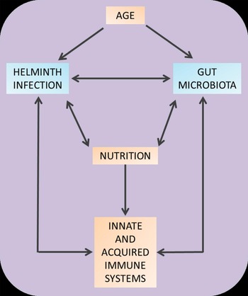
However, other pathogens can co-exist in the gut with the gut microbiota. Of particular importance in tropical countries are helminths. Helminths are eukaryotic, multi-cellular organisms which parasitize a wide range of hosts. Several species are able to cause disease in humans including soil-transmitted helminths such as roundworms, whipworms and hookworms; the schistosomes and food-borne pathogens such as the pork and beef tapeworms. Several mechanisms exist by which these helminths could interact with the microbiota (see Fig. 1), with multi-way interactions between host factors (such as age-related physiological changes and diet), helminth effects (e.g. effects on nutrition and host immune system) and finally microbe effects (also on host immune status and nutrition) and vice versa which may shape both the microbiome landscape and host health.
Potential interactions between the gut microbiota and helminth infection.

Helminths are able to modulate the host immune response to allow their survival and also reduce immunopathology (McSorley and Maizels, Reference McSorley and Maizels2012). However, unlike the gut flora, helminth infection often has a negative impact upon host nutrition (Stephenson et al. Reference Stephenson, Latham and Ottesen2000; Lwanga et al. Reference Lwanga, Kirunda and Orach2012). This is a particular problem in children where malnutrition associated with helminths can lead to fatigue, growth stunting, decreased cognitive development and thereby a general failure to thrive (King and Dangerfield-Cha, Reference King and Dangerfield-Cha2008).
If possible, the manipulation of the host's microbiota to a composition favourable to nutrition (greater nutrient availability to the host) or helminth clearance could help reduce helminth-related pathology and morbidity. This would have the advantage of being non-invasive and relatively inexpensive as the microbiota is able to be manipulated by nutritional or probiotic supplementation which could accompany standard treatment. Several countries where helminth infections are common already follow the WHO recommendations on supplementation of micronutrients such as vitamin A (see http://www.who.int/nutrition/publications/micronutrients/guidelines//vas_6to59_months/en/), meaning that nutritional supplementation for a healthy microbiome is a feasible consideration for improving paediatric health. Thus, associations between helminths and gut bacteria and their collective impact on human health present an urgent area of research. Harnessing the knowledge from studies on the human gut bacteria and helminths has the potential of providing/cultivating immune therapeutics as well as growth promoters in children exposed to helminthic infection. However, there is still a significant knowledge gap that has to be closed before dietary or immunological therapeutics that target the gut microbiome can be developed and translated to human disease. To this end, the work presented here reviews the current knowledge of the interactions between the gut bacteria and helminths and their potential for manipulation to improve host health in helminth endemic areas, while also highlighting important knowledge gaps and emphasizing the difficulties inherent in microbiome research.
DIVERSITY OF THE HUMAN GUT BACTERIA
A healthy or ‘normal’ microbiota has yet to be defined and may most likely not be a useful reference point due to the high geographical and temporal variability of bacterial communities between individuals and populations. The search for a ‘core microbiome’ which most people share has led to the discovery of only 18 bacterial species which were found in all 124 European participants in an initial MetaHIT (Metagenomics of the Human Intestinal Tract project) study (Qin et al. Reference Qin, Li, Raes, Arumugam, Burgdorf, Manichanh, Nielsen, Pons, Levenez, Yamada, Mende, Li, Xu, Li, Li, Cao, Wang, Liang, Zheng, Xie, Tap, Lepage, Bertalan, Batto, Hansen, Le Paslier, Linneberg, Nielsen, Pelletier, Renault, Sicheritz-Ponten, Turner, Zhu, Yu, Li, Jian, Zhou, Li, Zhang, Li, Qin, Yang, Wang, Brunak, Doré, Guarner, Kristiansen, Pedersen, Parkhill, Weissenbach, Bork, Ehrlich and Wang2010). Even within human communities which are closely genetically related and share similar diets, such as the Old Order Amish, there is still variability between individuals (Zupancic et al. Reference Zupancic, Cantarel, Liu, Drabek, Ryan, Cirimotich, Jones, Knight, Walters, Knights, Mongodin, Horenstein, Mitchell, Steinle, Snitker, Shuldiner and Fraser2012). In particular, Prevotella, which are able to metabolize complex plant carbohydrates, are more abundant in the gut microbiota of Amish farmers than Amish of different occupations, which could be explained by their closeness to livestock whose gut microbiotas tend to be dominated by this bacterial group (Stevenson and Weimer, Reference Stevenson and Weimer2007; Kim and Yu, Reference Kim and Yu2012). The microbiota does not easily lend itself to categorization and variation within even closely related populations is often high at the sub-phylum level (Jeffery et al. Reference Jeffery, Claesson, O'Toole and Shanahan2012).
What studies attempting to define a ‘healthy’ microbiome have suggested is that it is the ‘function’ rather than the diversity/composition of the microbiota that ultimately influences the host's health. This function has been elucidated by clustering microbiotas in one of three distinct ‘enterotypes’ based upon their ratios of Bacteroides, which primarily derive their energy from fermentation of carbohydrates and proteins, and Prevotella and Ruminococcus which are able to degrade host mucins (Arumugam et al. Reference Arumugam, Raes, Pelletier, Le Paslier, Yamada, Mende, Fernandes, Tap, Bruls, Batto, Bertalan, Borruel, Casellas, Fernandez, Gautier, Hansen, Hattori, Hayashi, Kleerebezem, Kurokawa, Leclerc, Levenez, Manichanh, Nielsen, Nielsen, Pons, Poulain, Qin, Sicheritz-Ponten, Tims, Torrents, Ugarte, Zoetendal, Wang, Guarner, Perderson, de Vos, Brunak, Doré, Weissenbach, Ehrlich and Bork2011). This ‘classification’ into enterotypes has proved useful in simplifying the otherwise diverse characteristics of the individual gut microbiome, but the clustering methodologies to identify the different enterotypes are still being developed (Koren et al. Reference Koren, Knights, Gonzalez, Waldron, Segata, Knight, Huttenhower and Ley2013).
Thus, for therapeutic interventions, these early studies suggest that focusing on manipulating the function of the gut microbiome could overcome some of the confounding effects of host heterogeneities, as reviewed in Bäckhed et al. (Reference Bäckhed, Fraser, Ringel, Sanders, Sartor, Sherman, Versalovic, Young and Finlay2012). Nonetheless, it is important to understand the factors influencing the composition of the gut microbiome. There is now cumulative evidence that the gut microbiome is influenced by several environmental factors including mode of delivery, antibiotic exposure, neonatal nutrition, parent nutrition, stress, age, degree of hygiene, nutrition/diet and co-infections (see review by Brown et al. Reference Brown, DeCoffe, Molcan and Gibson2012) as detailed below. Understanding the relative impact of these factors on the gut microbiome structure, composition and function is essential to allow predictions of the effects of any intervention aimed at altering the ecology of the gut.
FACTORS GIVING RISE TO THE DIVERSITY OF THE HUMAN GUT BACTERIA
Nutrition/diet
Experimental studies in mice suggest that diet accounts for a larger proportion of the structural variation in gut bacteria compared with genetic factors (57 vs 12%; Zhang et al. Reference Zhang, Zhang, Wang, Han, Cao, Hua, Mao, Zhang, Pang, Wei, Zhao, Chen and Zhao2010) and several experimental studies (some using gut communities from humans in humanized or germ-free mice) have demonstrated diet-induced dysbiosis in gut bacterial structure (see review by Brown et al. Reference Brown, DeCoffe, Molcan and Gibson2012). The recent studies demonstrating that the human gut microbiota clusters into functional groups (enterotypes), regardless of host age, nationality, gender and body mass index, suggest that these enterotypes may respond differently to diet.
Gut microbiomes from people consuming a Western diet and from people in rural communities consuming a predominantly plant-based diet were recently compared from a broad range of age groups amongst three populations from rural Venezuela, rural Malawi and the urban USA (Yatsunenko et al. Reference Yatsunenko, Rey, Manary, Trehan, Dominguez-Bello, Contreras, Magris, Hidalgo, Baldassano, Anokhin, Heath, Warner, Reeder, Kuczynski, Caporaso, Lozupone, Lauber, Clemente, Knights, Knight and Gordon2012). The differences found in the gut microbiota compositions between the two rural communities were found to be far less than the differences between the urban and rural populations. This indicates that nutritional factors are more important in influencing the composition of the gut microbiota than geographical or genetic differences. A previous study by this group also highlighted the importance of environment over genetics by showing that the gut microbiomes of monozygotic twins were as similar to each other as those of dizygotic twins (Turnbaugh et al. Reference Turnbaugh, Hamady, Yatsunenko, Cantarel, Duncan, Ley, Sogin, Jones, Roe, Affourtit, Egholm, Henrissat, Heath, Knight and Gordon2009).
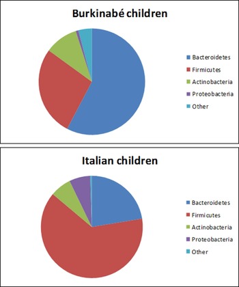
In another study, comparing the gut microbiomes of Italian children and children from rural Burkina Faso (De Filippo et al. Reference De Filippo, Cavalieri, Di Paola, Ramazzotti, Poullet, Massart, Collini, Pieraccini and Lionetti2010), Burkinabé children were found to have significantly different microbiotas from children living an Italian urban lifestyle. This included a decrease in the proportion of Bacteroidetes spp. and an increase in Firmicutes spp. in the Italian group (Fig. 2). Obesity has been correlated with changes in the ratios of these two phyla where individuals with low Bacteroidetes to Firmicutes ratios were found to be more likely to be obese (Ley et al. Reference Ley, Turnbaugh, Klein and Gordon2006b ). The children from rural Burkina Faso had diets containing higher amounts of starch, fibre and plant polysaccharides and lower amounts of fat and animal proteins than the Italian children. This was thought to be the main reason for gut bacterial differences as the gut microbiomes in children who were still being breast-fed in both groups were relatively similar. Changes were also found between the two populations in the ability of their gut microbiomes to harvest energy from food. Unlike the children fed a Western diet, the Burkinabé children were found to harbour the genera Xylanibacter, Prevotella, Butyrivibrio and Treponema which are able to degrade xylan and cellulose and are therefore able to utilize more energy from a plant-based diet.
The composition of the gut microbiota in African (Burkinabé) and European (Italian) children. Data adapted from De Filippo et al. (Reference De Filippo, Cavalieri, Di Paola, Ramazzotti, Poullet, Massart, Collini, Pieraccini and Lionetti2010) showing the major bacterial phyla composing the gut microbiotas of urban European and rural African children displayed as a pie-chart representation of the total bacterial microbiome.

Interestingly, very short-term changes in diet can still impact the composition of the microbiome. Subjects who ate a diet consisting either only of plants or only of meat/dairy foods over a 5-day period showed significant changes in the proportions of several groups of bacteria in their gastrointestinal tracts (David et al. Reference David, Maurice, Carmody, Gootenberg, Button, Wolfe, Ling, Devlin, Varma, Fischbach, Biddinger, Dutton and Turnbaugh2014). The meat/dairy group showed more changes in microbiome composition and more closely mirrored the Western urban groups in the above studies than the plant-based diet group, which showed fewer changes and more similarities to the rural groups. This demonstrates that altering the microbiome through diet to treat disease would not necessarily need to take place over a lengthy period of time.
Recently, the relationship between malnutrition and the gut microbiota has been addressed in a study of Malawian children suffering from kwashiorkor (a form of acute malnutrition caused by a lack of dietary protein but sufficient calorie intake) (Smith et al. Reference Smith, Yatsunenko, Manary, Trehan, Mkakosya, Cheng, Kau, Rich, Concannon, Mychaleckyj, Liu, Houpt, Li, Holmes, Nicholson, Knights, Ursell, Knight and Gordon2013). Significant differences were found between the gut microbiomes in twins where one twin was diagnosed with kwashiorkor and the other twin was healthy. The faecal microbiotas of several pairs of twins were transplanted into gnotobiotic mice (microbe-free mice which are then colonized by a specified microbial community) that were supplied with a Malawian-style diet followed by a standard peanut-based ready to use therapeutic food (RUTF) often used to treat malnutrition in children (Yang et al. Reference Yang, Van den Broeck and Wein2013). Upon returning the kwashiorkor mice to a Malawian-style diet, dietary metabolites were found to fall to pre-treatment levels in ‘kwashiorkor’ mice while those in ‘healthy’ mice were higher. This suggests that those with an originally kwashiorkor-associated gut microbiome would benefit less from RUTF treatment than those with a normal microbiome.
There are also significant differences between the gut microbiotas of children who are breast-fed and those who are not (Harmsen et al. Reference Harmsen, Wildeboer-Veloo, Raangs, Wagendorp, Klijn, Bindels and Welling2006; Fallani et al. Reference Fallani, Young, Scott, Norin, Amarri, Adam, Aguilera, Khanna, Gil, Edwards and Doré2010). Breast-fed infants develop a less diverse gut microbiota dominated by Bifidobacteria while formula-fed infants develop a more complex bacterial community which more closely resembles that of adults.
Several nutritional supplements have previously been shown to increase the abundance of bacterial species such as Bifidobacterium spp. and Lactobacillus spp. in humans, which are regularly used as probiotic supplements in their own right. These supplements include the plant polysaccharide inulin (Ramirez-Farias et al. Reference Ramirez-Farias, Slezak, Fuller, Duncan, Holtrop and Louis2009), oligosaccharides from human breast milk (Yu et al. Reference Yu, Chen, Kling, Liu, McCoy, Merighi, Heidtman and Newburg2013) and pomegranate extracts (Bialonska et al. Reference Bialonska, Ramnani, Kasimsetty, Muntha, Gibson and Ferreira2010). Bacterial species which have been associated with colon cancer initiation have also been found to be reduced in humans by the consumption of polydextrose (Costabile et al. Reference Costabile, Fava, Röytiö, Forssten, Olli, Klievink, Rowland, Ouwehand, Rastall, Gibson and Walton2012).
Caution must be taken when introducing new nutritional strategies as dietary supplementation does not always produce a beneficial change in the microbiota. For example, the impact of iron supplementation upon the gut microbiota of rural children from Côte d'Ivoire (Zimmermann et al. Reference Zimmermann, Chassard, Rohner, N'Goran, Nindjin, Dostal, Utzinger, Ghattas, Lacroix and Hurrell2010) was shown to increase the Gram-negative Enterobacteriaceae family of bacteria which contains potentially pathogenic species such as Salmonella spp., Shigella spp. and Escherichia coli while a decrease in the Lactobacilli family was found. This could be explained by the presence of siderophores which are often present amongst the Enterobacteriaceae (Reissbrodt and Rabsch, Reference Reissbrodt and Rabsch1988) but are absent amongst the Lactobacilli, which do not require iron to achieve growth (Archibald, Reference Archibald1983; Pandey et al. Reference Pandey, Bringel and Meyer1994). Siderophores are secreted by microorganisms in order to sequester iron from the environment and transform it into soluble complexes which can more easily be absorbed into the bacterial cells by active transport (Ferguson et al. Reference Ferguson, Hofmann, Coulton, Diederichs and Welte1998). Indeed recent studies also show that diet can cause dysbiosis which can lead to inflammatory conditions detrimental to human health, and the possibility of promoting microbes that can prevent or control these inflammatory-mediated diseases, through the manipulation of host diet, is already being discussed (Brown et al. Reference Brown, DeCoffe, Molcan and Gibson2012).
Age
Microbial colonization of individuals commences from birth with maternal, childhood and environmental conditions establishing a lifelong effect on the individual's gut microbiome (Collado et al. Reference Collado, Isolauri, Laitinen and Salminen2010; Gareau et al. Reference Gareau, Sherman and Walker2010; Manco et al. Reference Manco, Putignani and Bottazzo2010). In general, there is heterogeneity in the infant gut microbiome within the first months, stabilizing with a mix of the major bacterial phyla as previously described; the next changes occur when the infant is weaned, with the microbiome composition remaining relatively stable for life after the age of about 3 (Palmer et al. Reference Palmer, Bik, DiGiulio, Relman and Brown2007; Yatsunenko et al. Reference Yatsunenko, Rey, Manary, Trehan, Dominguez-Bello, Contreras, Magris, Hidalgo, Baldassano, Anokhin, Heath, Warner, Reeder, Kuczynski, Caporaso, Lozupone, Lauber, Clemente, Knights, Knight and Gordon2012). A study comparing the gut microbiotas of infants (3 weeks to 10 months), adults (25–45 years) and the elderly (70–90 years) eating a Western diet (Mariat et al. Reference Mariat, Firmesse, Levenez, Guimarăes, Sokol, Doré, Corthier and Furet2009) found significant differences between the three age groups. The average ratios of Firmicutes to Bacteroidetes in the elderly and infants were found to be more similar to each other than when either group was compared with adults. It has also been found that when comparing the diversity between children and adults, the variation between children's microbiotas within the same population tends to be larger than that of adults regardless of geography (Yatsunenko et al. Reference Yatsunenko, Rey, Manary, Trehan, Dominguez-Bello, Contreras, Magris, Hidalgo, Baldassano, Anokhin, Heath, Warner, Reeder, Kuczynski, Caporaso, Lozupone, Lauber, Clemente, Knights, Knight and Gordon2012).
Environment
One of the few studies conducted in an African population recently showed that different species of hosts sharing the same environment shared the different bacterial taxa present in their guts, as sampled by faecal samples collected from humans, cattle and semi-captive chimpanzees (Ellis et al. Reference Ellis, Bruce, Jenkins, Stothard, Ajarova, Mugisha and Viney2013). As already discussed, environmental factors are thought to significantly influence the gut microbiome structure. As more research is conducted it will become clearer which environmental factors can be more readily manipulated for improved host health.
Co-infections
In a healthy individual, the gut microbiota is able to protect the host against several gut pathogens due to colonization resistance which is maintained by multiple factors. Any invading gut pathogen must compete with the gut microbiota for available nutrients. This has been shown to be a significant factor in the inability of pathogens such as Clostridium difficile to cause disease in a non-antibiotic treated host (Britton and Young, Reference Britton and Young2012). The microbiota is also able to metabolize polysaccharides present in the gut environment into short chain fatty acids (Breznak and Kane, Reference Breznak and Kane1990; Louis and Flint, Reference Louis and Flint2009; Hosseini et al. Reference Hosseini, Grootaert, Verstraete and Van de Wiele2011) whose production can lead to reduced host deaths during E. coli infection (Fukuda et al. Reference Fukuda, Toh, Hase, Oshima, Nakanishi, Yoshimura, Tobe, Clarke, Topping, Suzuki, Taylor, Itoh, Kikuchi, Morite, Hattori and Ohno2011) and the down-regulation of virulence genes located in the Salmonella pathogenicity island 1 (Gantois et al. Reference Gantois, Ducatelle, Pasmans, Haesebrouck, Hautefort, Thompson, Hinton and Van Immerseel2006). The production of these fatty acids also leads to a decreased pH which is less well tolerated by pathogens than by the normal gut flora (Cherrington et al. Reference Cherrington, Hinton, Pearson and Chopra1991). The overall reduction in available oxygen by microbial metabolism in the gut can also lead to an unfavourable environment for facultatively anaerobic pathogens such as the Enterobacteriaceae (Altier, Reference Altier2005), in comparison to the majority of the gut flora which are obligate anaerobes (Marteau et al. Reference Marteau, Pochart, Doré, Béra-Maillet, Bernalier and Corthier2001). Apart from other bacteria, there are several parasites ranging from viruses, fungi, protozoans and helminths that can share the gastro-intestinal niche with bacteria (McKenna et al. Reference McKenna, Hoffmann, Minkah, Aye, Lackner, Liu, Lozupone, Hamady, Knight and Bushman2008; Parfrey et al. Reference Parfrey, Walters and Knight2011; Hoffmann et al. Reference Hoffmann, Dollive, Grunberg, Chen, Li, Wu, Lewis and Bushman2013). Their life history and clinical management can affect the composition and structure of the gut microbiome (Khoruts et al. Reference Khoruts, Dicksved, Jansson and Sadowsky2010; Dethlefsen and Relman, Reference Dethlefsen and Relman2011). This review focuses on helminth parasites.
HELMINTHS OCCUPYING THE GASTROINTESTINAL TRACT AND SURROUNDING TISSUES
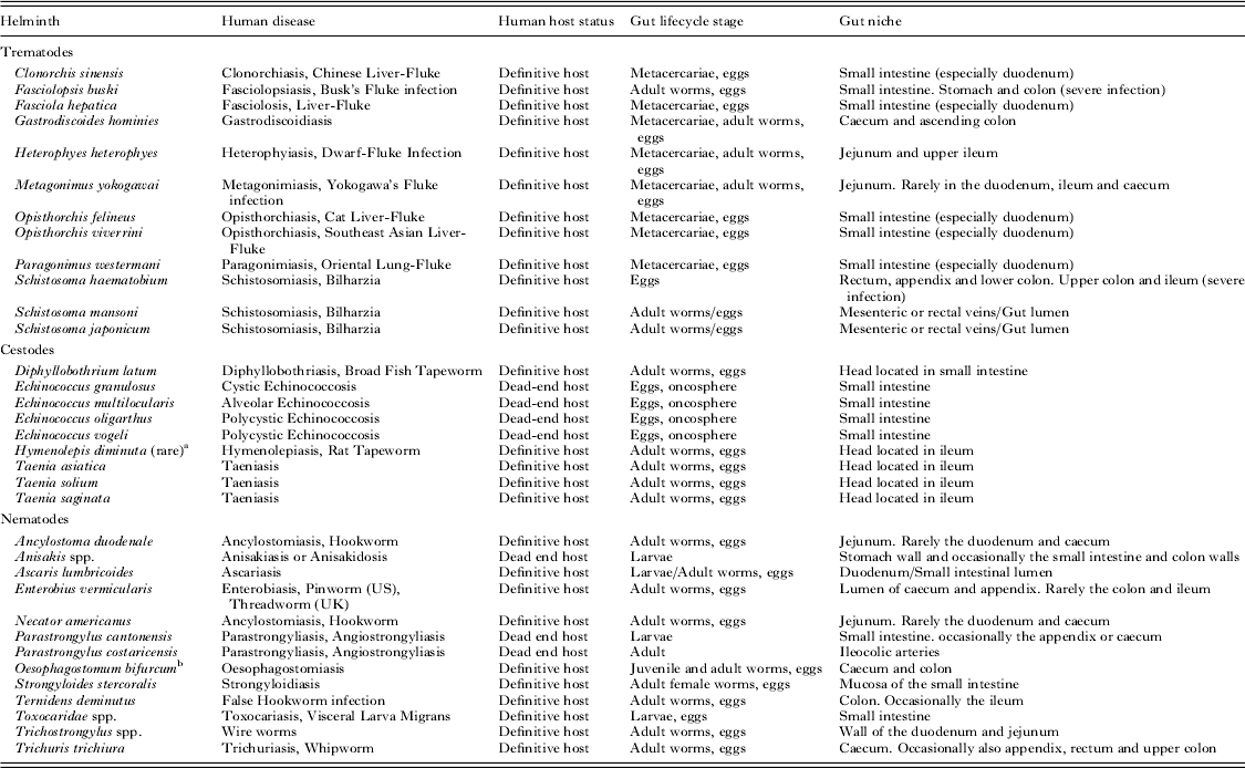
Helminths from two main phyla, the Nematoda and Platyhelminthes, occupy different niches within the gut for all or part of their life cycle, as is detailed in Table 2. The most important members of the Trematode ‘fluke’ class of platyhelminths are the schistosomes (Muller, Reference Muller2001), which cause the disease schistosomiasis. In humans this disease can be caused by several species, predominantly Schistosoma haematobium whose adults reside in the bladder venous plexus and Schistosoma mansoni and Schistosoma japonicum whose adults reside in the mesentery arteries of the intestines. The disease takes either a urogenital form as is the case with S. haematobium or a gastrointestinal form in the case of the other two species (Mahmoud, Reference Mahmoud2001).
Helminths which are able to cause infection in human hosts and share a niche with the gut microbiota
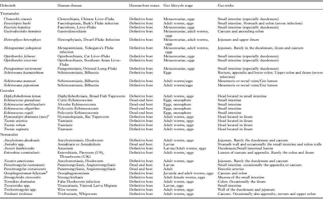
a Hamrick et al. (Reference Hamrick, Bowdre and Church1990).
b Polderman et al. (Reference Polderman, Krepel, Baeta, Blotkamp and Gigase1991).
The Cestode ‘tapeworm’ class of platyhelminths also play an important role in human health and several are highly important food-borne pathogens (Dorny et al. Reference Dorny, Praet, Deckers and Gabriel2009). The most important species to human health belong to the genus Taenia, which includes several pork and beef tapeworms, and the genus Echinococcus.
Several nematodes also occupy the human intestinal tract including the soil-transmitted nematodes (geohelminths) which include roundworms, whipworms and hookworms, all of which cumulatively currently infect over 1·5 billion people (World Health Organization, 2012). The most prevalent nematodes are Trichuris trichiura (whipworm), Ascaris lumbricoides (round worms), Necator americanus (hookworms) and Ancylostoma duodenale (hookworms) all of which occupy the human intestinal tract (Hotez et al. Reference Hotez, Brindley, Bethony, King, Pearce and Jacobson2008). While infection with these nematodes is often asymptomatic they are also associated with diarrhoea, abdominal pain and malaise during heavier infections and anaemia (hookworm) and intestinal obstruction (Ascaris spp.) in extreme cases.
EPIDEMIOLOGY OF HELMINTH INFECTION AND GUT BACTERIA
As already discussed, the relative effects of the host and environmental factors on the human gut microbiome structure and function are currently under investigation. In comparison, the effects of host and environment on the health impact of helminth infection have been studied for several decades (Gazzinelli et al. Reference Gazzinelli, Correa-Oliveira, Yang, Boatin and Kloos2012). Several features of the epidemiology of helminth infections are of relevance in the establishment and maintenance of the gut bacterial structure as well as overall health of the host.
As is detailed above, nutrition/diet is important in the ecology of the gut bacteria. While there is no clear evidence that diet/nutrition makes children susceptible to helminth infection, malnourishment has been suggested to increase susceptibility to disease caused by helminths in children, due to impaired immune responses and a decreased ability to repair damage caused by the parasites (Hall et al. Reference Hall, Zhang, MacArthur and Baker2012). Helminthic diseases have also been linked to generalized malnutrition (Stephenson et al. Reference Stephenson, Latham and Ottesen2000) and reduced host micronutrients such as iron (Gilles et al. Reference Gilles, Williams and Ball1964; Friedman et al. Reference Friedman, Kanzaria and McGarvey2005) and vitamin A (Friis et al. Reference Friis, Ndhlovu, Kaondera, Sandström, Michaelsen, Vennervald and Christensen1996; Haque et al. Reference Haque, Ahmed, Wahed, Mondal, Rahman and Albert2010). Several mechanisms by which helminths may cause malnutrition have been suggested including damage to the gut epithelium and anorexia (Symons, Reference Symons1985). Thus as previously suggested, it may be possible to manipulate the gut microbiome to avoid these pathologies as the gut flora composition is highly linked to nutrition. With more research it may be possible to manipulate the host microbiome through cheap, non-complex nutritional strategies maintained in at-risk human populations. For example, if malnourishment, associated with helminth infection, induces dysbiosis leading to a reduced ability to extract nutrients from food for host absorption, supplementation would be a useful therapeutic tool to accompany standard helminth treatment, particularly in children.
Host age is an important factor in the epidemiology of helminth infection, where infections are typically acquired in childhood and rise to peak in late childhood/early adulthood (Anderson and May, Reference Anderson and May1992). Levels of environmental contamination influence the rate and level of infection. In some cases, children's first exposure to helminth infection occurs before their microbiotas stabilize and reflect adult gut microbial communities, at around 3 years of age (Yu et al. Reference Yu, Jiang and Xu1995; Odogwu et al. Reference Odogwu, Ramamurthy, Kabatereine, Kazibwe, Tukahebwa, Webster, Fenwick and Stothard2006; Stothard et al. Reference Stothard, Sousa-Figueiredo, Betson, Bustinduy and Reinhard-Rupp2013). This provides the potential of the already established helminth infections to affect significantly the structure of the microbial community, as detailed below.
In the case of the schistosomes and some of the nematodes, there is a decline in infection levels which has been attributed to the gradual development of protective acquired immunity (Woolhouse, Reference Woolhouse1998). The effector responses induced during helminth infection are a complex combination of inflammatory and anti-inflammatory responses. Protective anti-helminth immunity is characterized by a balance between regulatory and effector responses. However, bacterial infections may alter this balance and may indeed skew the responses towards a different effector phenotype (see below). Thus, the time of gut colonization by bacteria relative to helminth colonization may be of importance in determining the health outcome for the host. Already human studies have begun to suggest that gut colonization by nematode infection (A. lumbricoides) may be associated with dysbiosis of the gut microbiome (Cooper et al. Reference Cooper, Walker, Reyes, Chico, Salter, Vaca and Parkhill2013). Of particular interest are those experimental studies suggesting that both history of helminth infection (even in animals that have cleared infection) and helminth infection intensity can affect the gut microbiome ecology (Broadhurst et al. Reference Broadhurst, Ardeshir, Kanwar, Mirpuri, Gundra, Leung, Wiens, Vujkovic-Cvijin, Kim, Yarovinsky, Lerche, McCune and Loke2012; Wu et al. Reference Wu, Li, Li, Beshah, Dawson and Urban2012).
THE EFFECT OF HELMINTH INFECTION ON GUT BACTERIAL POPULATIONS
The effect of helminth infections on the composition of the gut microbiota has mainly been studied in animal models, specifically Heligmosomoides polygyrus bakeri infection in mice and Trichuris suis infection in pigs. Significant differences have been observed between the ratios of bacterial families present in the proximal colon of pigs infected with T. suis in comparison with controls (Li et al. Reference Li, Wu, Li, Navarro, Couch, Hill and Urban2012). Several genera demonstrated significant decreases in prevalence upon infection including Oscillibacter which is the second most abundant genus in the porcine colon. In contrast, the three genera Mucispirillum, Paraprevotella and Desulfovibrio all significantly increased in prevalence, although the overall diversity of the porcine colon remained unchanged. These changes were related to differences in the metabolic potential of the gut microbiota between the two groups. Amongst the changes in genes associated with bacterial metabolism were significant decreases in genes relating to carbohydrate metabolism and the biosynthesis of the amino acids lysine, cysteine and methionine upon helminth infection. This finding indicates that during infection the proximal colonic bacteria are less able to utilize carbohydrates effectively. It has also previously been suggested that there is a need for increased sulphur-containing amino acids during infection with intestinal parasites in order to combat parasitic infection (MacRae, Reference MacRae1993), potentially due to decreased cysteine and methionine production by the microbiome.
The changes in the gut microbiome following helminth infection and subsequent clearance were still apparent 53 days post-infection (Wu et al. Reference Wu, Li, Li, Beshah, Dawson and Urban2012), with significantly higher proportions of Campylobacter occurring in pigs that were infected but did not clear infection compared with those that had been infected but had low worm burdens or were worm free. This agrees with previous findings that T. suis infection can exacerbate campylobacteriosis (Rutter and Beer, Reference Rutter and Beer1975; Mansfield and Urban, Reference Mansfield and Urban1996; Shin et al. Reference Shin, Gardiner, Deitel and Kandel2004).
In contrast to the above studies where helminths are shown to increase the quantity of a pathogenic bacterial species, investigations in H. polygyrus bakeri infection have shown an increase in commensal bacterial species. Heligmosomoides polygyrus bakeri infection was examined in two duplicate experiments to attempt to explain the ability of the helminth to ameliorate the symptoms of IBDs in model IL-10 deficient mice inducing chronic colitis (Walk et al. Reference Walk, Blum, Ewing, Weinstock and Young2010). While no significant difference was found in the caecum of infected mice the group did note a significant 1·8-fold increase in bacterial numbers within the ileum where the parasites were located. Infection was also associated with a change in the families of bacteria occupying the ileum, with members of the Lactobacillaceae family increasing in both experiments. The Lactobacillaceae group has been found to produce reactive oxygen species which inhibit activation of the transcription factor NF-κB in neonatal mouse intestines (Lin et al. Reference Lin, Myers, Ray, Song, Nasr, Berardinelli, Kundu, Murthy, Hansen and Neish2009), reverse intestinal injury (Mañé et al. Reference Mañé, Lorén, Pedrosa, Ojanguren, Xaus, Cabré, Domènech and Gassull2009) and offer protection against graft-versus-host disease (Jenq et al. Reference Jenq, Ubeda, Taur, Menezes, Khanin, Dudakov, Liu, West, Singer, Equinda, Gobourne, Lipuma, Young, Smith, Ghosh, Hanash, Goldberg, Aoyama, Blazar, Pamer and van den Brink2012). This may lead to a symbiotic relationship between infecting helminths and the microbes to reduce intestinal inflammation. However at this level of bacterial taxonomic hierarchy the mouse gut microbiota is significantly different from the human microbiota (Ley et al. Reference Ley, Bäckhed, Turnbaugh, Lozupone, Knight and Gordon2005) and the study was conducted in a mouse disease model which cannot easily be compared with healthy humans.
It is not necessarily the case that helminths would only influence the microbiota occupying the same niche as them. While Opisthorchis viverrini primarily occupies the bile ducts it has been shown to alter the microbiome of hamster colorectal faeces (Plieskatt et al. Reference Plieskatt, Deenonpoe, Mulvenna, Krause, Sripa, Bethony and Brindley2013). There have been indications that the gut microbiota is also affected by schistosomiasis infection in both mice and humans as the urinary host metabolites associated with the gut microbiota are changed upon infection (Wang et al. Reference Wang, Holmes, Nicholson, Cloarec, Chollet, Tanner, Singer and Utzinger2004; Balog et al. Reference Balog, Meissner, Göraler, Bladergroen, Vennervald, Mayboroda and Deelder2011). It has also been shown that the absence of a gut microbiome during S. mansoni infection in mice significantly reduces the amount of intestinal granulomas and gut inflammation, potentially influencing the ability of the helminth to excrete eggs into the gut lumen (Holzscheiter et al. Reference Holzscheiter, Layland, Loffredo-Verde, Mair, Vogelmann, Langer, Wagner and Prazeres da Costa2014).
The effect of the gut microbiota on the establishment, maintenance and pathology of helminth infections in the gut has mainly been addressed in studies investigating gnotobiotic/germ-free animals (Wescott, Reference Wescott1968; Chang and Wescott, Reference Chang and Wescott1972; Johnson and Reid, Reference Johnson and Reid1973) and in relation to egg-hatching of Trichuris muris (Hayes et al. Reference Hayes, Bancroft, Goldrick, Portsmouth, Roberts and Grencis2010).
Hayes et al. compared T. muris hatching in eggs incubated with explant from a mouse caecum (upper large intestine) with eggs incubated with separate cultures of E. coli, Staphylococcus aureus, Salmonella typhimurium, Pseudomonas aeruginosa and Saccharomyces cerevisiae (Hayes et al. Reference Hayes, Bancroft, Goldrick, Portsmouth, Roberts and Grencis2010). All of the tested microbial cultures produced hatching at 37 °C after 2 h, at similar levels to hatching in the mouse caecum, while filtered cultures failed to cause hatching. The importance of bacterial contact with eggs was established with the use of transwells with E. coli and S. cerevisiae cultures, which when used failed to elicit hatching when pores were too small for passage of cells through to the eggs (0·4 μm). This indicated that the factor affecting egg hatching was unlikely to be a microbially derived part of the supernatant. The study also established the necessity of having an intact bacterial structure in the case of E. coli as clustering of green-fluorescent tagged E. coli around the poles of the helminth eggs where worms emerge was observed (Preston and Jenkins, Reference Preston and Jenkins1985), demonstrating that a component of the bacterial cell surface played a role.
The group hypothesized that the Type 1 fimbriae of E. coli which bind the cells to surfaces in a mannose-dependent manner may play a role in T. muris hatching. While they found that purified Type 1 fimbriae did not elicit hatching, the addition of mannose decreased hatching and several other experiments pointed to it playing a key role. However, it is unlikely that this is the only mechanism at play as S. aureus and P. aeruginosa do not contain these proteins but are still able to induce T. muris hatching.
To establish whether the microbiota had an effect in vivo the team treated a group of mice with antibiotics and measured the worm-burden 21 days after infection. The worm-burden was found to be significantly decreased and the host's Th2 response was significantly increased with elevated amounts of IL-4 and IL-13. However, the decrease in worm numbers was found not to be caused by this increase in the Th2 response due to decreased worm numbers still occurring in severe combined immunodeficient mice. This study indicates that the absence of the gut microbiota causes a decrease in the helminth's ability to establish infection but does not necessarily decrease its ability to survive within the host.
While the above study did show that the presence of bacterial species played a role in initiating egg-hatching and germ-free/gnotobiotic studies have indicated a relationship between the presence of a microbiome and the ability of helminths to establish chronic infection, the contribution of specific commensal bacterial groups has not currently been addressed. Limited information can therefore be drawn from these studies about how the microbiota interacts with helminth pathogens although they do indicate that colonization resistance by the gut microbiome may not play a role in preventing these helminth infections.
THE EFFECTS OF GUT BACTERIA AND HELMINTHS ON THE HOST IMMUNE SYSTEM
A potential mechanism by which helminth infection could alter the gut microbiota composition is its effect upon the host immune system, which could disrupt the homeostatic relationship established between the gut flora and the host.
The majority of bacteria present within the intestines are commensals which despite their large numbers do not cause disease, nor do they generate a predominantly pro-inflammatory environment (Chinen and Rudensky, Reference Chinen and Rudensky2012). This is true despite the presence of pattern recognition receptors (PRRs) within the intestinal epithelium, which recognize broadly distributed bacterial antigens and are able to initiate an inflammatory response to bacterial cells (Philpott and Girardin, Reference Philpott and Girardin2004). Pathogenic bacteria also often produce molecular factors which are not present in commensal microbes, which can induce specific immune responses (Franchi et al. Reference Franchi, Kamada, Nakamura, Burberry, Kuffa, Suzuki, Shaw, Kim and Nunez2012; Larsen et al. Reference Larsen, Steen-Jensen, Laursen, Søndergaard, Musavian, Butt and Brix2012; Rizzetto et al. Reference Rizzetto, Buschow, Beltrame, Figdor, Schierer, Schuler and Cavalieri2012).
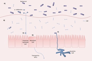
The host gut is able to create a degree of separation between its immune cells and the gut flora by maintaining a dense mucus layer which separates most of the commensal bacteria from the gut epithelium (Fig. 3). However, this mechanism is insufficient to explain the lack of a pathology associated with commensal bacteria as cells lining the epithelium are constantly sampling microbes from within the lumen. The gut microbiota have been found to inhibit NF-κB activation (Kelly et al. Reference Kelly, Campbell, King, Grant, Jansson, Coutts, Pettersson and Conway2004; Kumar et al. Reference Kumar, Wu, Collier-Hyams, Hansen, Li, Yamoah, Pan, Jones and Neish2007; Kaci et al. Reference Kaci, Lakhdari, Doré, Ehrlich, Renault, Blottière and Delorme2011; Lakhdari et al. Reference Lakhdari, Tap, Béguet-Crespel, Le Roux, de Wouters, Cultrone, Nepelska, Lefèvre, Doré and Blottière2011) and in a non-diseased state it has been noted that intestinal epithelial cells express few lipopolysaccharide recognizing molecules such as Toll-like receptor (TLR) co-receptor CD14, TLR2 and TLR4 (Abreu et al. Reference Abreu, Vora, Faure, Thomas, Arnold and Arditi2001). This suggests that the immune response to gut commensal bacteria is low when pathogenic bacteria are not present or that there is a reduced ability to initiate responses through innate receptors.
The interaction of the microbiota with the host immune system. The first layer of protection for the gut epithelium is a double layer of mucus comprising an easily invaded loose outer layer with low quantities of IgA where the majority of the gut microbiome resides (A) and a denser layer containing larger amounts of IgA which lies next to the epithelium and is less conducive to microbial growth (B) (McAuley et al. Reference McAuley, Linden, Png, King, Pennington, Gendler, Florin, Hill, Korolik and McGuckin2007). Intestinal epithelial cells also secrete a large amount of antimicrobial peptides called defensins (C) which broadly act upon bacteria, fungi and some enveloped viruses to increase the permeability of their membranes (Ganz, Reference Ganz2003). The gut constantly samples the surrounding microbial communities via microfold cells (D), contained within specialized Peyer's patches, and lymphoid follicles which sample the surrounding antigens and microbes and transport them to the sub-epithelial dome which contains many antigen-presenting cells. Dendritic cells sample microbes from within the gut lumen (E) (Niess and Reinecker, Reference Niess and Reinecker2005), and contain pattern recognition receptors that can detect a wide variety of microbe-associated molecular patterns.

Individual bacterial species also possess mechanisms to induce an anti-inflammatory response which may help to reduce immunopathology related to the gut bacterial flora. These include the human commensal bacteria Bacteroides fragilis (Round and Mazmanian, Reference Round and Mazmanian2010), Faecalibacterium prausnitzii (Sokol et al. Reference Sokol, Pigneur, Watterlot, Lakhdari, Bermúdez-Humarán, Gratadoux, Blugeon, Bridonneau, Furet, Corthier, Grangette, Vasquez, Pochart, Trugnan, Thomas, Blottière, Doré, Marteau, Seksik and Langella2008), several Lactobacillus spp. (Jenq et al. Reference Jenq, Ubeda, Taur, Menezes, Khanin, Dudakov, Liu, West, Singer, Equinda, Gobourne, Lipuma, Young, Smith, Ghosh, Hanash, Goldberg, Aoyama, Blazar, Pamer and van den Brink2012; Shimazu et al. Reference Shimazu, Villena, Tohno, Fujie, Hosoya, Shimosato, Aso, Suda, Kawai, Saito, Makino, Ikegami, Itoh and Kitazawa2012; van Baarlen et al. Reference van Baarlen, Wells and Kleerebezem2013) and Bifidobacterium spp. (Imaoka et al. Reference Imaoka, Shima, Kato, Mizuno, Uehara, Matsumoto, Setoyama, Hara and Umesaki2008; Khokhlova et al. Reference Khokhlova, Smeianov, Efimov, Kafarskaia, Pavlova and Shkoporov2012).
It has also been suggested that diet-dependent nutrients and metabolites produced by the gut microbiota can have an immunomodulatory effect. This has been reviewed recently in some detail (Brestoff and Artis, Reference Brestoff and Artis2013). The microbiota has been implicated in the production of secondary bile acids (Sayin et al. Reference Sayin, Wahlström, Felin, Jäntti, Marschall, Bamberg, Angelin, Hyötyläinen, Orešič and Bäckhed2013) which interact with monocytes and macrophages to produce an anti-inflammatory response via the G protein-coupled bile acid receptor 1 (Wang et al. Reference Wang, Chen, Yu, Forman and Huang2011b ) and the nuclear receptor subfamily 1, group H, member 4 (Attinkara et al. Reference Attinkara, Mwinyi, Truninger, Regula, Gaj, Rogler, Kullak-Ublick and Eloranta2012). Various other compounds produced by the microbiota influence the development and/or regulation of the immune system such as essential amino acids, short chain fatty acids and vitamins (Jose and Good, Reference Jose and Good1973; Chandra, Reference Chandra1992; Gill et al. Reference Gill, Pop, DeBoy, Eckburg, Turnbaugh, Samuel, Gordon, Relman, Fraser-Liggett and Nelson2006; Maslowski et al. Reference Maslowski, Vieira, Ng, Kranich, Sierro, Yu, Schilter, Rolph, Mackay, Artis, Xavier, Teixeira and Mackay2009; LeBlanc et al. Reference LeBlanc, Milani, de Giori, Sesma, van Sinderen and Ventura2013).
The avoidance of inflammation by the microbiota helps maintain the homoeostasis of the gut and helminths reinforce this stability by excreting/secreting anti-inflammatory molecules. As a result, helminths and their products are being investigated as potential therapies for inflammatory bowel diseases (IBDs). Several trials of T. suis treatment have been carried out for IBDs in humans (Summers et al. Reference Summers, Elliott, Urban, Thompson and Weinstock2005a , Reference Summers, Elliott, Urban, Thompson and Weinstock b ) which have shown positive results. The hookworm N. americanus has also been tested as a therapy for IBDs (Croese et al. Reference Croese, O'Neil, Masson, Cooke, Melrose, Pritchard and Speare2006; Feary et al. Reference Feary, Venn, Mortimer, Brown, Hooi, Falcone, Pritchard and Britton2010; Daveson et al. Reference Daveson, Jones, Gaze, McSorley, Clouston, Pascoe, Cooke, Speare, Macdonald, Anderson, McCarthy, Loukas and Croese2011) and has been separately shown to alter the gut microbiota in Syrian hamsters (Wang et al. Reference Wang, Xiao, Xue, Singer, Utzinger and Holmes2009).
Mechanisms by which helminths decrease the immune response to bacterial antigens
Several mechanisms have been suggested for how helminths are able to reduce the immune response to bacterial antigens translocating across the gut epithelium during infection. Schistosomiasis infections have been linked to an increase in B-cell expression of Toll-like receptors specific for Gram-negative (TLR4) and Gram-positive (TLR2) bacteria where TLR4 in this case causes an anti-inflammatory response in humans (Onguru et al. Reference Onguru, Liang, Griffith, Nikolajczyk, Mwinzi and Ganley-Leal2011). This heightened expression of TLR2 and TLR4 has also been observed in epithelial cells in rats infected with the tapeworm Hymenolepis diminuta (Kosik-Bogacka et al. Reference Kosik-Bogacka, Wojtkowiak-Giera, Kolasa, Salamatin, Jagodzinski and Wandurska-Nowak2012). TLR mediated targeting of bacterial antigens has also been studied during nematode infection, with an up-regulation of TLR2, TLR4 and TLR9 observed in H. polygyrus bakeri infection in mice (Friberg et al. Reference Friberg, Little, Ralli, Lowe, Hall, Jackson and Bradley2013).
It is also possible that helminths could impact the immune response to gut bacterial antigens through their induction of specific T-cell responses. Helminth infection is usually characterized by a strong T helper 2 cell (Th2) type response characterized by an increase in IL-4, IL-5 and IL-13; the expansion of CD4+ Th2 cells, eosinophils, mast cells, basophils and alternatively activated macrophages and an increased secretion of IgE (Anthony et al. Reference Anthony, Rutitzky, Urban, Stadecker and Gause2007). In contrast, antibacterial immune responses tend to be dominated by a T helper 1 (Th1) cell type response characterized by increased levels of IFN-γ and IL-2 (Lorvik et al. Reference Lorvik, Haabeth, Clancy, Bogen and Corthay2013). Helminths also decrease the amount of bacterial translocation across the gut epithelium through the induction of a Th2 type response which leads to faster tissue healing (Chen et al. Reference Chen, Liu, Wu, Rozo, Bowdridge, Millman, Van Rooijen, Urban, Wynn and Gause2012) and thereby fewer bacterial antigens coming into contact with immune cells.
An antibacterial immune response has been observed during several helminth infections including a model of hookworm infections in mice (Nippostrongylus brasiliensis) which has been shown to stimulate mast cells to increase their innate responses to bacteria by the induction of IL-4 (Sutherland et al. Reference Sutherland, Xu, Kim, Seeley, Caughey and Wolters2011). In T. muris infection in mice, expression of mouse angiogenin 4 (a goblet cell derived antimicrobial peptide) was found to be increased (Forman et al. Reference Forman, deSchoolmeester, Hurst, Wright, Pemberton and Else2012), indicating a targeted immune response to bacteria during infection.
IL-22 has also been implicated in the induction of an antimicrobial response during helminth infections (Leung and Loke, Reference Leung and Loke2013). This cytokine is produced by a variety of immune cells, both innate and adaptive (Sabat et al. Reference Sabat, Witte, Witte, Wolk, Quesniaux, Ryffel and Padova2013), and regulates the production of numerous antimicrobial peptides (Wolk et al. Reference Wolk, Kunz, Witte, Friedrich, Asadullah and Sabat2004; Liang et al. Reference Liang, Tan, Luxenberg, Karim, Dunussi-Joannopoulos, Collins and Fouser2006). The lack of IL-22 has been implicated in more severe pathologies of several bacterial infections including infections with Citrobacter rodentium (Zheng et al. Reference Zheng, Valdez, Danilenko, Hu, Sa, Gong, Abbas, Modrusan, Ghilardi, de Sauvage and Ouyang2008) and Salmonella enterica (Schulz et al. Reference Schulz, Köhler, Schütze, Knauer, Straubinger, Chackerian, Witte, Wolk, Sabat, Iwakura, Holscher, Müller, Kastelein and Alber2008). It has been noted that during helminth treatment for inflammatory bowel diseases, both N. americanus excretory proteins (NaES) (McSorley et al. Reference McSorley, Gaze, Daveson, Jones, Anderson, Clouston, Ruyssers, Speare, McCarthy, Engwerda, Croese and Loukas2011) and T. trichiura (Broadhurst et al. Reference Broadhurst, Leung, Kashyap, McCune, Mahadevan, McKerrow and Loke2010) caused an increase in IL-22 expression, although no difference was observed between IL-22 deficient and wild-type mice infected with S. mansoni (Wilson et al. Reference Wilson, Feng, Barber, Yarovinsky, Cheever, Sher, Grigg, Collins, Fouser and Wynn2010), indicating that IL-22 does not play a significant role during schistosomiasis.
This evidence points to a significant increase in the immune response to bacterial antigens upon helminth infection, which may potentially lead to changes in the gut microbiota composition in infected individuals. While this would not necessarily negatively affect host health, depending upon the bacterial species affected it could cause a decrease in host nutrition or lead to bacterial disease.
CONCLUSIONS/FUTURE DIRECTIONS
Experimental studies show that helminths influence the composition of the microbiota during infection and that this is mediated through different mechanisms including alteration of immunological and metabolic pathways. Helminth infection may result in changes in the ability of the host to extract nutrients from their diet, particularly in malnourished children, and could also affect the numbers of potentially pathogenic bacteria within the gut, causing bacterial co-infection.
Currently there is a paucity of data on the interactions between the microbiota and helminths in humans, but this should soon change with increasing technologies and tools to carry out more large-scale and detailed human studies.
With more understanding of how modern living and medical interventions (e.g. use of antibiotics) have affected the human immune system and the role played by helminths and the gut microbiome (and the mechanisms/pathways involved) in maintaining immune and metabolic homoeostasis, it may be possible to manipulate the microbiota in order to reduce morbidity (e.g. malnutrition) and pathology caused by helminth infection and metabolic diseases arising from dysbiosis of the gut microbiome. Although the realization of this prospect is far from immediate, it presents an exciting prospect that warrants further investigation.
ACKNOWLEDGEMENTS
The authors would like to thank the Parasite Immuno-epidemiology Group and Julien Sindt, both at the University of Edinburgh, for their feedback during the writing of this review.
FINANCIAL SUPPORT
The review was prepared as part of laboratory studies which were funded by the Thrasher Research Fund (www.thrasherresearch.org) and the University of Edinburgh.