Introduction
Squamous cell carcinoma (SCC) of the tonsil is the most common form of oropharyngeal malignancy.Reference Weatherspoon, Chattopadhyay, Boroumand and Garcia1 As tonsillar SCCs are typically asymptomatic in the early course of the disease, the majority of cases are diagnosed in the later stages when symptoms become apparent.Reference Seiden, Tami, Pensak, Cotton and Gluckman2 Metastases to cervical lymph nodes are a common finding; however, distant metastases occur less frequently and most often in the later stages of disease.Reference de Bree, Mehta, Snow and Quak3 The most common sites of distant metastasis include the lungs and bone.
Whilst metastases to the brain and skull base have been reported, they are rare and most commonly a feature of advanced disease.Reference Barrett, Gill, Miles, Iloreta, Bakst and Fowkes4,Reference Dobelbower, Nabell, Markert, Carroll, Said-Al-Naief and Meredith5 We report a case of an isolated pituitary fossa metastasis from a primary tonsillar SCC, which presented as an ipsilateral oculomotor nerve palsy and partial Horner's syndrome. We evaluate our clinical approach towards this patient in order to identify diagnostic improvements and make recommendations that may aid early recognition of similar presentations.
Case report
Background
This case report concerns a 64-year-old male ex-smoker, with a history of significant alcohol intake and two previous right-sided oral cavity malignancies (floor of mouth SCC and mandible SCC). Both tumours were managed definitively with surgery alone.
He presented with tenderness in the right temporomandibular joint. Following biopsy and staging, he was diagnosed with a p16-negative primary SCC of the right tonsil (tumour–node–metastasis staging of T3N0M0). Subsequently, he was treated with a six-week course of chemotherapy (synchronous platinum and carboplatinum) and radiotherapy (60 Gy) with curative intent.
An F-fluoro-2-deoxy-D-glucose positron emission tomography/computed tomography (FDG-PET/CT) was performed six months after completion of treatment to assess response. This revealed complete resolution of the tumour, with no evidence of metastatic disease. The patient was placed on routine follow up at two-monthly intervals.
Presentation
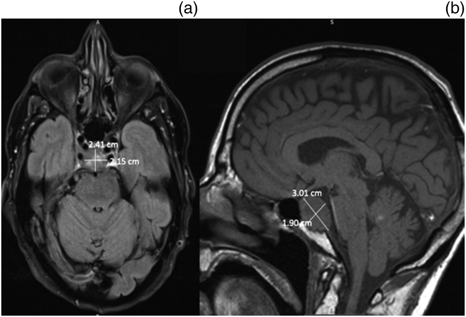
Two months into his follow up, the patient presented acutely to hospital with a two-week history of worsening right-sided ptosis and associated horizontal diplopia. Additionally, he was found to have mydriasis and reduced pupil reactivity, but no relative afferent pupillary defect or papilloedema were present. This indicated a right-sided IIIrd cranial nerve palsy and ipsilateral partial Horner's syndrome (lid ptosis). Magnetic resonance imaging (MRI) of the brain revealed a new lesion within the pituitary fossa (Figure 1).
Axial, T2-weighed (a) and sagittal, T1-weighted (b) contrast-enhanced magnetic resonance imaging scans of the head, showing an enhancing mass with central tumour necrosis arising from the sella turcica. The pituitary gland is being displaced superiorly and anteriorly. Lateral expansion into the cavernous sinuses, with encasement of the right internal carotid artery, can be seen.

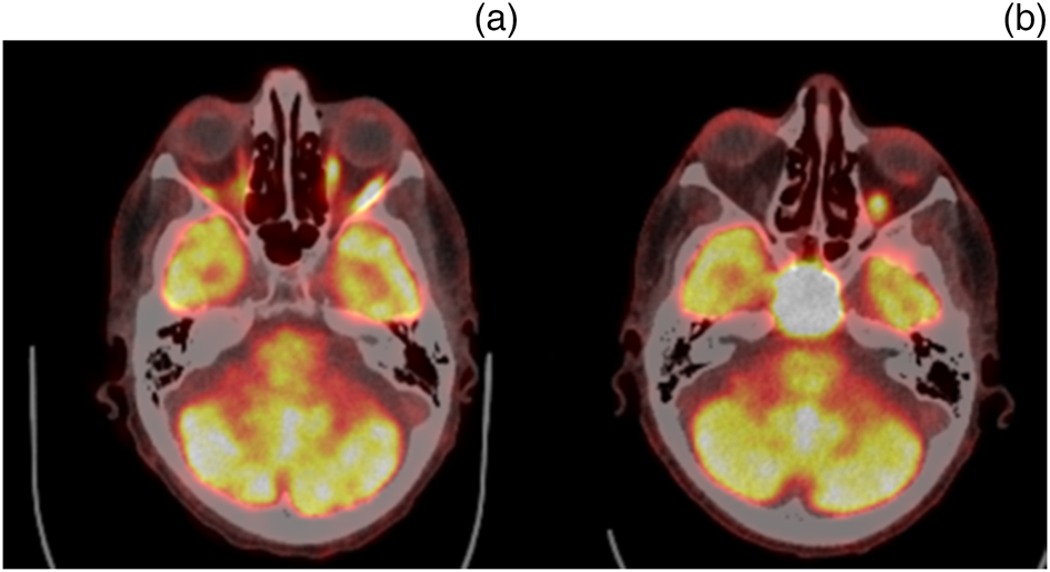
A trans-sphenoid biopsy of the mass was subsequently performed. Histology revealed a p63-positive, p16-negative SCC. The histology differed from the two previous oral cavity malignancies, but was consistent with his previously treated tonsillar SCC, and was hence a metastasis. Further FDG-PET/CT scanning (Figure 2) revealed a previously undetected metastasis. No other distant metastases were seen.
Axial F-fluoro-2-deoxy-D-glucose positron emission tomography/computed tomography image comparison at (a) five months before acute presentation (4 April 2018) and (b) two months following acute presentation (8 November 2018). The latter image shows an avid mass in the pituitary fossa that was not present on the previous scan. No other pathological intracranial activity was identified.

Baseline pituitary function measurements were also carried out; these included tests of thyroid function, serum follicle stimulating and luteinising hormone levels, and testosterone and cortisol levels. These were all found to be within normal limits.
Approach and follow up
The patient was referred to the skull base multidisciplinary team (MDT) for consideration of surgical debulking and stereotactic radiotherapy. However, he was deemed unsuitable for further surgery or stereotactic radiotherapy given the close proximity of the metastasis to the internal carotid artery, and the extent of the recurrence. The head and neck MDT recommended a course of palliative radiotherapy with the option of adding palliative chemotherapy.
Two months after diagnosis, the patient was admitted to a hospice for symptom control following the development of episodic frontal headaches and vomiting. He underwent a course of palliative radiotherapy and daily dexamethasone, which resolved these symptoms without any complications. Follow up after four months revealed he had not experienced any further intracranial symptoms, other than right-sided ptosis, whilst receiving palliative care under the community hospice team.
Discussion
Metastatic breast and lung cancer are the two most common causes of pituitary fossa metastases.Reference Altay, Krisht and Couldwell6 Whilst there have been reported cases of atypical metastases to the central nervous system and clivus from primary tonsillar SCCs, there has been no published case of an isolated pituitary fossa metastasis.Reference Marchese-Ragona, Ferraro, Marioni, Staffieri, Manara and Restivo7 Our patient, therefore, represents the first reported case of a pituitary fossa metastasis from a tonsillar SCC. Given that pituitary fossa metastases carry a poor prognosis,Reference Lin, Huang, Chung and Chang8 prompt investigation and recognition is needed so that effective management remains a viable option once the diagnosis is established.
Pituitary fossa metastases represent 1 percent of pituitary fossa masses surgically excised; the majority of lesions are most commonly caused by primary tumours such as pituitary adenomas.Reference Altay, Krisht and Couldwell6,Reference Valassi, Biller, Klibanski and Swearingen9 Differentiation between these two causes is therefore needed in order to determine both management and prognosis. However, this can be difficult given the scarcity of symptoms that can distinguish metastases from primary adenomas.
According to Al-Aridi et al.,Reference Al-Aridi, El Sibai, Fu, Khan, Selman and Arafah10 and as in our case presentation, features suggestive of a pituitary fossa or sellar metastasis include headaches, visual field defects and abnormal eye movements. The presence of cranial nerve palsies, especially of the IIIrd and VIth cranial nerves, are also associated with metastases due to invasion of the tumour into the cavernous sinus. Though invasion of the cavernous sinus may also occur with pituitary adenomas, a slower rate of growth is usually seen.
• Brain and skull base metastases from oropharyngeal malignancies are rare, most commonly a feature of advanced disease
• This patient represents the first reported case of an isolated pituitary fossa metastasis from tonsillar squamous cell carcinoma
• Pituitary fossa metastases carry a poor prognosis
• Patients presenting with acute unilateral cranial nerve palsies following treatment of locally advanced tonsillar malignancies should be investigated with high suspicion
• A low threshold for pituitary fossa biopsy is warranted in such cases
• The role of F-fluoro-2-deoxy-D-glucose positron emission tomography/computed tomography in identifying developing brain metastases during follow up is limited
Although distinguishing between benign and malignant disease may be clinically possible, the role of imaging can be limited.Reference Aaberg, Kay and Sternau11 This is because of the lack of pathognomonic features to reliably differentiate a metastasis from a benign lesion.Reference Al-Aridi, El Sibai, Fu, Khan, Selman and Arafah10 Biopsy and histological assessment of a pituitary fossa mass thereby represents a definitive method of diagnosis, as suggested by the literature.Reference Lin, Huang, Chung and Chang8,Reference Al-Aridi, El Sibai, Fu, Khan, Selman and Arafah10,Reference Goulart, Upadhyay, Ditzel Filho, Beer-Furlan, Carrau and Prevedello12 Consequently, the decision to proceed with a trans-sphenoid biopsy represents a key step in the approach towards such cases.
Although FDG-PET/CT has been recommended in the follow up of head and neck malignancies to assess treatment response and exclude distant metastases,Reference Tantiwongkosi, Yu, Kanard and Miller13 its ability to detect brain metastases is limited.Reference Kitajima, Nakamoto, Okizuka, Onishi, Senda and Suganuma14,Reference Bochev, Klisarova, Kaprelyan, Chaushev and Dancheva15 In particular, Lee et al.Reference Lee, Lee, Kim, Cho, Lee and Yi16 found PET/CT to have a sensitivity of 24 per cent in detecting symptomatic brain metastases and 19 per cent for detecting asymptomatic metastases, when compared against contrast-enhanced MRI. This limitation can be explained by the high physiological uptake of FDG by the cerebral cortexReference Galldiks, Langen, Albert, Chamberlain, Soffietti and Kim17 under normal conditions, which can mask the presence of a developing metastatic lesion. This is especially the case with microscopic lesions.
It is possible that a developing metastasis within the pituitary fossa was missed on the post-treatment follow-up FDG-PET/CT scan, and was not visible until large enough to produce symptoms five months later. As invasion into nearby structures, such as the cavernous sinus and the internal carotid artery, had already taken place at the time of diagnosis, the management options available were limited. Detection of this mass prior to the development of symptoms may have offered the opportunity for more aggressive treatment options, and a more favourable prognosis.
Another important consideration in this case was the fact that the primary tumour was p16-negative. P16 is tumour suppressor protein that is often over-expressed in human papilloma virus related oropharyngeal SCCs.Reference Kida, Terada, Uwa, Omori, Fujii and Tomita18 The presence of p16 in such malignancies has been correlated with a more favourable prognosis.Reference Weinberger, Yu, Haffty, Kowalski, Harigopal and Sasaki19 Studies have suggested this is because of lower recurrence rates and better responses to treatment.Reference Fischer, Zlobec, Green, Probst, Storck and Lugli20 The absence of p16 in our patient thereby served as a poor prognostic indicator.
Conclusion
Our patient represents the first reported case of an isolated pituitary fossa metastasis from a primary tonsillar SCC. We recommend a high degree of clinical suspicion in such patients with unusual new symptoms, particularly in the presence of p16 negativity. Furthermore, we suggest prompt neuroimaging in patients presenting with acute unilateral cranial nerve palsies in the context of previously treated malignancy, with a low threshold for pituitary fossa biopsies in similar recurrent presentations. Finally, given the limitations in identifying developing brain metastases, cautioned use of FDG-PET/CT is proposed when following up patients with locally advanced tonsillar SCC post-treatment.
Competing interests
None declared