INTRODUCTION
Whether depression is conceptualized as a disorder of the mind or of the brain, we desire meaningful measures of clinically relevant phenomena to guide diagnosis and treatment. An ambition of brain-based biomarker research is to augment psychometric and cognitive assessment with neural measures. The hope is that biomarkers will be informative where symptom measures are not, and also instruct novel prevention or treatment strategies. These applications have been slow to arrive, but we can observe a steady evolution of the idea and anticipate emerging technologies with some optimism. To explore these developments, we anchor our review in one line of inquiry; a personal discovery narrative that highlights several critical themes in depression biomarker research.
We begin our narrative in neurology when clinical observation of patients with brain lesions first linked depression and frontal lobe functioning (Starkstein & Robinson, Reference Starkstein and Robinson1993). We then introduce a proposed model of limbic-cortical pathology, which summarized these foundational discoveries (Mayberg, Reference Mayberg1997). This proposal was prescient because it emphasized reciprocal changes in separate divisions of the frontal lobe as an individual moves in and out of negative emotion. This nod to dynamic change suggested that depression might be poorly represented by a linear “symptom-to-substrate” map. Contemporary models preserve this insight, recognizing depression as an emergent property of broadly distributed functional networks (e.g., Badcock, Davey, Whittle, Allen, & Friston, Reference Badcock, Davey, Whittle, Allen and Friston2017; Chanes & Barrett, Reference Chanes and Barrett2016; Tang & Bassett, Reference Tang and Bassett2017; Tucker & Luu, Reference Tucker and Luu2012; Williams, Reference Williams2016).
A clear next step for the model of reciprocal dynamics was to understand whether successful treatment would reverse the pattern observed in depression, and to use these neural indices to optimize treatment for individuals (Mayberg, Reference Mayberg2003). One approach was to compare and contrast effects of various treatment modalities: cognitive therapy or antidepressant medications. Researchers also sought to identify features of pre-treatment brain activity that would predict positive outcomes, and importantly, which treatment modality would best serve a given individual. While still a growing area of inquiry, several putative biomarkers have been identified in frontal limbic activity (Crane et al., Reference Crane, Jenkins, Bhaumik, Dion, Gowins, Mickey and Langenecker2017; Fu, Steiner, & Costafreda, Reference Fu, Steiner and Costafreda2013; McGrath et al., Reference McGrath, Kelley, Holtzheimer, Dunlop, Craighead, Franco and Mayberg2013; Siegle et al., Reference Siegle, Thompson, Collier, Berman, Feldmiller, Thase and Friedman2012), and brain-based treatment selection algorithms are now being tested prospectively in clinical populations (Dunlop & Mayberg, Reference Dunlop and Mayberg2014).
In parallel with this treatment research was a similar push to use the tools of neuroscience to deconstruct specific cognitive and affective features of depression (e.g., Davidson, Irwin, Anderle, & Kalin, Reference Davidson, Irwin, Anderle and Kalin2003; Siegle, Carter, & Thase, Reference Siegle, Carter and Thase2006). Here, we focus our case study on findings from research on self-evaluative cognition (Lemogne, Delaveau, Freton, Guionnet, & Fossati, Reference Lemogne, Delaveau, Freton, Guionnet and Fossati2012; Waters & Tucker, Reference Waters and Tucker2016). Work in this area elaborated on a model of dynamic reciprocity in frontal networks by contextualizing brain changes in psychological theory. It also reflected a desire for biomarkers that have clear relevance to clinical phenomenology. Findings that meet this criterion apply not only to the measurement of symptom severity, but might also instruct the development of more integrative psycho-biological treatment modalities (Ressler & Mayberg, Reference Ressler and Mayberg2007; Williams, Reference Williams2016).
Lastly, we explore one example of a translational advance, deep brain stimulation to the ventral cingulate of the frontal lobe, a treatment modality that emerged directly from the line inquiry under examination (Mayberg et al., Reference Mayberg, Lozano, Voon, McNeely, Seminowicz, Hamani and Kennedy2005). In the context of this treatment modality, we point to yet another variation in the applied goals of biomarker research. Here, patient-specific anatomic and functional brain features are used to optimize treatment parameters for individuals (Riva-Posse et al., Reference Riva-Posse, Choi, Holtzheimer, McIntyre, Gross, Chaturvedi and Mayberg2014).
We conclude by considering central themes in our discovery narrative and draw future directions for biomarker research. A key to success in this line of inquiry was the early articulation of a model of depression neuropathology, which provided context for new findings and perspectives from neighboring disciplines. In this way, past insight was preserved as more complex methods and technologies became available. A growing appreciation of the brain’s dynamical nature also shifted applied goals from discretized, disease-substrate mapping to the measurement and targeted modulation of distributed, domain-general networks. By extension, this shift in attitude and application may begin to dissolve boundaries between psychiatric disorders, as well as mental health disciplines.
A MODEL OF DEPRESSION NEUROPATHOLOGY: FROM LOCALIZATION TO NETWORK DYNAMICS
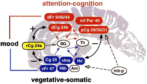
In the 1980s and 1990s, concepts of depression as a disease of both mind and brain were beginning to coalesce across disciplines. The notion of depression biomarkers was also developing; imagined then as a diagnostic ancillary or map of symptom-substrate correlates. An innovation at that time was to consolidate findings into an adaptable, whole-brain framework from which to devise and test hypotheses. The model proposed by Mayberg (Reference Mayberg1997) integrated key findings from neurological depression, as well as clinical and cognitive science, and was later elaborated with research on the neural mechanisms of treatment efficacy (Figure 1). The model conceptualized depression as a disruption in functionally interactive brain networks; a shift in thought from localization of function in specific neural regions to understanding symptoms as an emergent property of distributed networks.
Depression model originally proposed in 1997. The schematic highlights three compartments (dorsal, red; ventral, blue; rostral, yellow), each characterized by patterns of change with mood induction, treatment, and correspondence with symptom features of psychiatric or neurological disorders. Colored arrows depict connectivity. Solid black lines emphasize connectivity and reciprocal dynamics with induced sadness and treatment remission. Stippled lines posit effects of antidepressant medication. Numbers refer to Brodmann areas. (red) dFr=dorsolateral prefrontal; infr Par=inferior parietal; dCg=dorsal anterior cingulate; pCg= posterior cingulate. (blue) Cg25=sugenual (infralimbic) cingulate; vIns=ventral anterior insula, Hc=hippocampus; vFr=ventral frontal; Hth=hypothalamus. (yellow) rCg=rostral anterior cingulate. (white) mb-p=midbrain-pons; BG=basal ganglia; Th=thalamus; Am=amydgala. Reprinted from Mayberg (Reference Mayberg1997) with permission.

At the core of this initial proposition were early observations from neurology that linked symptoms of depression to damage in the frontal lobe. As empirical approaches to lesion-deficit correlation emerged, it became clear that when depression followed a neurological insult, the lesion was most likely to be located in the frontal lobe (Robinson, Kubos, Starr, Rao, & Price, Reference Robinson, Kubos, Starr, Rao and Price1984) or to disrupt connections to the prefrontal cortex via the basal ganglia (Starkstein & Robinson, Reference Starkstein and Robinson1993; Starkstein, Robinson, & Price, Reference Starkstein, Robinson and Price1987).
There was also early evidence that localized symptoms in dorsal and ventral (superior and inferior) divisions of the frontal cortex (Blumer & Benson, Reference Blumer and Benson1975; Stuss & Benson, Reference Stuss and Benson1986). Dorsal lesions, particularly to the motor system, were more often associated with what was then called “pseudo-depression”: apathy, indifference, and lack of motivation. Ventral limbic lesions, in contrast, caused behavioral dysinhibition, perhaps reflecting a failure to generate negative, and thus adaptive, emotion from aversive events in the social or physical world. This close phenotypic association with depression and anxiety troubled the division between neurological and psychiatric disorders.
The seminal work of Baxter and colleagues (Reference Baxter, Schwartz, Phelps, Mazziotta, Guze, Selin and Sumida1989) demonstrated common abnormalities in the dorsolateral prefrontal cortex across various neurological conditions that also produced depressive symptoms. Mayberg (Reference Mayberg1994) sought to better understand and expand on these commonalities as well as potential differences between neurological and psychological depressions. Despite the variety and complexity of disease-specific pathologies, all of the depressed patients, regardless of etiology, showed an additional common feature: decreased metabolism in the ventral cortices of the frontal lobe. This was a clue that negative emotion in depression might extend to dysfunction of the ventral corticolimbic system (Alexander, DeLong, & Strick, Reference Alexander, DeLong and Strick1986), independent of disease etiology.
Building further on earlier work with emotion elicitation, another key insight came from studies that strategically manipulated states of sadness (Liotti, Mayberg, McGinnis, Brannan, & Jerabek, Reference Liotti, Mayberg, McGinnis, Brannan and Jerabek2002; Mayberg et al., Reference Mayberg, Liotti, Brannan, McGinnis, Mahurin, Jerabek and Lancaster1999). When healthy individuals used the recall of memories to induce sadness, activity increased in ventral limbic structures, such as subgenual cingulate and insula, which regulate visceral homeostasis. In contrast, the opposite pattern, decreased activity relative to the neutral baseline, was observed in dorsal structures, including those associated with cognitive control (e.g., dorsal lateral prefrontal cortex). Importantly, this reciprocal pattern in dorsal and ventral compartments appeared to reverse when depressed patients were successfully treated with antidepressant medication (Mayberg, Brannan, Tekell, Silva, Mahurin, McGinnis, & Jerabek, Reference Mayberg, Brannan, Tekell, Silva, Mahurin, McGinnis and Jerabek2000).
The model proposed in Mayberg (Reference Mayberg1997) consolidated this body of evidence that associated various symptoms of depression with either superior or inferior (dorsal or ventral) “compartments” of the brain (Figure 1: dorsal, red; ventral, blue). Borrowing from the neurological literature reviewed above, the dorsal aspect (i.e., dorsal cingulate cortices, dorsal lateral cortices of the frontal lobe) was implicated in the cognitive aspects of depression, such as attention and executive function. Decreased metabolism in this upper network was also associated with psycho-motoric deficits, such as apathy, low positive arousal and physical slowness. Ventral limbic functions, such as the regulation of hypothalamus by amygdala, insula and subcortical structures, were tied to bodily symptoms of depression: sleep disturbance and lack of appetite. Ventral midline and orbital frontal cortices were involved in the production and regulation of negative emotion, particularly sadness (Mayberg et al., Reference Mayberg, Liotti, Brannan, McGinnis, Mahurin, Jerabek and Lancaster1999).
Early versions of this model also included a rostral medial aspect, thought to mediate between dorsal and ventral compartments (Figure 1, yellow). Curiously, depressed individuals who showed greater pre-treatment metabolism in this region, at the median between dorsal and ventral compartments (i.e., rostral cingulate; BA24), were more likely to remit with treatment (Mayberg et al., Reference Mayberg, Brannan, Mahurin, Jerabek, Brickman, Tekell and Martin1997). Individuals with low pretreatment metabolism in this area, however, failed to respond. The implication of this finding was that the reciprocal dynamics between cortico-limbic systems might be indirect and non-linear, such that treatment mechanisms are moderated by other compensatory factors and individual differences.
Although couched in the modular thinking of its time, this proposed model of depression neuropathology anticipated contemporary views of brain function and dysfunction, which emphasize domain-general circuitry (Chanes & Barrett, Reference Chanes and Barrett2016; Drevets, Price, & Furey, Reference Drevets, Price and Furey2008), integrate large scale networks, such as a ventral “salience” network, dorsal lateral “executive control” network, medial “default mode” (reviewed in Williams, Reference Williams2016), and propose mechanisms by which the brain self-organizes (Badcock et al., Reference Badcock, Davey, Whittle, Allen and Friston2017; Tang & Bassett, Reference Tang and Bassett2017; Tucker & Luu, Reference Tucker and Luu2012).
TREATMENT MECHANISMS AND BIOMARKERS OF TREATMENT EFFICACY
The conceptualization of depression as a failure of coordination in these distributed frontal networks instructed a new direction for biomarker research. It provided a road map for predicting what should happen in the brain when a treatment is successful, and initiated an area of productive, contemporary research into the neural mechanisms of treatment efficacy (e.g., Crane et al., Reference Crane, Jenkins, Bhaumik, Dion, Gowins, Mickey and Langenecker2017; DeRubeis, Siegle, & Hollon, Reference DeRubeis, Siegle and Hollon2008). Ideally, findings will coalesce on a mechanistic understanding of adaptive coordination in distributed neural networks that best promotes wellness.
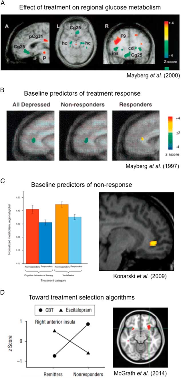
Early evidence pointed to a mechanism by which treatment modalities normalized pathological inactivity in dorsal structures, perhaps related to cognitive slowing observed in depression, and also reduced ventral limbic hyperactivity (i.e., subcallosal cingulate cortex, SCC), which had been previously related to negative mood (Figure 2A, green). Curiously, responders in a placebo group (i.e., spontaneous remitters) also showed this reciprocal pattern of change with wellness (Mayberg et al., Reference Mayberg, Brannan, Tekell, Silva, Mahurin, McGinnis and Jerabek2000). This prompted a hypothesis to refute: based on the observation that divergent forms of disruption to the frontal lobe may be associated with convergent outcomes, there may also be a common neural pathway from depression to wellness.
Sequence of advances toward treatment selection algorithms. A: Change in regional glucose metabolism in responders after 6 weeks of antidepressant medication (fluoxetine): ventral brain decreases (green) and dorsal brain increases (red). Cg25=subcallosal cingulate cortex, pCg31=posterior cingulate/Brodmann area (BA) 31, p=putamen, hc=hippocampus, F9=BA9, Ins=insula. Adapted from Mayberg et al. (Reference Mayberg, Brannan, Tekell, Silva, Mahurin, McGinnis and Jerabek2000). B: Glucose metabolism in rostral anterior cingulate differentiates eventual responders to medication (fluoxetine) from non-responders. Adapted from Mayberg et al. (Reference Mayberg, Brannan, Mahurin, Jerabek, Brickman, Tekell and Martin1997). C: Pre-treatment glucose hypermetabolism in ventral anterior cingulate cortex characterizes non-responsers to both Cognitive Behavioral Therapy (CBT) and medication (venlafaxine). Adapted from Konarski et al. (Reference Konarski, Kennedy, Segal, Lau, Bieling, McIntyre and Mayberg2009). D: Treatment by outcome interaction indicates a potential treatment selection biomarker: brain glucose hypometabolism in anterior insula for CBT; hypermetabolism in eventual responders to medication (escitalopram). Adapted from McGrath et al. (Reference McGrath, Kelley, Dunlop, Holtzheimer, Craighead and Mayberg2014).

However, evidence also pointed to divergent mechanisms across treatment modalities. Goldapple and colleagues (Reference Goldapple, Segal, Garson, Lau, Bieling, Kennedy and Mayberg2004), for example, associated cognitive behavioral therapy (CBT) with decreased resting metabolism in both dorsal and ventral cortices of the frontal lobe but increased metabolism in dorsal limbic components (e.g., hippocampus). In contrast, a near opposite pattern was observed following treatment with antidepressant medication (Figure 1A: decrease, green vs. increase, red).
Building on these observations, Kennedy and colleagues (Reference Kennedy, Konarski, Segal, Lau, Bieling, McIntyre and Mayberg2007) set out to clarify discrepant findings using a randomized control design. They found some comparable effects of CBT and antidepressant medication (i.e., venlafaxine): not previously appreciated, responders to both treatments showed decreased resting metabolism in the orbital ventral cortices of the frontal lobe. However, the trial also indicated some complex differences between treatment modalities. For example, resting metabolism in the posterior cingulate, a region associated with mentation and autobiographical memory, was decreased following CBT but increased with venlafaxine. This was thought to be consistent with a central goal of CBT, which is to alter dysfunctional cognitive processes. In contrast, venlafaxine, but not CBT, decreased resting metabolism in ventral limbic components (i.e., subgenual cingulate), perhaps reflecting a reduction in autonomic reactivity (i.e., stress response).
Critically, these studies provided evidence that falsified the hypothesis of a final common pathway of changes mediating antidepressant response, independent of modality-specific mechanisms. Nonetheless, the common involvement of many regions, but in opposite directions for medication and psychotherapy, provided the critical foundation for considering differential baseline states as potential biomarkers of treatment-specific subtypes.
A significant literature has since developed to clarify these effects. Several recent reviews provide a more comprehensive summary of results, challenges and emerging methods (DeRubeis et al., Reference DeRubeis, Siegle and Hollon2008; Savitz, Rauch & Drevets, Reference Savitz, Rauch and Drevets2013; Siegle et al., Reference Siegle, Thompson, Collier, Berman, Feldmiller, Thase and Friedman2012; Treadway & Leonard, Reference Treadway and Leonard2016). Significant advances were made with inclusion of task-related neural activity, a shift to measures of cerebral blood flow in PET research, and the application of functional magnetic resonance imaging (fMRI). The summary provided by DeRubeis et al. (Reference DeRubeis, Siegle and Hollon2008) suggests that cognitive therapies tend to increase adaptive functionality of dorsal networks (i.e., executive control, dorsal attention network). Antidepressant medications and, to a lesser extent, cognitive therapies, both tend to reduce hyper-reactivity of ventral cortico-limbic structures, particularly the amydgala, for both depression and anxiety disorders.
Importantly, DeRubeis et al. (Reference DeRubeis, Siegle and Hollon2008) explore the possibility that the context of resting and task-oriented measures may explain some variance across studies. For example, a decrease in resting dorsal metabolism with successful treatment (regardless of modality) might represent an adaptive readiness for cognitive signaling; whereas increased metabolism in the context of a cognitive task reflects adaptive engagement of network functionality (Mayberg, Reference Mayberg2003).
A theme that emerges from this still growing literature is the complexity of the depressive phenotype, suggesting subtypes (Trivedi et al., Reference Trivedi, McGrath, Fava, Parsey, Kurian, Phillips and Cooper2016). These may be diversely described as dysfunction in cognitive processes (e.g., reward processes), mood state (e.g., anhedonia), or homeostatic regulation (e.g., stress reactivity). Despite a common phenotypic outcome (i.e., wellness), the combination of compensatory and normalization effects are non-linear and also shows complex temporal dynamics (Mayberg, Reference Mayberg2003; Mayberg et al., Reference Mayberg, Brannan, Tekell, Silva, Mahurin, McGinnis and Jerabek2000; Treadway & Leondard, Reference Treadway and Leonard2016). This indicates a need for complementary approaches to the enhancement of treatment with brain-based biomarkers.
PREDICTIVE BIOMARKERS OF TREATMENT RESPONSE
One approach, which complements research into the mechanism of treatment efficacy, is to instead discover baseline predictors of positive outcome in a given modality; however, complex the trajectory to wellness, the biomarker would instruct optimal treatment selection for individuals (e.g., Fu et al., Reference Fu, Steiner and Costafreda2013; Siegle et al., Reference Siegle, Carter and Thase2006). A technological outcome would be brain-based biomarkers that predict which treatment will be most effective for whom. Such optimization strategies could amount to a significant cost savings in health care, but more importantly, a lifesaving reduction in time to wellness.
An early application of this approach sought pre-treatment predictors of response to antidepressant medication in resting state metabolism (Mayberg et al., Reference Mayberg, Brannan, Mahurin, Jerabek, Brickman, Tekell and Martin1997). Relative to controls, rostral cingulate (BA24) at baseline was found to be higher for responders and lower for non-responders (Figure 2B). Given its anatomical location and known structural connections, it was thought that activity in this region might represent enhanced capacity to mediate between competing drives from dorsal and ventral networks. Later, Saxena et al. (Reference Saxena, Brody, Ho, Zohrabi, Maidment and Baxter2003) also showed a statistically significant correlation of pretreatment metabolism in the medial cingulate and treatment response: higher metabolism predicted greater improvement. Importantly, predictive biomarkers from frontal midline have also been identified in electrophysiological, theta band oscillations, which have been shown to signal cognitive control processes (Pizzagalli, Reference Pizzagalli2011). Although the mechanism is still unclear, these results suggest that the pre-treatment functioning in the frontal midline enhances an individual’s ability to acquire and maintain treatment gains.
Ventral limbic structures, including the SCC (BA25), amygdala, and anterior insula, have also emerged as candidate predictors of positive treatment response (Saxena et al., Reference Saxena, Brody, Ho, Zohrabi, Maidment and Baxter2003; Siegle et al., Reference Siegle, Carter and Thase2006). Although these structures are broadly implicated in neural functioning, the ventral limbic system is essential for integrating need states with environmental demands to maintain bodily homeostasis. The most consistent predictor of poor treatment response is exaggerated baseline functioning of the SCC (Figure 2C), previously linked to negative emotionality (Konarski et al., Reference Konarski, Kennedy, Segal, Lau, Bieling, McIntyre and Mayberg2009; McGrath et al., Reference McGrath, Kelley, Dunlop, Holtzheimer, Craighead and Mayberg2014; Siegle et al., Reference Siegle, Carter and Thase2006, Reference Siegle, Thompson, Collier, Berman, Feldmiller, Thase and Friedman2012). Using path-modeling analysis, Seminowicz et al. (Reference Seminowicz, Mayberg, McIntosh, Goldapple, Kennedy, Segal and Rafi-Tari2004) suggested that, in eventual responders to medication, SCC is highly regulated by other cortical and subcortical regions. Interestingly, this quantitative approach suggested a different profile for responders to CBT, one that largely excluded subcortical structures and instead emphasized pathways within the ventral prefrontal cortex.
This finding raised the important question of whether biomarkers could provide a prediction of outcome that would also contrast different treatment modalities. If one considers the ultimate goal of precision medicine, it is to develop a biomarker that will match individuals to the treatment that is optimal for them. For example, McGrath et al., (Reference McGrath, Kelley, Holtzheimer, Dunlop, Craighead, Franco and Mayberg2013; Figure 2D) observed hypometabolism in the anterior insula in eventual remitters to CBT as well as patients who failed to remit with antidepressant medication (esitalopram). In contrast, insula hypermetabolism was seen in eventual remitters with medication and patients who failed to remit with CBT. Metabolic activity in this region is now being prospectively tested as a possible guide to treatment selection (Clinical Trials.gov # NCT02137369).
While a central goal of this research is to identify the optimal treatment for an individual, this research illustrates what is perhaps an even more urgent goal: to identify which first line treatments will be unlikely to help an individual. With this need in mind, there is growing evidence that high SCC metabolism may predict treatment resistance, regardless of the treatment modality (Konarski et al., Reference Konarski, Kennedy, Segal, Lau, Bieling, McIntyre and Mayberg2009; McGrath et al., Reference McGrath, Kelley, Dunlop, Holtzheimer, Craighead and Mayberg2014). However, following the observation by Semonowicz et al. (Reference Seminowicz, Mayberg, McIntosh, Goldapple, Kennedy, Segal and Rafi-Tari2004) of different frontal lobe path models for responders to CBT or drug, a more explicit functional connectivity approach is now being explored (Dunlop et al., Reference Dunlop, Rajendra, Craighead, Kelley, McGrath, Choi and Mayberg2017; Fox, Buckner, White, Greicius, & Pascual-Leone, Reference Fox, Buckner, White, Greicius and Pascual-Leone2012).
Recently, Dunlop et al. (Reference Dunlop, Rajendra, Craighead, Kelley, McGrath, Choi and Mayberg2017) used pretreatment SCC connectivity as the exploratory method to define treatment specific predictive discriminators. Authors report that positive connectivity of the SCC to the medial and ventrolateral frontal cortices and midbrain predicted a positive response to CBT. In contrast, absent or negative connectivity of the SCC to these same regions was associated with a good therapeutic response to antidepressant medication. Combined with the previously described insula findings derived using FDG PET, these imaging studies provide mutually corroborative evidence of brain-based depression subtypes with potential relevance to selecting the optimal treatment for an individual, a classifier not captured in psychometric measures of negative emotional bias.
A COGNITIVE NEUROSCIENCE APPROACH TO DEPRESSION BIOMARKERS
A complementary approach to biomarker research uses the tools of neuroscience to study specific cognitive and affective features relevant to depression pathology and treatment (e.g., Crane et al., Reference Crane, Jenkins, Bhaumik, Dion, Gowins, Mickey and Langenecker2017; Davidson et al., Reference Davidson, Irwin, Anderle and Kalin2003; Phillips, Drevets, Rauch, & Lane, Reference Phillips, Drevets, Rauch and Lane2003; Tenke et al., Reference Tenke, Kayser, Pechtel, Webb, Dillon, Goer and Parsey2017; Treadway & Leonard, Reference Treadway and Leonard2016; Trivedi et al., Reference Trivedi, McGrath, Fava, Parsey, Kurian, Phillips and Cooper2016). Negative self-focus, for example, is prominent in the clinical phenomenology of depression; so emblematic of the illness that self-report measures of mood state are a gold standard for assessing symptom severity and remission. Some form of self-processing is also an explicit target of many psychotherapies, including meditation training (Farb et al., Reference Farb, Segal, Mayberg, Bean, McKeon, Fatima and Anderson2007). Consistent with themes of the discovery narrative thus far, a considerable metabolic imaging literature implicates dorsal–ventral reciprocity in the emergence and maintenance of negative self-schema.
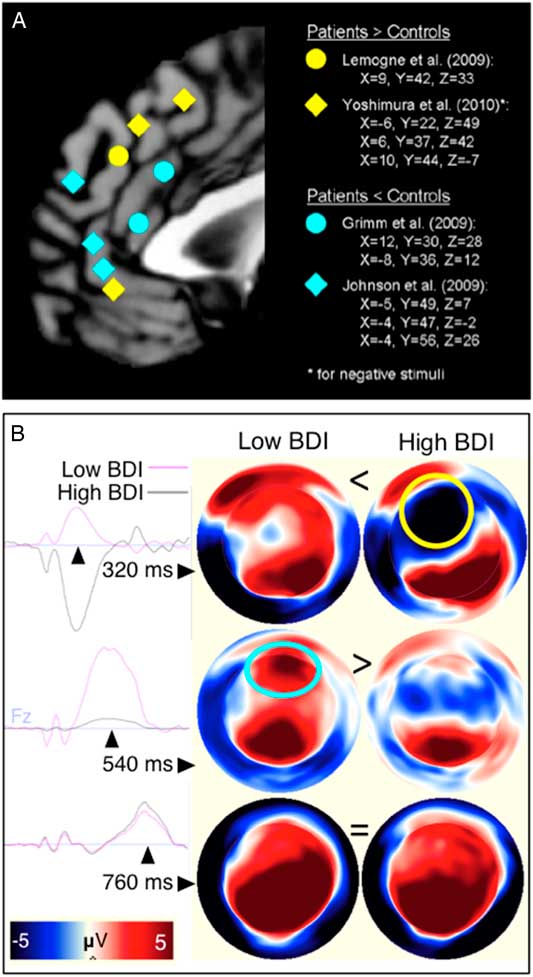
Lemogne et al. (Reference Lemogne, Delaveau, Freton, Guionnet and Fossati2012) synthesize key findings from metabolic research into self-evaluative cognition. The authors suggest that tonic elevation of ventral metabolism reflects automatic self-associations, particularly with negative information (Figure 3A, cyan). Phasic activation of dorsal metabolism, in contrast may be related to specific evaluative processes (Figure 3A, yellow). Depression is also associated with abnormal deactivation of the midline cortical network (i.e., default mode), which has been characterized by its involvement in self-processing (Northoff et al., Reference Northoff, Heinzel, De Greck, Bermpohl, Dobrowolny and Panksepp2006). A proposal by Northoff, Wiebking, Feinberg, and Panksepp (Reference Northoff, Wiebking, Feinberg and Panksepp2011) posits that depression involves a failure of reciprocal inhibition between the default mode network and processes engaged when interfacing with the environment, including dorsal lateral cognitive control networks. Aberrant connectivity profiles during self-evaluation in depression, which, notably, also implicate the SCC region, are consistent with this hypothesis (Lemogne et al., Reference Lemogne, Delaveau, Freton, Guionnet and Fossati2012; Yoshimura et al., Reference Yoshimura, Okamoto, Matsunaga, Onoda, Okada, Kunisato and Yamawaki2017).
Cortico-limbic engagement during self-evaluation: reciprocal contributions to the depressive phenotype. A: Summary of findings from four independent studies using fMRI shows location of increased (yellow) and decreased (cyan) activity during self-referential processing. Adapted from Lemogne et al. (Reference Lemogne, Delaveau, Freton, Guionnet and Fossati2012). B: Principal components of the event-related potential (left) over frontal cortex differentiate high and low symptom severity groups (at electrode Fz). Topomaps (right) show electrical potentials across the head surface as if nose is pointing to the top of the page (positive/negative voltage: red/blue). Sequential components of the event-related potential show exaggerated frontal negativity (yellow circle; black time series, peak at 320 ms), followed by an attenuated frontal positivity (cyan circle, black line, peak at 540 ms) in individuals reporting high symptom severity. Topomaps adapted from Waters and Tucker (Reference Waters and Tucker2016).

Although a smaller literature, electroencephalographic approaches to the study of self-evaluative cognition offers the requisite temporal specificity to disentangle neural processes and network-level integration. For example, Waters and Tucker (Reference Waters and Tucker2016) used factor analysis to observe latent components of the event-related potential (ERP) during self-evaluative decisions. Depressed-anxious individuals were differentiated from controls on two components. The first was exaggerated in depression, and captured variance in a fronto-medial ERP related to discrepancy detection and negative affect (Figure 3B, top row). The second was attenuated in the depressed group and captured variance in ERPs associated with dorsal and default mode functions, such as autobiographical memory consolidation and positive affect (Figure 3B, bottom row). These measures may also differentiate cognitive features associated with high negative and low positive emotionality in comorbid anxiety and depression. Although preliminary, this and other studies of self-evaluation provide an example of how cognitive approaches with high temporal resolution may give way to brain-based biometrics in the vein of psychometric dimensionality (Waters & Tucker, Reference Waters and Tucker2013) and assessment (Auerbach et al., Reference Auerbach, Bondy, Stanton, Webb, Shankman and Pizzagalli2016).
What begins to emerge is a mechanistic view of maladaptive frontal lobe functioning that can be related to broader psychological theories. For example, one theory posits that negative affect arises from dissonance between self-schema and the social valuation of personality traits (Tucker & Luu, Reference Tucker and Luu2007). The medial frontal negativity (MFN) is an ERP feature that reflects detection of discrepancy from expectations, and is thus useful for exploring this theory. Medial negativities have been associated with cingulate functioning and individual differences in negative emotionality (Luu, Tucker, & Makeig, Reference Luu, Tucker and Makeig2004). In a clinical sample of depressed individuals, the MFN was attenuated specifically when negative words were considered self-descriptive (Poulsen, Luu, Crane, Quiring, & Tucker, Reference Poulsen, Luu, Crane, Quiring and Tucker2009). This effect was interpreted as evidence that maladaptive self-schema in depression, associating the self with negative traits, would fail to evoke a strong discrepancy response. One hypothesis is that alterations in electrophysiological signaling efficacy are related to the observation of enhanced metabolic activation of BA25 during negative word reading (Siegle et al., Reference Siegle, Thompson, Collier, Berman, Feldmiller, Thase and Friedman2012).
Combined, these indices might reflect the psychological theory of learned helplessness, wherein repeated failure is associated with eventual withdrawal. Perhaps consistent with this view, Tucker, Luu, Frishkoff, Quiring, and Poulsen (Reference Tucker, Luu, Frishkoff, Quiring and Poulsen2003) reported a non-linear association of MFN responsiveness with symptom severity; amplitude increased with moderate depression but then attenuated with more severe symptomology. Similarly, Waters and Tucker (Reference Waters and Tucker2016) showed attenuation in a right anterior ventral lateral ERP, with modeled sources in right ventral lateral prefrontal cortex, as symptom severity moved from moderate to severe. Notably, experienced meditators showed increased metabolism in this region during experiential self-reference, as if engaging inhibitory control over a narrative tendency associated with depressive rumination (Farb et al., Reference Farb, Segal, Mayberg, Bean, McKeon, Fatima and Anderson2007). Taken together, these findings point to a complex temporal trajectory in both pathological and compensatory process as depression worsens or remits with treatment.
What the cognitive neuroscience approach offers is the prospect of biomarkers with precision relevance to learning mechanisms and psychological theory (Ressler & Mayberg, Reference Ressler and Mayberg2007; Tucker & Luu, Reference Tucker and Luu2007). For example, an interesting question explored by Pizzagalli, (Reference Pizzagalli2011) and others is how predictive biomarkers of positive treatment response, specifically midline frontal theta, might promote the disruption of negative self-schema. In this way, understanding the neural mechanisms related to self-processing may suggest new ways to target and tune cognitive intervention (Ressler & Mayberg, Reference Ressler and Mayberg2007), and integrate neural measures more directly with clinical practice.
DEEP BRAIN STIMULATION TO SCC (BA25) FOR TREATMENT RESISTANT DEPRESSION
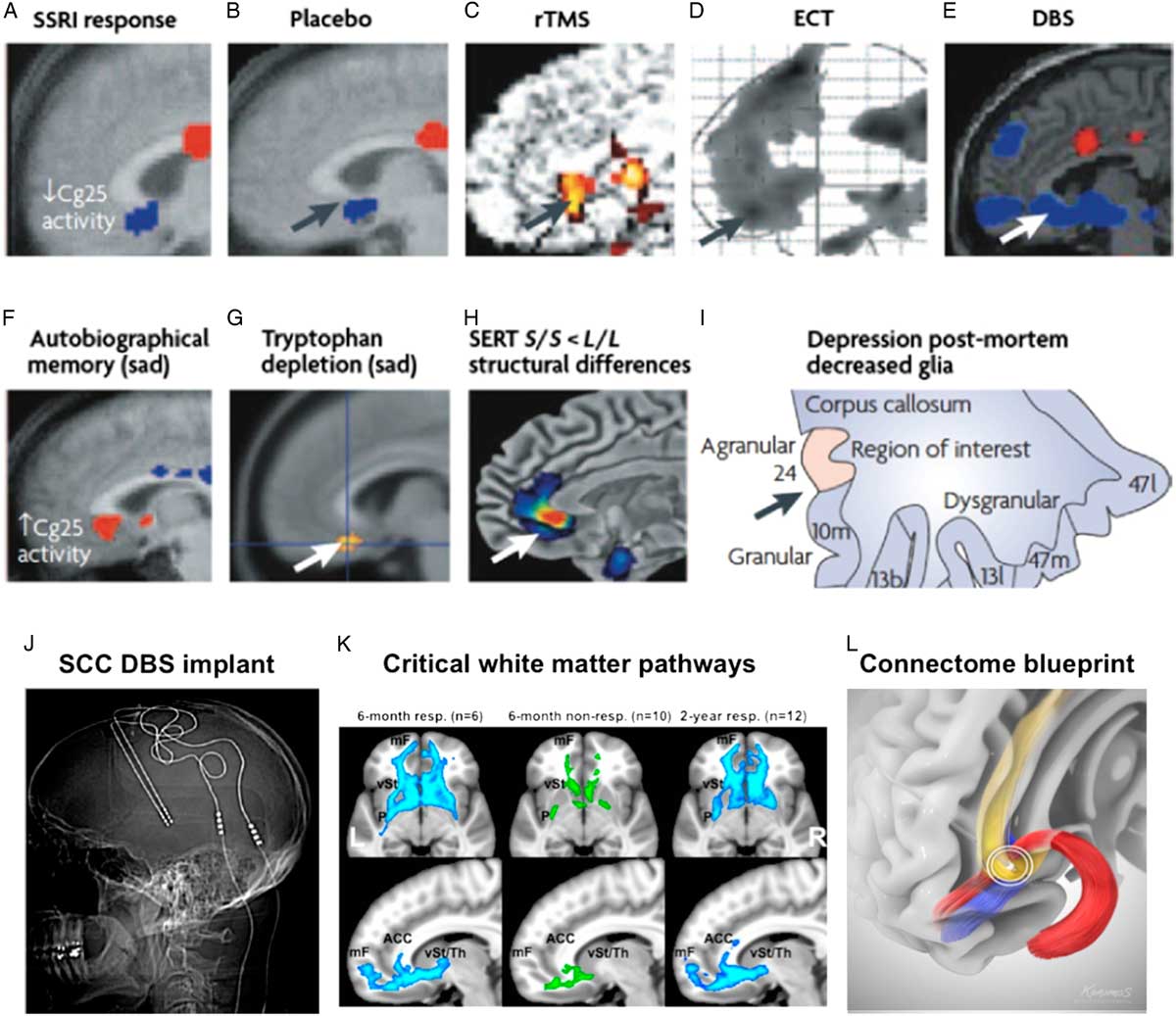
Translational advances in depression biomarker research have been largely theoretical; in practice, direct application is rare. One exception is the development of a novel treatment, deep brain stimulation (DBS) of the SCC, shown in Figure 4J. Evidence implicating the SCC region was fundamental to early models of depression neuropathology (Mayberg, Reference Mayberg1997, Drevets et al., Reference Drevets, Price, Simpson, Todd, Reich, Vannier and Raichle1997). Hamani et al. (Reference Hamani, Mayberg, Stone, Laxton, Haber and Lozano2011) compiled this seminal evidence, which included: observations of increased metabolism in the SCC following sad mood induction or tryptophan depletion (Figure 4F,G), as well as anatomic and structural abnormalities (e.g., atrophy; decreased oligodendroglial cells) in individuals with depression or at risk for depression (Figure 4H,I). Another key observation, summarized in Figure 4A–E, was the reversal of pathological activity in the SCC with various interventions (Mayberg, Reference Mayberg2003; Siegle et al., Reference Siegle, Thompson, Collier, Berman, Feldmiller, Thase and Friedman2012).
Subcallosal cingulate (SCC) deep brain stimulation for treatment resistant depression: the development of an idea. Panels (A–I) unfold key points of evidence that implicate and validate SCC as a target for neuromodulation. Panels (J–L; gray) show efforts to optimize therapeutic engagement of distributed cortical networks by targeting white matter fiber bundles within the SCC. Row 1: Change relative to baseline for treatment responders following (A) antidepressant medication, (B) placebo response, (C) repetitive transcranial magnetic stimulation, (D) electroconvulsive shock therapy, and (E) deep brain stimulation. Row 2: Links to depression pathology include: decreased metabolic activity in healthy subjects with induced sadness using (F) autobiographical memories, and (G) tryptophan depletion; (H) structural abnormalities in individuals with genetic risk for depression and (I) decreased glial cell number. Panels (A–I) adapted from Hamani et al. (Reference Hamani, Mayberg, Stone, Laxton, Haber and Lozano2011). Item (K) adapted from Riva-Posse et al. (Reference Riva-Posse, Choi, Holtzheimer, McIntyre, Gross, Chaturvedi and Mayberg2014).

That a variety of treatment modalities could reduce SCC reactivity suggested that the effect was mediated by a more complex and distributed neural response. For some individuals, an estimated 10–20% of depressed patients, treatment efficacy was somehow lost in this complexity (reviewed in Hotzheimer & Mayberg, Reference Holtzheimer and Mayberg2011a). SCC-DBS was proposed as an alternative for these treatment resistant individuals; directly targeting the pathological node with electrical current (Mayberg et al., Reference Mayberg, Lozano, Voon, McNeely, Seminowicz, Hamani and Kennedy2005). Early efficacy trials of SCC-DBS showed the expected reversal of SCC pathology over time (Lozano et al., Reference Lozano, Mayberg, Giacobbe, Hamani, Craddock and Kennedy2008; Mayberg et al., Reference Mayberg, Lozano, Voon, McNeely, Seminowicz, Hamani and Kennedy2005) and sustained remission of symptoms for many patients in the earliest cohorts. SCC-DBS has now been tested in approximately 200 people world-wide.
Outcome reports from open label trials range from 40 to 70% of individuals showing at least a 50% reduction in symptom severity (reviewed in Holtzheimer & Mayberg, Reference Holtzheimer and Mayberg2011a), whereas a recent clinical trial was halted, ostensibly because effect sizes fell below a reasoned threshold. Although several factors may exaggerate effect size estimates at the open label stage of testing, reported long-term efficacy rates are notable given that eligible patients are treatment refractory, many having failed response to even electroconvulsive therapy. The uncertainty in these measures also underscores the need for a protocol that involves objective biomarkers to standardize patient selection and surgical procedures.
For example, a “connectomic strategy” is now being proposed as a method for standardizing procedures (Riva-Posse et al., Reference Riva-Posse, Choi, Holtzheimer, Crowell, Garlow, Rajendra and Mayberg2017). It was recognized early on that the treatment mechanism involved not only local effects, but effects on a distributed cortical regions. As depicted in Figure 4K,L, this hypothesis was later confirmed by the observation of a tractography blueprint; a profile of white matter activation that was associated with positive outcomes (Riva-Posse et al., Reference Riva-Posse, Choi, Holtzheimer, McIntyre, Gross, Chaturvedi and Mayberg2014). Meanwhile, there is a growing convergence in connectivity-based targeting: functional connectivity to SCC also predicts outcomes using transcranial magnetic stimulation (TMS) (Fox et al., Reference Fox, Buckner, White, Greicius and Pascual-Leone2012). In this instance, understanding of the precise mechanism of treatment efficacy has matured to a point where individualized biomarkers (i.e., profiles of white matter connectivity) are playing a central role in the treatment process, and may be essential for effective testing and dissemination of this treatment on a larger scale.
TOWARD MEASUREMENT AND MODULATION OF DYNAMICAL SYSTEMS
The goals of biomarker research have evolved with a changing conceptualization of brain function, in general. We can appreciate how the steady accumulation of evidence shifted our understanding of regional contributions to emotional experience. For example, the SCC was first conceptualized as the seat of negative affect, but later as a key node in distributed brain networks. When disrupted, as in SCC-DBS or TMS, the emergent subjective experience can be altered dramatically (Fox et al., Reference Fox, Buckner, White, Greicius and Pascual-Leone2012; Hotzheimer & Mayberg, Reference Holtzheimer and Mayberg2011a). This points to clear potential for advances in non-invasive brain circuit-modulation to treat both symptoms and underlying pathology, directly.
In parallel, the applied goal of biomarker research shifted from a means to differentiate sick from well (i.e., diagnostic) to identifying dynamic changes with treatment, predictors of treatment outcome, and treatment selection algorithms. At the same time, cognitive and affective neuroscience pursued biomarkers with clearer psychological significance. Contextualized in psychological and learning theories, these biomarkers might also shape the approach of some cognitive therapies. Overall, this discovery narrative illustrates a complementary alternative to direct, symptom-substrate mapping, or approaches that take cognitive and psychological symptoms as a starting place (e.g., Snyder, Reference Snyder2013). Future research will be needed to associate predictive biomarkers, which are instead derived from the observation of neural abnormality, more directly with features of the clinical presentation (Trivedi et al., Reference Trivedi, McGrath, Fava, Parsey, Kurian, Phillips and Cooper2016).
While there is obviously no one strategy to deconstruct the network structure and dynamical characteristics of a putative depression circuit, a key to success in biomarker research was the early articulation of a working model of depression neuropathology. This model was data driven and organic, evolving with the steady accommodation of findings and perspectives from neighboring disciplines. It also preserved past observations and theoretical constructs as more complex methods and technologies became available. Also in this discovery narrative, we see how counter-intuitive evidence, discrepancies with the initial conceptualization, were drivers of innovation. One such insight was an appreciation of the brain’s dynamical nature, as well as the distribution of functional capacity across whole-brain networks.
The notion of dynamic variation also extends to non-linearity of change in brain systems during emotion regulation, and over time in treatment (Holtzheimer & Mayberg, Reference Holtzheimer and Mayberg2011b; Treadway & Leonard, Reference Treadway and Leonard2016). As we move toward the measurement of dynamical systems, we seek more integrative methods and computational approaches (e.g., Ramirez-Mahaluf, Roxin, Mayberg, & Compte, Reference Ramirez-Mahaluf, Roxin, Mayberg and Compte2017). A goal will be to develop algorithms that make use of variance between individuals to intervene on pathological neural dynamics and drive the system to a more adaptive homeostatic state (Lozano & Mayberg, Reference Lozano and Mayberg2015; Tang & Bassett, Reference Tang and Bassett2017). In this sense, it is possible to imagine some clinical constructs replaced with a more general notion of problematic biases in neural self-regulation. We are beginning to see that a consequence of this reframing is the breakdown of classical diagnostic categories and disciplinary boundaries.
Acknowledgments
We thank our many colleagues who contributed to the concepts and studies reviewed, including Don M. Tucker and Phan Luu, Robert Robinson, Sergio Starkstein, Stephen Brannan, Mario Liotti, Sidney Kennedy, Zindel Segal, Andres Lozano, Paul Holtzheimer, Boadie Dunlop, Callie McGrath, Patricio Riva Posse and Kisueng Choi. Past and current work is supported by grants from the National Institute of Mental Health (NIMH), Canadian Institute for Health Research (CIHR), National Alliance for Research on Schizophrenia and Depression (NARSAD), Dana Foundation, Stanley Medical Research Institute, the Woodruff Foundation and the Hope for Depression Research Foundation. Dr. Mayberg has a consulting agreement with St Jude Medical, Inc., which has licensed her intellectual property to develop SCC DBS for the treatment of severe depression. Dr. Waters has no financial disclosures.