Introduction
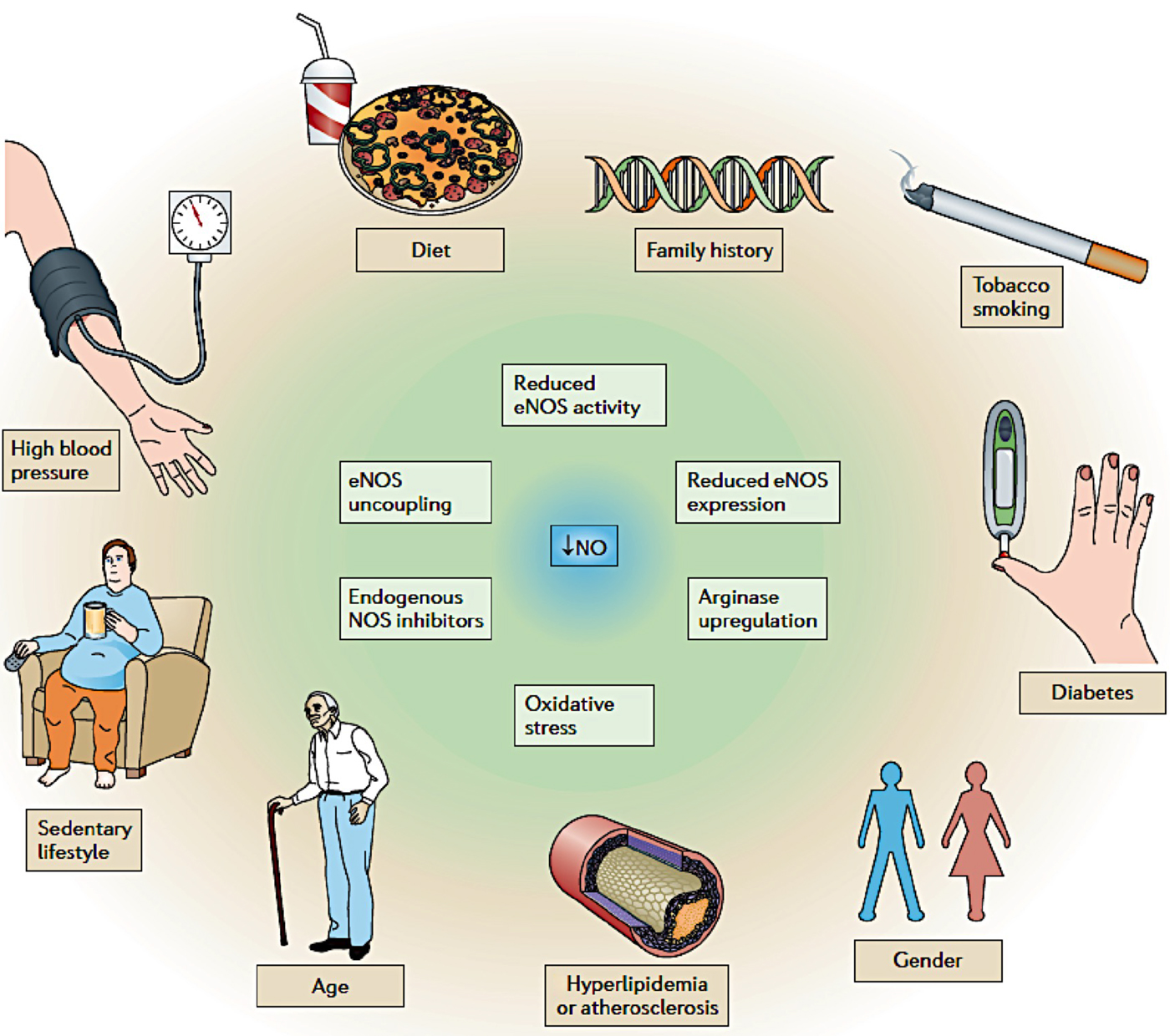
Nitric oxide (NO) is a key signaling messenger that can be found in almost all tissues of the body and throughout cardiovascular, immune, and central nervous systems.[Reference Lowenstein, Dinerman and Snyder1] It was first identified as an endothelium-derived relaxing factor in 1980. In 1992, it was titled as “Molecule of the Year” by Science, which later was selected as the subject of the Nobel Prize in Physiology or Medicine 1998. Indeed, accumulating evidence supports the pivotal role for NO as an efficient inhibitor of restenosis, a simple key molecule involved in accelerated wound repair and an anticancer and antibacterial agent.[Reference Seabra, Justo and Haddad2] Various disease and lifestyle factors for cardiovascular diseases (CVDs) are associated with NO-signaling pathways (Fig. 1),[Reference Lundberg, Gladwin and Weitzberg3] and benefits from the mostly acceptable lifestyles and cardiovascular therapies are due to the release of NO. Therapeutic approaches for CVDs emerging over the past 10 years have been proposed to target both the upstream and downstream of NO-signaling pathways. As such, NO becomes the limelight for many therapeutic activities due to its pivotal roles in both physiological and pathological processes that affect neurotransmission,[Reference Garthwaite4] cardiovascular homeostasis,[Reference Ignarro5] immune regulation,[Reference Hibbs6] and inhibition of platelet aggregation.[Reference Azuma, Ishikawa and Sekizaki7] However, precise NO delivery is the challenging issue for regenerative therapy, as predetermined effects must occur at the correct agent amount with spatiotemporal regulation. Although this makes clinical administration of NO difficult due to its transient chemical nature, the combined approaches of NO and biomaterials has provided advanced delivery systems to overcome the difficulties related to NO administration.
Diseases and lifestyle factors associate with NO-signaling pathways [Adapted with permission from Ref. Reference Lundberg, Gladwin and Weitzberg3. Copyright 2015 Macmillan Publishers Limited].

In this review, we summarized the current understanding on the physiological roles of NO and discuss different NO delivery systems, including film layers, hydrogels, and particle-based scaffolds in regulating vascular functionality. We will attempt to uncover the mechanisms underlying these three-dimensional (3D) material strategies to impact NO bioactivity in terms of correct doses, localization, and sustention. We also make an effort to insight recent developments in this field and propose future perspectives for precise NO delivery toward the regeneration of expected tissue functionality.
NO in blood vessels
NO production
The physiological production of NO is catalytically from a five-electron oxidation of the terminal guanidine nitrogen of the amino acid L-arginine, which forms L-citrulline and the NO-free radical. Molecular oxygen[Reference Leone, Palmer, Knowles, Francis, Ashton and Moncada8] and nicotinamide adenine dinucleotide phosphate-oxidase (NADPH)[Reference Nathan9] are involved in the reaction as the co-substrates. The reaction relies on three NO synthase (NOS) isoforms: endothelial (eNOS), neuronal (nNOS), and inducible (iNOS) with different regulation and activities (Reaction mechanisms for these NOS reviewed by Lundberg et al.[Reference Lundberg, Gladwin and Weitzberg3]). The nNOS and eNOS form a constitutive class that is regulated by calcium (Ca2+) and calmodulin, while iNOS mostly found in macrophages, neutrophils, fibroblasts, and hepatocytes, which is activated immunologically by exposure to bacterial endotoxin or cytokines,[Reference Loscalzo and Welch10] with less dependence on intracellular Ca2+ levels. Besides these factors, NO production is also regulated by the presence of agonists and stimuli like fluid shear stress and pathogens.[Reference Jen, Serrano, van Lith and Ameer11] It has been suggested that NO-signaling pathways can be up-regulated by both physical exercise and certain dietary components[Reference Lundberg, Gladwin and Weitzberg3].
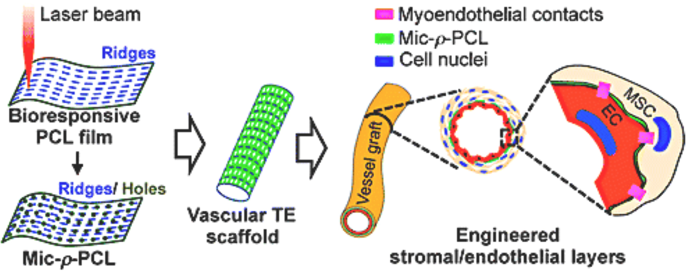
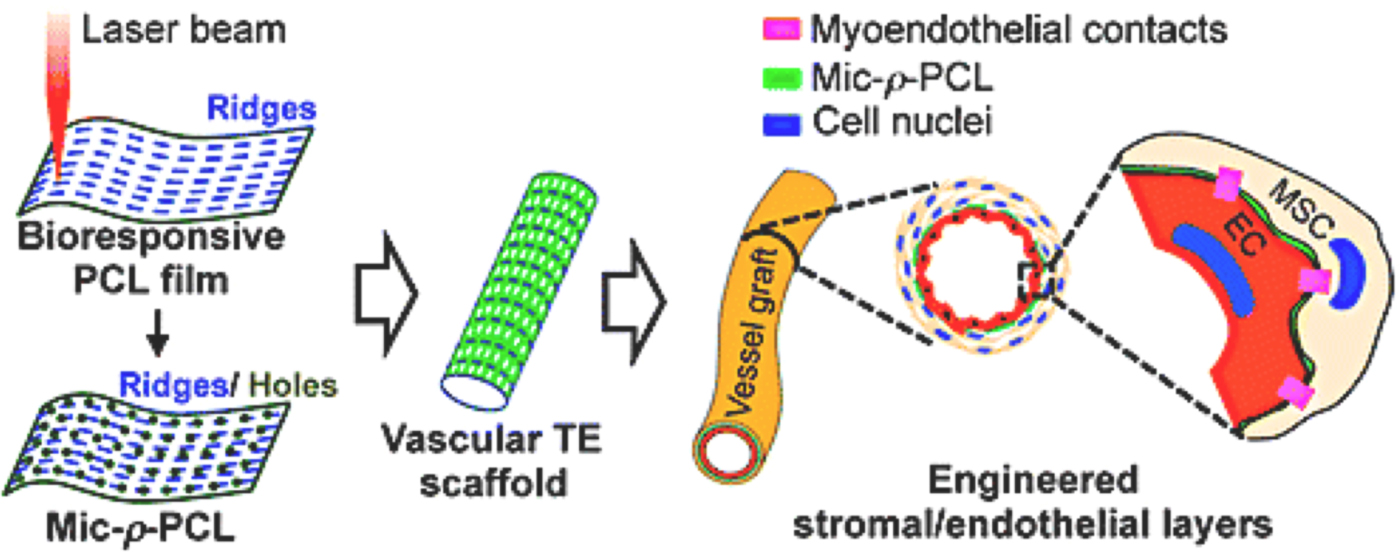
Straub et al. have demonstrated the existence of another pathway involving deoxygenated globins for NO production in human endothelial cells (ECs).[Reference Straub, Lohman, Billaud, Johnstone, Dwyer, Lee, Bortz, Best, Columbus and Gaston12] Under conditions of hypoxia, myoglobin in response to the low oxygen level in smooth muscle cells (SMCs) can react with nitrite, during the process the Fe2+ deoxymyoglobin transfers an electron to nitrite and generates NO and metmyoglobin. In addition to ECs, mesenchymal stem cells (MSCs) are also reported for NO production, and their co-culture with ECs has been found for synergistic increase in the NO generation from both the ECs and MSCs, through the formation of physical cell–cell contact.[Reference Wang, Teoh, Hong, Luo, Teo, Chan and Thian13] Forward research has shown that the vascular scaffold engineered with through-holes at an aspect ratio approximating to that of the endothelial basement membrane could result in vessel-like wall construction (Fig. 2).[Reference Wang, Du, Chan, Teoh, Thian and Hong14]
Schematic diagram illustrating physical cell–cell contact in engineering of vessel-like wall construction (Mic-ρ-PCL, laser-microperforated films; MSCs, mesenchymal stem cells; and ECs, ECs) [Adapted with permission from Ref. Reference Wang, Du, Chan, Teoh, Thian and Hong14. Copyright 2017 American Chemical Society].
NO diffusion
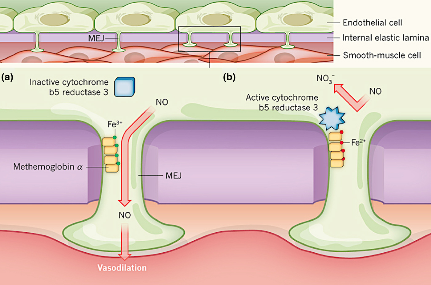
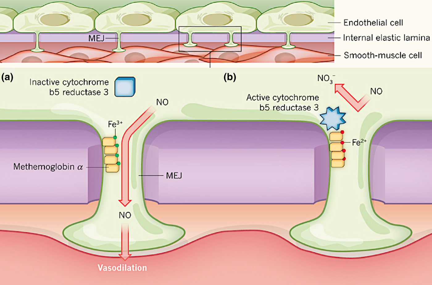
As the smallest bioactive messenger molecule, NO possesses free radical nature (due to the odd electrons) and is paramagnetic, reluctant to dimerise, and highly reactive with a myriad of atoms and free radicals (the high reactivity with a half-life of 2–30 s in the blood).[Reference Lowenstein, Dinerman and Snyder1, Reference Butler and Williams15] NO shows a very high water solubility at room temperature, and has been considered to diffuse freely across the cell membranes due to its small size and uncharged characteristics.[Reference Butler and Williams15] Unexpectedly, recent evidence demonstrates that a redox switch in the protein hemoglobin α within ECs regulates NO diffusion and further signaling pathways from endothelium to media layers in blood vessels.[Reference Gladwin and Kim-Shapiro16] Active hemoglobin α concentrated at myoendothelial junctions (MEJs) can block NO diffusion across the endothelial basement membrane, through the extremely fast dioxygenation reaction of NO with ferrous (Fe2+) oxyhemoglobin, while inactive hemoglobin α does not bind NO tightly and can enable NO diffusion through MEJs to approach SMCs quickly before conversion to inert species (Fig. 3).
(a) Fe3+-hemoglobin α enables NO diffusion. (b) Fe2+-hemoglobin α blocks NO diffusion [Adapted with permission from Ref. Reference Gladwin and Kim-Shapiro16. Copyright 2012 Macmillan Publishers Limited].
NO-signaling pathways
Three principle pathways of NO signaling are identified, which include the direct binding of NO radical to ferrous (Fe2+) hemoglobin (high affinity), the oxidative reactions with oxygen or related oxygen species, and other redox reactions such as to form S-nitrosothiols (RSNOs) through thiyl radicals, and chain termination for lipid peroxides.[Reference Lundberg, Gladwin and Weitzberg3] These pathways possess distinct reaction speeds, with only picomolar or nanomolar NO required for the activation of soluble guanylate cyclase (sGC) and triggering radical–radical reactions. However, the effects of NO on cell responses are dose-dependent. Quantitative studies with modeling and biosensors have suggested that the physiological concentration range of NO is 100 pM (or below) up to ~5 nM.[Reference Hall and Garthwaite17] Low NO concentrations (10 nM to 1 µM) result in anti-apoptotic effects, whereas high concentrations (>1 µM) induce additional necrotic cell death.[Reference Brookes, Salinas, Darley-Usmar, Eiserich, Freeman, Darley-Usmar and Anderson18] High NO levels also induce chemical reactions specifically with molecular oxygen, which generates nitrosating species being capable of mediating protein modifications and subsequent cell function.[Reference Gow, Farkouh, Munson, Posencheg and Ischiropoulos19] On the other hand, the oxidization of NO to dinitrogen trioxide requires higher NO concentration, which occurs as immune response during infection and inflammation.
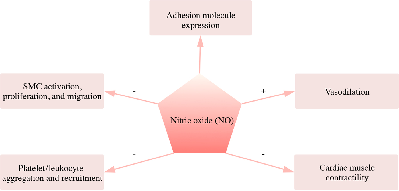
In physiological conditions, NO is generated from L-arginine continuously through the oxidation catalyzed by eNOS in vascular ECs, and it is implicated as a mediator in several physiological roles (Fig. 4).[Reference Loscalzo and Welch10] For the regulation of vascular tone, NO after diffusion across the cell membranes approaches to neighboring SMCs and targets sGC, during which NO activates sGC by binding to its heme moiety and induces accumulated cyclic guanylate monophosphate (cGMP).[Reference Jen, Serrano, van Lith and Ameer11] The increase in cGMP level activates a GMP-dependent pathway that results in a variety of downstream processes such as the vasodilation and antidepressant effect.[Reference Kaster, Rosa, Santos and Rodrigues20] By this route of signaling transduction, NO is considered as the most potent endogenous vasodilator.[Reference Schulz and Triggle21] In a similar manner, NO also reduces myocardial contraction via a cGMP-dependent mechanism. In addition to sGC, NO can target sulfhydryl-containing proteins[Reference Kim, Talanian and Billiar22] and certain protein kinases[Reference Kibbe, Li, Nie, Watkins, Lizonova, Kovesdi, Simmons, Billiar and Tzeng23] through cGMP-independent mechanisms. Another important role of NO is to regulate SMCs mitogenesis and proliferation. It helps maintain the quiescent status of SMCs by acting together with other mediators like transforming growth factor β1 and counteracting proliferative agents such as platelet-derived growth factor, and fibroblast growth factors.[Reference Sarkar and Webb24] Besides the direct inhibitive effect on SMC proliferation, NO also indirectly influences SMC proliferation through effects on other cell types such as ECs, which generally suppress SMC proliferation.[Reference Jeremy, Rowe, Emsley and Newby25]
Schematic diagram illustrating physiological regulations of NO in the cardiovascular system. “+” and “−” representing promoted and inhibiting effects, respectively.
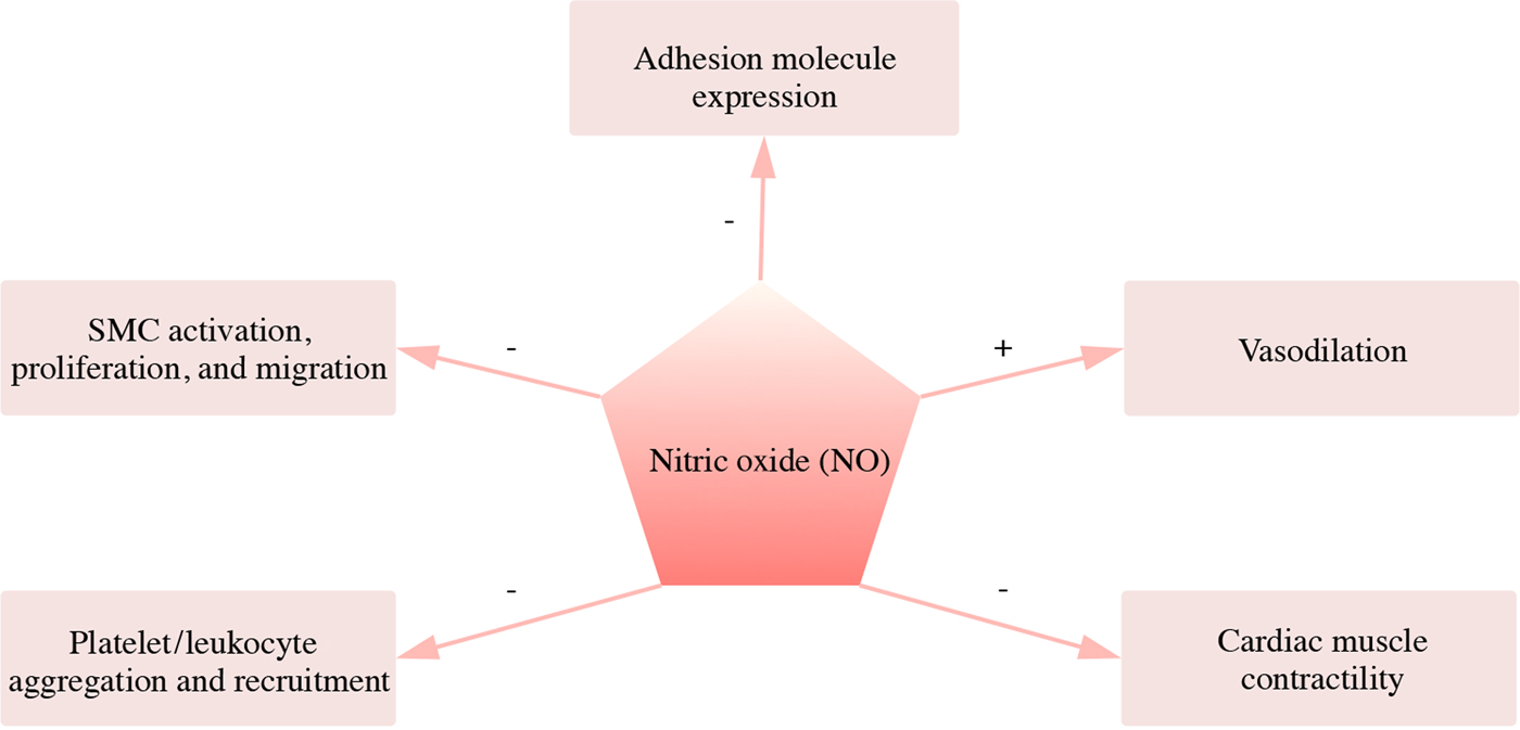
NO also functions to inhibit platelet and leukocyte responses, such as adhesion, aggregation, and recruitment to maintain the integrity of endothelium.[Reference Janero and Ewing26] The underlying mechanism of this inactivation effect relates to an increased cGMP level, which results in the impairment of shape change and granule secretion of the platelets. An attenuation of intracellular calcium flux is also associated, resulting in the inactivation of the receptors for adhesion on the platelet surface.[Reference Loscalzo27] In parallel with these, NO deficiency is involved in the development of several CVDs (e.g., hypertension and vascular stenosis).[Reference Cannon28] A decrease in the local production of NO caused by the impairment of endothelial lining in blood vessel is probably due to the loss of the eNOS-containing ECs, which are the prime source of vasoactive NO.[Reference Lafont, Guzman, Whitlow, Goormastic, Cornhill and Chisolm29] NO deficiency not only leads to the diminishment of vasodilation,[Reference Cannon28] but also causes the loss of antiplatelet and anti-inflammatory properties of the endothelial layer, causing vessel lumen prone to thrombotic events.[Reference Stemerman and Ross30] Moreover, NO deficiency impairs the inhibitive regulation of SMC proliferation and migration. As NO deficiency facilitates the development of CVDs, the local and systemic supplementation of NO may serve as an effective measure to control and treat those diseases.
NO donors for biomedical and therapeutic applications
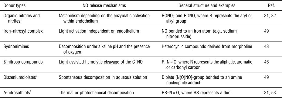
While NO plays a vital role in physiological activities, it is difficult to manipulate NO-signaling pathways with gaseous NO. As such, extensive research over the past decade has been done to explore chemical NO donors (Table I) for biomedical and therapeutic applications. These NO donors are with different NO donation mechanisms, and can be divided into following categories: organic nitrate/nitrites, iron–nitrosyl complexes, sydnonimines, C-nitroso compounds, diazeniumdiolates (NONOates), and RSNOs.[Reference Garrel, Fontecave, Favier, Cadet, Kalyanaraman, Fontecave and Pierre31]
Current NO generating components for biomedical and therapeutic applications.

a Representing as the most widely used NO donor systems.
Organic nitrate and nitrites
Organic nitrate and nitrite esters are the first generation of nitrovasodilators applied in clinical for NO-targeting therapies. The general formulae for organic nitrates and nitrites are RONO2 and RONO, respectively, where R is an aryl or alkyl group. Organic nitrates and nitrites are primarily used for the treatment of angina symptoms because they can produce NO from the metabolism within the endothelium.[Reference Garrel, Fontecave, Favier, Cadet, Kalyanaraman, Fontecave and Pierre31] These organic nitrates and nitrites can include amyl nitrite, glyceryl trinitrate (GTN), isosorbide dinitrate, and isosorbide 5-mononitrate.[Reference Nossaman, Nossaman and Kadowitz32] Amyl nitrite is used as a therapeutic agent for angina initially, and superseded a decade later by organic nitrates such as GTN, due to the ease of administration and longer duration of action.[Reference Omar, Artime and Webb33] Organic nitrates have been investigated extensively for numerous CVDs ranging from the heart failure to anal fissures.[Reference King34] Rapid-acting nitrates such as GTN are used to treat acute attacks like angina as they can relieve the symptoms within minutes, while intermediate- to long-term nitrates such as isosorbide dinitrate are valuable for the prevention of exercise angina.[Reference Omar, Artime and Webb33, Reference Jerie35]
Despite of the extensive usage the past, the effectiveness of organic nitrates and nitrites are limited for prolonged continuous treatment due to the development of pharmacologic tolerance and endothelial dysfunction.[Reference Gori and Parker36] This is because the NO donors rely on enzymatic activation to release NO, particularly for inflammatory conditions. In addition, in vivo studies have shown that endothelial dysfunction and oxidative stress probably aggravate the tolerance, and increase the risk in cardiovascular events.[Reference Munzel, Li, Mollnau, Hink, Matheis, Hartmann, Oelze, Skatchkov, Warnholtz, Duncker, Meinertz and Forstermann37] Other side effects related to organic nitrates and nitrites include headache, vomiting, and increased ischemia-reperfusion injury,[Reference Omar, Artime and Webb33] and thus scheduled nitrate-free breaks are required to prevent the development of tolerance. It has been suggested that to a certain degree co-administration of organic nitrates with other vasodilators, for example captopril and hydralazine, can avoid the development of nitrate tolerance.[Reference Fung38] The sustained-release tablets of isosorbide 5-mononitrate have also been reported to be effective with long-term use without inducing tolerance.[Reference Jerie35]
Iron–nitrosyl complexes
Iron–nitrosyl complex comprises NO bonded to an iron atom. Sodium nitroprusside (SNP) belongs to this category, and is the most used vasodilator in clinic to treat hypertensive crises. It is also known as gold standard for the NO-dependent and endothelium-independent vasodilators.[Reference Miller and Megson39] SNP does not spontaneously liberate in the physiological conditions, and it requires either light activation or tissue-specific release model for a slow release of NO.[Reference Garrel, Fontecave, Favier, Cadet, Kalyanaraman, Fontecave and Pierre31, Reference Grossi and D'Angelo40] However, the major concern on SNP is with potential release of cyanide groups, and there exist cases of cyanidosis with the long-term SNP usage have been reported.[Reference Butler and Glidewell41] Some other limitations related to SNP include the restriction to intravenous delivery, the photolability in solution, and the difficulty in dose titration.[Reference Megson42]
Sydnonimines
Sydnonimines are heterocyclic compounds deriving from morpholine, and decompose to liberate NO under the alkaline pH conditions in presence of oxygen.[Reference Garrel, Fontecave, Favier, Cadet, Kalyanaraman, Fontecave and Pierre31, Reference Feelisch, Ostrowski and Noack43] Linsidomine (3-morpholinosydnonimine or SIN-1) belongs to sydnonimines a metabolite of the anti-anginal drug molsidomine, and functions as a vasodilator and anti-aggregator through NO release occurring spontaneously in the blood.[Reference Feelisch, Ostrowski and Noack43] Molsidomine is an orally active, long-acting vasodilator. It is metabolized and converted to SIN-1 in liver, which then releases NO. As possible tolerance-free alternatives to organic nitrates, sydnonimines have received a lot of attention in angina, heart failure, and coronary vasospasm.[Reference Reden44] However, one problem associated with sydnonimines is the stoichiometric production of superoxide radicals (ONOO−), which is a highly toxic agent.[Reference Megson and Webb45]
C-nitroso compounds
C-nitroso compounds are with alkyl, aryl, or acyl that contains a nitroso group (–N = O) directly attached to an aliphatic, aromatic or carbonyl carbon, and follow a general formula of R–N = O.[Reference King, Wang, Cai and Taniguchi46] C-nitroso compounds can response to light exposure and undergo a first-order hemolytic cleavage of the C–NO bond for NO generation.[Reference Garrel, Fontecave, Favier, Cadet, Kalyanaraman, Fontecave and Pierre31] Therefore, C-nitroso compounds as the potential source of neutral NO are able to display robust NO bioactivity. It has been reported that appropriately substituted C-nitroso compounds can release neutral NO with up to 88% efficiency.[Reference Chakrapani, Bartberger and Toone47] C-nitroso compounds can exist as stable dimers in pure form for a significant period of time, making them with a great potential in therapeutic treatment as a NO donor.[Reference Chakrapani, Bartberger and Toone47] However, a major problem with C-nitroso compounds is their preparation due to high reactivity of these compounds, particularly with regard to the yield of desired product.[Reference Gowenlock and Richter-Addo48]
Diazeniumdiolates
Diazeniumdiolates (NONOates) are the chemical species containing a diolate [N(O)NO]− group to bond to an amine nucleophile adduct. They can be synthesized by exposing the nucleophiles to NO at several atmospheres pressure with protection from aerobic conditions. NONOates are stable in solid form when stored at 4 °C under argon or dry nitrogen. However, they possess excellent aqueous solubility, and produce NO spontaneously without requirements of redox or light activation.[Reference Lam, Sviri, Ilett and van Heerden49] NONOates have received attention for their clinically relevant NO donor properties since 1990s.[Reference Maragos, Morley, Wink, Dunams, Saavedra, Hoffman, Bove, Isaac, Hrabie and Keefer50] They are favored because of their stability in solid form and prolonged release of NO in vivo. Parameters, including pH, temperature, and chemical nature of the nucleophile affect the decomposition rate of NONOates, which presents a wide range of 1 min to 1 day under physiological conditions.[Reference Fitzhugh and Keefer51] The lack of tachyphylaxis is another advantage of NONOates as compared to organic nitrates, due to their tissue-independent decomposition mechanism.
Though numerous studies have demonstrated that NONOates can be a reliable source of NO for therapeutic applications, no diazeniumdiolates compounds have been clinically approved to-date. One concern with NONOates is that they are powerful mutagens.[Reference Garrel, Fontecave, Favier, Cadet, Kalyanaraman, Fontecave and Pierre31] The subsequent reactions among the decomposed by-products may also induce the formation of carcinogenic nitrosamines.[Reference Maragos, Morley, Wink, Dunams, Saavedra, Hoffman, Bove, Isaac, Hrabie and Keefer50] However, it has been suggested that the incorporation of NONOates into polymeric matrix may prevent the leaching of the by-products and improve the long-term safety of NONOates.[Reference Mowery, Schoenfisch, Saavedra, Keefer and Meyerhoff52]
S-nitrosothiols
RSNOs can be organic compounds or functional groups that contain a nitroso group to bond to the sulfur atom of a thiol, and form a general formula of RS–N = O.[Reference Al-Sa'doni and Ferro53] It has been assumed that RSNOs liberate NO through thermal or photochemical decomposition, during which the S-N bond undergoes a homolytic cleavage to generate a thiyl radical together with NO.[Reference Garrel, Fontecave, Favier, Cadet, Kalyanaraman, Fontecave and Pierre31] The presence of transition metal ions (Cu2+) or reducing agents can greatly accelerate the reaction.[Reference Holmes and Williams54] RSNOs occur decomposition through a reaction with nucleophilic species, where NO+ binds to the nucleophile. The biologic activity of RSNOs is influenced by their parent thiol.[Reference Friedman and Friedman55] Some of the RSNOs such as S-nitrosoglutathione (GSNO), S-nitrosoalbumin and S-nitrosocystein are naturally existing in blood and tissues.[Reference Jen, Serrano, van Lith and Ameer11] They act as the NO transporter in biologic system, and take part in both physiological and pathophysiological processes.[Reference Al-Sa'doni and Ferro53] The endogenous occurrence of the biologic RSNOs may explain the limited cytotoxicity shown by RSNOs, such as GSNO at pharmacologically relevant concentrations as compared to other NO donors.
There are numerous animal and clinical related studies demonstrating the advantageous features of RSNOs, although they are still not in clinical application. Some ex vivo studies on rat thoracic aorta rings reveal that RSNOs have a lower incidence of inducing tolerance and cross-tolerance with long-term usage as compared to organic nitrates.[Reference Hanspal, Magid, Webb and Megson56] In parallel, in vivo study reveals a significant antiplatelet effect of certain RSNOs with only a small decrease in blood pressure.[Reference Ramsay, Radomski, De Belder, Martin and Lopez-Jaramillo57] Moreover, the rates of NO production from RSNOs can be varied within a wide range by simple chemical modifications of the R group,[Reference Garrel, Fontecave, Favier, Cadet, Kalyanaraman, Fontecave and Pierre31] and some S-nitrosated dipeptides have also been reported with enhanced tissue selectivity.[Reference Al-Sa'doni, Khan, Poston, Fisher and Ferro58] Since RSNO can be obtained by treating thiols with a variety of nitrosating agents,[Reference Roy, du Moulinet d'Hardemare and Fontecave59] they hold a great potential as NO donor for therapeutic applications.
Spatiotemporal control of NO delivery in 3D vascularizable tissue engineering
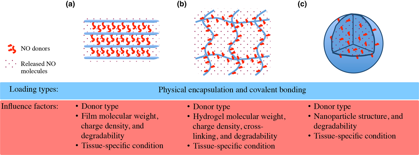
Therapeutic consequence of NO release from donors relies on the concentration, duration, and location of NO generation. As such, precise delivery of NO is required to avoid the dosage-dependent side effects of NO on regulating vascular tone. The extremely short half-life (seconds) of NO and limited diffusion distance from its source (~100 µm) also highlight the necessity of precise NO delivery. In addition, to build a 3D microenvironment suitable for blood vessel tissue formation stimuli from NO-signaling pathways needs coordinated support from 3D materials that deliver a variety of physical cues, such as mechanical force and space constraint. While the development of NO donors solves issues related to its storage, increasing attentions and efforts has been directed to explore precise NO delivery in 3D materials, with control over parameters, including concentration, rate, and location toward vascularizable tissue regeneration. Generally, there are two main categories of methods to enable spatiotemporal control of NO release. One approach is to incorporate NO donors physically into a host material, of which the release profile of NO depends on the type of NO donor,[Reference Hrabie and Keefer60] the characteristics of host materials (e.g., the water uptake rate for polymers),[Reference Mowery and Meyerhoff61] and the interaction between the additive and host materials.[Reference Batchelor, Reoma, Fleser, Nuthakki, Callahan, Shanley, Politis, Elmore, Merz and Meyerhoff62] The other approach is to immobilize NO donors or NO-donating groups on a host material via covalent bonding. A slow and localized release of NO can also be achieved depending on the type of functional immobilization and the properties of the host material.[Reference Bohl and West63] In this section, a variety of NO-releasing 3D materials such as film layers, hydrogels, and particle-based scaffolds (Fig. 5) to regulate vascular functionality in sizable tissue reconstruction will be reviewed.
Spatiotemporal NO delivery and related influence factors in advanced 3D materials. (a) Layered films. (b) Hydrogels. (c) Nano/microparticles.

NO-releasing polymeric films
NO-releasing polymeric films represent a significant portion in the development of NO-releasing 3D materials, as they are widely used in the development of biomedical devices such as synthetic vascular grafts. These 3D materials are layer-by-layer assembled generally through electrostatic attraction for charge bearing films and chemical reaction (e.g., plasma pretreatment) for universal films. Such simple procedures can allow for the layered assembly with enhanced drug delivery efficacy for localized targeting, decreased toxicity, and timely and more sustainable release. The physicochemical features of the films such as molecular weight, charge density and degradability under tissue-specific conditions affect the release profiles of drugs. Therefore, understanding the properties of each assembled film component is necessary for the design and fabrication of 3D-layered assembly, and numerous biocompatible polymer systems have investigated for effective NO delivery toward spatiotemporal control.
Non-biodegradable polymers
Saavedra et al.[Reference Saavedra, Southan, Davies, Lundell, Markou, Hanson, Adrie, Hurford, Zapol and Keefer64] made polyurethane (PU) membranes using the solvent cast method with a NONOate adduct (PROLI/NO) as the additive. This membrane exhibited a stable NO release for 7 weeks under physiological conditions (pH 7.4 and 37 °C) at a rate of 0.2–0.7 pM/(min·mg). Mowery et al.[Reference Mowery, Schoenfisch, Saavedra, Keefer and Meyerhoff52] reported poly(vinyl chloride) (PVC) membranes with the addition of another NONOate adduct (N,N′-dimethyl-1,6-hexanediamine or DMHD/NO), a plasticizer and ion-sensing additives. The PVC membranes showed NO release for up to 72 h, resulting in a significant improvement in thromboresistivity to platelet adhesion. However, there was a leaching of a toxic degradation by-product being detected.
Biodegradable polymers
Smith et al.[Reference Smith, Chakravarthy, Pulfer, Simmons, Hrabie, Citro, Saavedra, Davies, Hutsell, Mooradian, Hanson and Keefer65] incorporated low molecular weight NONOate adducts into molten poly(ε-caprolactone) (PCL) or poly(ethylene glycol) (PEG), and made into solid membranes. The PCL membranes showed gradual NO release for 7 days at physiological conditions by inhibiting the acid-catalyzed decomposition. However, the PEG membranes dissolved instantly in physiological fluids. There was also a concern on the technique for NO donor incorporation as the polymer melt may induce thermal decomposition of the NO donor, and result in decreased total NO release. Seabra et al.[Reference Seabra and de Oliveira66] prepared solid polymeric membranes from the blend of poly(vinyl alcohol)/poly(vinyl pyrrolidone) (PVA/PVP) with GSNO addition. During the thermal decomposition of GSNO, the PVA/PVP–GSNO membranes exhibited an improved stabilization. The first-order rate constants of the NO release profile for PVA/PVP–GSNO were 8–16-fold less than those for the aqueous GSNO solutions. The NO release under physiological conditions could last for 24 h, with 90% of the total incorporated NO released. In a later study, they found that both NO and GSNO were released from PVA/PVP–GSNO to aqueous solution through diffusion.[Reference Seabra, Da Rocha, Eberlin and De Oliveira67]
In addition to physical incorporation of NO donors as additives, polymers can be also modified covalently with NO donors or NO-donating functional groups. Saavedra et al.[Reference Saavedra, Booth, Hrabie, Davies and Keefer68] modified PVC with O 2-methoxymethylated piperazine diazeniumdiolates, where chloride in the polymer (1–2%) could be replaced by NONOate piperazine analogues. In a later study, films comprising diazeniumdiolated PVC blended with plasticizers and ionophores were proven to be thromboresistant and have a potential as a coating for blood-contacting devices.[Reference Mowery, Schoenfisch, Saavedra, Keefer and Meyerhoff52] In another study, Smith et al.[Reference Smith, Chakravarthy, Pulfer, Simmons, Hrabie, Citro, Saavedra, Davies, Hutsell, Mooradian, Hanson and Keefer65] modified 1,4-butanediol diglycidyl ether to poly(ethylenimine) (PEI), and then treated it with pressurized NO for 3–6 days. The crosslinked diazeniumdiolated PEI could release NO stably for 5 weeks under physiological conditions. Such NO-releasing material could also provide a flexible film coating on the PTFE vascular graft that reduced platelet adhesion and SMC proliferation.
Another major category of biodegradable polymeric films is based on poly(methyl methacrylate) (PMMA). de Oliveira et al.[Reference de Oliveira, da Silva, Seabra and de Oliveira69] synthesized polynitrosated polyesters (PNPE) from diols and mercaptosuccinic acid through polycondensation and subsequent nitrosation. They blended PNPE with PMMA to form membranes, and the membranes showed significant effect in decreasing platelet adhesion. Li and Lee[Reference Li and Lee70] modified poly(vinyl methyl ether-co-maleic anhydride) with GSNO, and blended it with PVP to form interpolymer complexes through intermolecular hydrogen bonding. The obtained membranes exhibited enhanced stability in the solid state than free GSNO, and could release NO for more than 10 days.
NO-releasing hydrogels/xerogels
Hydrogels are a network of hydrophilic polymer chains dispersed in water that are sometimes colloidal, whereas xerogels are solids formed from a gel by drying with unhindered shrinkage. Because of the complex and interconnected porous network within the matrix, both hydrogels and xerogels allow exchange of solvent and small solute molecules, making them good candidates for localized drug delivery.
Hydrogels
Shishido et al.[Reference Shishido, Seabra, Loh and Ganzarolli de Oliveira71] developed F127 hydrogel with the incorporation of NO donor GSNO and S-nitroso-N-acetylcysteine (SNAC), which retained a fluidic status at refrigerator temperature (~5 °C) and showed a temperature response as rigid semi-solid clear gels in the body (~37 °C). The resultant hydrogels could release NO through thermal and photochemical decomposition. The F127 hydrogel matrices exhibited a stabilizing effect on both thermal and photochemical NO release during the initial 2 h, and resulted in a rate range of 2–45 µM/L/min depending on the NO donor type and irradiation condition. This effect was attributed to a cage recombination mechanism. The study suggested that F127 hydrogels might be used as the storage and controlled release material for S-nitrosothiols. A similar study was done by Seabra et al.,[Reference Seabra, Fitzpatrick, Paul, De Oliveira and Weller72] in which GSNO and SNAC were incorporated into Synperonic F-127 hydrogels [a triblock copolymer of poly(ethylene oxide) and poly(propylene oxide)]. When applied on human forearm skin, both GSNO- and SNAC-containing hydrogels increased blood flows over the 3 h study. The dermal nitrite concentration was measured simultaneously and found directly correlated with blood flow. In contrast, high levels of blood flow associated with increased blood scavenging and therefore a reduction in dermal nitrite. In a recent study done by Simoes and de Oliveira, PVA hydrogels with GSNO addition were prepared using the freezing–thawing (F/T) technique.[Reference Simoes and de Oliveira73] It was found that the pore size of PVA hydrogels decreased with increasing F/T cycles, whereas the crystallinity rose. NO diffusion coefficient depended not only on the pore size of the hydrogels, but also on the interconnectivity of the pores. The vasodilating effect of NO-releasing PVA hydrogels was also confirmed with tests on human forearm skin.
In addition, there are also many studies reported to produce NO-releasing hydrogels/xerogels via covalent modification. Masters et al.[Reference Masters, Leibovich, Belem, West and Poole-Warren74] developed a NO-releasing PVA hydrogel dressings via amination of PVA and subsequent diazeniumdiolation. The diazeniumdiolated PVA (PVA-NO) was photocrosslinked with Irgacure initiator to form hydrogel sheets. The PVA-NO hydrogels could release almost 80% of the total incorporated NO over 48 h at pH 6.0, which is the pH level of most wound environment. In order to prevent neointima hyperplasia, Lipke and West[Reference Lipke and West75] developed a NO-releasing PEG-based hydrogel (PEG-Cys-NO) via covalent binding cysteine to a PEG-based copolymer and subsequent nitrosation. The PEG-Cys-NO hydrogels could undergo rapid photopolymerization in situ, and generated NO for up to 50 days. In vitro test of the PEG-Cys-NO hydrogels proved that the hydrogels could effectively inhibit SMC proliferation and platelet adhesion, and increase EC proliferation. The perivascular utilization of PEG-Cys-NO hydrogels in a rat carotid balloon-injury model revealed that could inhibit intimal thickening and improve endothelialization, leading to approximately 80% reduced neointima formation 2 weeks post-injury compared with control groups. Using a similar rat balloon-injury model, Bohl and West[Reference Bohl and West63, Reference Masters, Lipke, Rice, Liel, Myler, Zygourakis, Tulis and West76] observed reduced neointima formation by approximately 75% 14 days post-injury. Agreed with the finding Taite and West[Reference Taite and West77] in a rat balloon-injury model showed an approximately 90% decrease in intimal thickening with the application of NO-releasing PEG hydrogels 28 days post-injury. Recently, Kim et al.[Reference Kim, Lee, Singha, Kim, Shin, Jo, Han and Kim78] reported a NO-conjugated gel system (F127-BPEI-NONOates) through composition of thermosensitive Pluronic F127 and branched PEI (BPEI) followed with NONOates conjugation. The F127-BPEI-NONOates system accounted for a much longer half-life of NO release (1.68–2.0 h) as compared with the control group without F127 (<1 min), which was attributed to the cage recombination efficiency of F127. The longevity of NO release for F127-BPEI-NONOates ranged from 32 to 54 h depending on the composition. F127-BPEI-NONOates also showed inhibitive and promoted effect on in vitro SMC and EC proliferation, respectively.
Xerogels
Marxer et al.[Reference Marxer, Rothrock, Nablo, Robbins and Schoenfisch79] reported the method to incorporate NO donors within a cross-linked silica xerogel matrix. Highly cross-linked amine-containing networks could be produced using sol–gel chemistry with amine-functionalized alkoxysilanes (aminosilanes) and alkyltrimethoxysilanes (alkylsilanes). Upon diazeniumdiolation, the xerogels could release NO from several hours to 20 days under physiological conditions depending on the type and amount of aminosilane precursor. For example, a xerogel matrix at 50 µm in thickness could have an average NO fluxes from 8.0 × 10⁻⁶ to 5.6 × 10⁻⁵ µM/s/cm2 during the initial 10 h.[Reference Marxer, Rothrock, Nablo, Robbins and Schoenfisch79] In vitro study showed a flux-dependent platelet adhesion on the xerogels, suggesting the NO-releasing xerogels might be useful for short-term applications.
NO-releasing particle-based scaffolds
The idea of using particles as scaffolds for NO release was first attempted by Forslund and Sundqvist in 1997.[Reference Forslund and Sundqvist80] They incubated the heat-killed yeast particles for 1 h in a hydrophobic NO-releasing system to absorb the agent, and used them to study the effect of NO on neutrophil functions in vitro. Along with the extensive applications of micro/nanoparticles (NPs) in different research fields, a variety of NO-releasing particles have been developed either by entrapping NO donors or derivatizing the particle surface with NO-releasing functional groups. NO-releasing particles are usually used as fillers, and they can be blended into various polymeric matrixes without altering the fundamental chemistry of polymers. NO release can be modulated by changing the amount of particle blended or changing the physicochemical properties of polymers (e.g., molecular weight and structure).[Reference Reynolds, Frost and Meyerhoff81] In addition, for derivatized NO-releasing particles, the risk of by-product leaching from NO donor decomposition can be eliminated as they are covalently modified and physically entrapped in the polymer matrix.[Reference Frost and Meyerhoff82] However, more attention on toxicity is suggested when introducing particles for NO delivery, especially when nanoscale materials are applied. The smaller size of such materials increases their contacting area with biologic membrane, which may lead to an alteration of toxicity.
Silica particles
The first NO-releasing particles with covalent modification was reported by Zhang et al.[Reference Zhang, Annich, Miskulin, Stankiewicz, Osterholzer, Merz, Bartlett and Meyerhoff83] Non-toxic amorphous fumed silica particles (200–300 nm in diameter) were tethered with amine-containing silylation reagents followed by diazeniumdiolation. The diazeniumdiolated fumed silica particles released NO under physiological conditions via primarily the proton-driven dissociation associated with negligible thermal dissociation. The fumed silica particles also exhibited a stabilizing effect on the NONOate groups anchored on the surface characterized by a longer half-life (43 min) than that of the solution phase analog NONOate compound (2.8 min). The NO-releasing particles bended into PU matrices were coated on lumen side of an extracorporeal PVC tube and evaluated in a rabbit extracorporeal circulation (ECC) model. Prolonged NO release was detected for at least 4 h, with a NO flux comparable with that from ECs under stimulated conditions (4.1 × 10⁻¹⁰ M/cm2/min).[Reference Vaughn, Kuo and Liao84] Fewer thrombi and much lower platelet activation and consumption for NO-releasing particle coating were observed compared to the control surfaces, suggesting a good potential of the NO-releasing fumed silica particles in the development of more blood-compatible devices.
A similar study was carried out by Carpenter et al.,[Reference Carpenter, Slomberg, Rao and Schoenfisch85] in which NO-releasing silica NPs were prepared from diazeniumdiolation of amine-functionalized silica NPs of distinct sizes (50, 100, and 200 nm). The diazeniumdiolated silica NPs could release NO instantaneously characterized by an initial burst of NO with the maximum concentration of NO ranging from 43.5 to 49.7 ppm/mg. The NPs showed a faster diffusion rate into the cells along with decreasing particle size. In another study done by Shin and Schoenfisch,[Reference Shin and Schoenfisch86] aminosilanes were first diazeniumdiolated and then mixed with tetraethoxysilane or tetramethoxysilane (TEOS or TMOS) to form organic/inorganic hybrid silica NPs via a sol-gel process. Such silica NPs were reported to improve NO storage with more sustainable release profiles over macromolecular NO donors. The NO payload could reach 11.26 µM/mg, and the release could last for 101 h with a half-life of 253 min. Recently, Zhang et al.[Reference Zhang, Wang, Wen, Ren, Li, Teoh and Thian87] reported a cytocompatible NO-releasing NP synthesized via thiolation of gelatin–siloxane NPs. The resultant NO-releasing NPs (GS-NO NPs) could release NO following a first-order exponential profile (~0.12 µM/mg within 7 days), and could realize rapid intracellularization within 2 h. In vitro study with human aortic SMCs and human umbilical vein ECs suggested that GS-NO NPs could exhibit a dose-dependent effect for inhibited SMCs proliferation and promoted ECs proliferation, with great potential of preventing vascular restenosis.
Gold nanoparticles (AuNPs)
Rothrock et al.[Reference Rothrock, Donkers and Schoenfisch88] functionalized monolayer-protected cluster AuNPs (MPCs) with amino groups followed by diazeniumdiolation with pressurized NO. The NONOate-modified MPCs were with an average core diameter of 2 nm. The total release amount of NO was found tunable through simply changing properties of the substituted amino ligands (e.g., the number and chemical structure). Increased amount of amino ligands or increased length of the alkyl chain could result in more NO released, ranging from 9750 to 87,000 pM/mg with NO release duration from 200 to 300 min. In another study done by the same research group, water-soluble NO-releasing AuNPs were synthesized from Tiopronin-protected MPCs (Tio-MPCs) via similar modifications.[Reference Polizzi, Stasko and Schoenfisch89] Despite the water solubility, the NO-releasing Tio-MPCs could only release modest amount of NO (<0.023 µM/mg) for short durations (<1.5 h). In the same study, they also demonstrated that the NO storage capacity and release stability could be improved by using polyamine-stabilized MPCs, which could release 0.386 µM/mg NO within up to 16 h after modifications. A concern with AuNPs is that AuNPs with small sizes (<2 nm) or certain surface chemistry (e.g., cationic AuNPs) may be cytotoxic.[Reference Alkilany and Murphy90] Therefore, more cautions should be taken in using AuNPs as NO-releasing particles.
Polymeric particles
Compared with silica and gold particles, polymeric particles may have some advantages for drug delivery as they can be synthesized from biodegradable polymers. In mice model biodegradable polymeric particles have been found to increase blood circulation time with reduced liver accumulation.[Reference Gref, Minamitake, Peracchia, Trubetskoy, Torchilin and Langer91] Kumar et al.[Reference Kumar, Hong, Maciag, Saavedra, Adamson, Prud'homme, Keefer and Chakrapani92] reported the synthesis of NO-releasing PEGylated NPs from different PEG-based block copolymers. The block copolymers were mixed with the NONOate adducts (PABA/NO or Double JS-K) and subjected to processes of a rapid micromixing and precipitation followed with a block-copolymer-directed encapsulation. The size of the resultant NPs was 195–240 nm depending on the combination of block copolymer and NONOate adduct, and it increased to 290–440 nm after 1–20 h due to slow ripening of the NPs. The soluble PEG blocks in the amphiphilic block copolymers provide steric barrier to protect and stabilize the encapsulated NONOates from glutathione deactivation, which was characterized by much longer half-life of PEG-protected NPs in glutathione solution (40 min–5 h) than that of the free NONOates (4.5–15 min).
Yoo et al.[Reference Yoo, Lee and Lee93] developed NO-releasing polymeric microparticles from poly(lactic-co-glycolic acid) (PLGA) and dethylenetriamine (DETA) NONOate. DETA NONOate was mixed with the PLGA solution and entrapped by the microparticles during their formation. The NO-releasing PLGA microparticles (19–83 µm) could entrap up to ~73% of the total DETA NONOate added with the loading content up to ~3.7 mg. DETA NONOate was stabilized in the microparticles characterized by longer decomposition time (2–6 h) of the microparticles than that of the intact DETA NONOate (20 min).
Recently, a new type of NO-releasing NPs was developed by Friedman et al.[Reference Friedman, Han, Navati, Chacko, Gunther, Alfieri and Friedman94] based on biocompatible hydrogel/glass composites. The NO-releasing composite NPs were produced from the sol–gel process of a mixture containing TMOS, PEG, chitosan, glucose, and sodium nitrite (NaNO2), followed by lyophilization or heat treatment and grinding. The composite NPs liberated NO via a different mechanism, in which NO is generated from the thermal reduction of added NaNO2 by glucose once exposed to moisture. The glassy matrices provided necessary condition for thermal reduction, while hydrogels retained the generated NO within the dry composite NPs. The release of NO was found regulated by the extent of hydration of the composite NPs. The composite NPs had an average size of 10 nm, and could release NO for over 1 month with sustained NO concentration more than 800 nM. Later studies showed that the NO-releasing composite NPs could effectively exert antimicrobial activity and accelerate wound healing process.[Reference Martinez, Han, Chacko, Mihu, Jacobson, Gialanella, Friedman, Nosanchuk and Friedman95] The circulation of these NPs also induced vasodilation and increased microvascular perfusion.[Reference Cabrales, Han, Roche, Nacharaju, Friedman and Friedman96]
NO-releasing NPs based 3D scaffolds
While NPs can be injected for the localized and temporal delivery of NO to regulate neovessel formation, the incorporation of these NPs into a variety of 3D scaffolds offers additional benefits, including capabilities of load bearing, force transmission, cell attachment, as well as guided neovessel formation. These incorporated NPs can also obtain improved spatiotemporal control and more efficiency of delivered NO for the precise therapy purpose over direct NPs administration. Current techniques that include physical blending (e.g., electrospinning and two-roll milling) and chemical modification (e.g., plasma treatment) can be used for scaffold to incorporate NO-releasing NPs.[Reference Tong, Wang, Lim, Wang and San Thian97] Such scaffolds can have more tailorable release profiles of vessel regulating agents, and can bridge critical-sized tissue gaps with physically constrained microenvironments for vasculature developing.[Reference Wang, Wen, Lim, Zhang, Konishi, Wang, Teoh and Thian98] In addition, physical cues delivered through the scaffold such as geometric features and mechanical stimuli can facilitate NO-signaling pathways for complex tissue regeneration, with desirable functionality related to proper vascularization, ordered tissue organization,[Reference Wang, Teo, Chong, Zhang, Lim, Zhang, Hong, Thian, Chan and Teoh99] and tissue-specific differentiation.[Reference Wang, Teoh, Johana, Chong, Teo, Hong, Chan and Thian100]
Concluding remarks and future prospects
NO has been evidenced as a vital molecule to play an important role for cardiovascular system in both physiological and pathophysiological processes. Despite the extensive applications of organic nitrates and inhaled NO in clinic, there exist concerns of drug tolerance and difficulty in handling gaseous NO, leading to more researches in recent efforts focusing on the localized delivery of NO. To explore potential of NO for cardiovascular therapy, physical laden of NO donors as additives in a 3D material is the most straightforward approach, of which NO release profiles depend on the types of NO donors, the host materials, and the amount of dopants. However, there exists a risk of NO donor or decomposition by-products leaching from the host material, which are often toxic and may cause serious problems. In construct, covalently modifying the host material with NO-releasing functional groups may be an improved promising approach.
Most of the reported NO delivery systems over the past two decades have been developed in membrane or gel form. However, interests in developing NO-releasing particles keep increasing. The NO-releasing particles are not only able to impose controlling effects on the NO liberation by themselves but could also be conveniently synchronized into a variety of 3D materials including hydrogels,[Reference Hoo, Sarvi, Li, Chan and Yue101] microcapsules,[Reference Chen, Yue, Moulton, Hayes, Cook and Wallace102] and electrospunfibers,[Reference Jayasinghe103, Reference Townsend-Nicholson and Jayasinghe104] which were used for drug delivery and cell encapsulation without losing their bioactivities, for the enhancement in spatiotemporal control of NO delivery. The NO release profiles of 3D materials could be tailored by varying the amount of NO-releasing particles incorporated in order to meet the requirements for specific applications.
The development of NO-releasing medical devices including stents and vascular grafts is thus promising to provide great therapeutic benefits and address problems during CVD treatment ranging from the infection to thrombosis, the foreign body response, and neointimal hyperplasia. Other than developing NO-releasing coatings on currently in-use devices, developing advanced NO delivery materials as alternative 3D devices are promising future options. For this purpose, more factors including mechanical compliance, the bioactivity of material surface, biodegradability, and the ease of storage and handling are necessary for consideration besides the NO release profiles. Nevertheless, a better understanding on physiological effects of NO in different tissues can facilitate the development of 3D NO-releasing materials for specific application. These efforts together may direct promising material strategies for the applications of NO in precise CVD treatment therapy.
Conflict of interest
The authors have no competing interests.
Acknowledgments
This work was supported by the Fundamental Research Funds for the Central Universities (People's Republic of China) Project number 531107050927. Z. W. received salary support from Hunan University for the Yuelu Young Scholars B (JY-Q/008/2016).