Introduction
Temporal bone defects can be acquired or congenital in origin. Acquired defects are either secondary to head trauma, otological surgery, tumour, irradiation or chronic otitis media, or spontaneous in onset.Reference Sanna, Fois, Russo and Falcioni1–Reference Feenstra, Sanna, Zini, Gamoletti and Delogu3 These defects can result in meningoencephalic herniation, and when associated with violation of meninges, the clinical presentation may be that of cerebrospinal fluid (CSF) otorrhoea or otorhinorrhoea. In addition, the defects provide a route for the spread of infection into the intracranial cavity, resulting in recurrent episodes of meningitis and brain abscess.Reference Feenstra, Sanna, Zini, Gamoletti and Delogu3–Reference Iurato, Ettore and Selvini5 Temporal bone defects within and around the bony labyrinth present only as CSF leak, whereas tegmen plate or posterior fossa plate defects manifest clinically as either meningoencephalic herniation or CSF leak.
There are no available guidelines for the management of temporal bone defects; most surgeons rely on their personal experience for the diagnosis and surgical closure of such defects. A variety of surgical techniques have been described over time.Reference Sanna, Fois, Russo and Falcioni1, Reference Glasscock, Jackson and Wiet4, Reference Jackson, Pappas, Manolidis, Glasscock, Von Doersten and Hampf6 The closure can be achieved either from the mastoid, middle cranial fossa, or with a combined approach in which the hernia is amputated from below and the defect sealed from above. According to Glasscock et al., a combined middle cranial fossa and mastoid approach provides a robust closure for defects larger than 1 cm.Reference Glasscock, Jackson and Wiet4 Sanna et al. described subtotal petrosectomy with middle-ear obliteration and cul de sac closure of the external auditory canal to obtain secure closure of the skull base.Reference Sanna, Fois, Russo and Falcioni1
In this article, we review the aetiology, clinical presentation, diagnostic challenges, surgical technique and post-operative outcomes of 16 patients with temporal bone defects managed at our centre. The relevant literature, available surgical options and post-operative concerns arising in such clinical situations are discussed.
Materials and methods
A retrospective review of the medical records of patients who presented with temporal bone defects at our centre between 1991 and 2016 was conducted. The inclusion criteria were: (1) all patients with meningoencephalic herniation involving the temporal bone; (2) CSF leak from the temporal bone requiring surgical correction; and (3) post-operative follow up of at least one year. We excluded all cases wherein surgical correction of the temporal bone defect was part of the primary procedure, as in the repair of petrous apex or cerebellopontine angle lesions. Data pertaining to aetiology, presenting complaints, audiological findings, surgical approach and repair technique, post-operative complications, and outcomes were extracted from medical records.
Results
Demographics
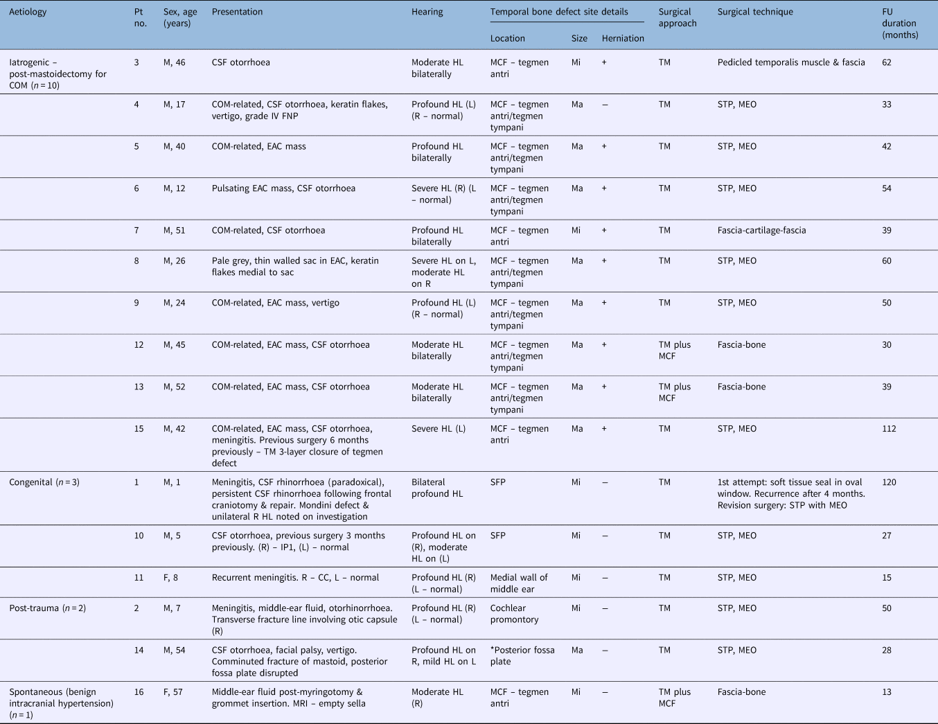
Overall, 16 patients with temporal bone defects who had presented during the stated period were included in the study. Of these, 14 were male and 2 were female. The median age of patients was 33 years (range, 1–57 years). Six patients had left-sided symptoms and 10 patients had right-sided temporal bone involvement. None of the patients had a bilateral temporal bone defect. Table 1 describes the temporal bone defects in terms of aetiology, site, size, surgical approach and repair technique adopted for management.
Demographic data, aetiology, presentation, surgical details and follow up of 16 patients with temporal bone defects

*Multiple defects. Pt no. = patient number; FU = follow-up; COM = chronic otitis media; M = male; CSF = cerebrospinal fluid; HL = hearing loss; MCF = middle cranial fossa; Mi = minor; ‘+’ = positive; TM = transmastoid; FNP = facial nerve palsy; L = left; R = right; Ma = major; ‘−’ = negative; STP = subtotal petrosectomy; MEO = middle-ear obliteration; EAC = external auditory canal; SFP = stapes footplate; F = female; MRI = magnetic resonance imaging; IP1 = incomplete partition type 1; CC = common cavity
Aetiology
Aetiology was: post-mastoidectomy (n = 10), congenital (n = 3), post-trauma (n = 2) or spontaneous (n = 1). Temporal bone defects were classified as major if the size was larger than 1 cm.
Post-mastoidectomy
The median age of patients in this group was 41 years (range, 12–52 years). Temporal bone defects of all patients in this group were suspected to be iatrogenic or secondary to chronic otitis media with cholesteatoma. All the defects in this group were single and located in the tegmen plate. Eight were classified as major and two were classified as minor. The diagnosis was evident pre-operatively, either clinically or radiologically, in all patients. In patients with minor defects (numbers 3 and 7), a small meningoencephalic herniation was identified intra-operatively.
All patients had a prior history of a canal wall down mastoidectomy performed at another institution, with the original diagnosis being cholesteatoma. With the exception of patients 7 and 15, all had undergone a single operative procedure. Both of these patients had undergone more than two mastoid surgical procedures since childhood for residual or recurrent cholesteatoma in the involved ear. The mean time of presentation since the last mastoid surgery was 57.1 months (±36.1 months).
At the time of surgery, co-existing cholesteatoma was found only in two patients (numbers 4 and 8). Patient 9 had bilateral chronic otitis media; however, the contralateral ear did not show similar findings. In all patients, the hearing threshold in the contralateral ear was either normal or better than in the involved ear.
Congenital
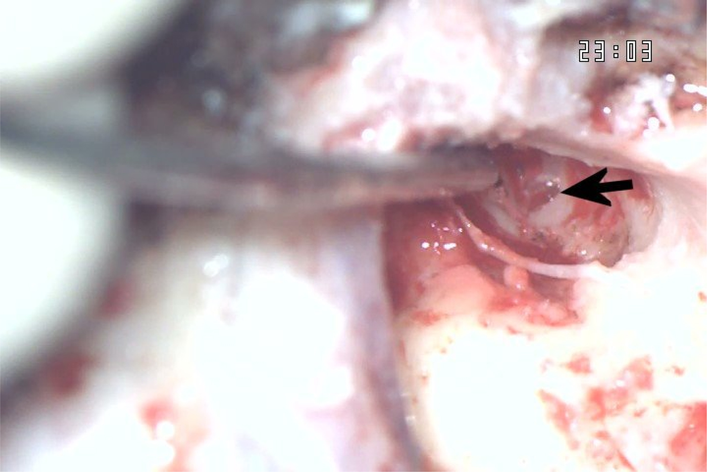
The median age of patients in this group was five years (range of one to eight years). The three patients in this group presented with: CSF otorrhoea (n = 1), CSF otorhinorrhoea (n = 1) and/or recurrent meningitis (n = 2). Patient 1 had 2 meningitis episodes and patient 11 had 3 episodes. All the patients in this group had translabyrinthine defects located either on the stapes footplate or the medial wall of middle ear (Figure 1). In patient 10, an attempt to locate and close the temporal bone defect had been made three months previously at another centre, wherein a soft tissue plug reinforced with incus had been used to seal the defect. The leak had recurred within a month of repair.
Intra-operative photograph of a patient with a congenital defect on the promontory (arrow).

Post-trauma
The two patients in this group were aged 7 and 54 years respectively. Patient 2 had sustained a fall from a height, after which he developed CSF rhinorrhoea and recurrent meningitis episodes. He underwent frontal craniotomy and repair, but remained symptomatic after the surgery. Subsequent radiological investigations confirmed the defect site as in the temporal bone. Patient 14 had a pellet injury of the temporal bone, which had resulted in comminuted fracture of the mastoid tip and disruption of the posterior fossa plate.
Spontaneous onset
There was only one patient with spontaneous symptom onset, aged 57 years. The patient had become symptomatic two years previously with unilateral ear fullness. She had undergone myringotomy and grommet insertion for management of persistent middle-ear fluid. She was investigated for a possible temporal bone defect in light of persistent watery otorrhoea in the involved ear.
Clinical presentation
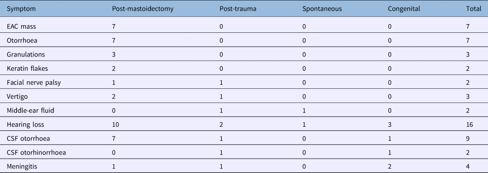
The symptoms and findings of all patients at the time of presentation are shown in Table 2. The most common symptoms in our series were: hearing loss, CSF otorrhoea and an external auditory canal mass. Recurrent episodes of meningitis were reported by four patients. The mean number of meningitis episodes was 4 (± 2.71). None of the patients in our series had a history of seizures or a co-existent brain abscess.
Common symptoms at time of presentation*

Data represent numbers of cases. *Total n = 16. EAC = external auditory canal; CSF = cerebrospinal fluid
Diagnosis
All patients except patient 1 (15 of 16 patients) underwent high-resolution computed tomography (CT) as a part of the diagnostic protocol. Cochlear anomalies were identified in patients 10 and 11. A fracture of the temporal bone was evident on high-resolution CT in patients 2 and 14. Patients 1 and 2 had a history of clear watery rhinorrhoea; both these patients had undergone a prior anterior skull base procedure (performed by neurosurgeons) for CSF leak closure, but the site of the leak had remained unidentified.
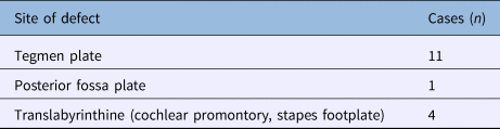
In patient 1, polytomography was conducted, which showed a Mondini-like deformity; subsequent radionuclide cisternography further confirmed the origin of the leak in the temporal bone. In patient 2, radiology identified fluid signals in the ethmoid sinuses and mastoid, and a fracture involving the otic capsule. The site of the leak was identified in the temporal bone on CT cisternography in this patient. The site of the defect had already been documented during previous surgery in patient 10, and no further investigation was considered. In patient 11, CT cisternography was not carried out as there was a strong clinical suspicion of a temporal bone defect causing recurrent meningitis, given the co-existent profound hearing loss and cochlear anomaly on the involved side. The tegmen plate was the most common site of defects (in 11 of 16 cases) in our series (Table 3).
Site of temporal bone defect*

*Total n = 16.
Gadolinium-enhanced magnetic resonance imaging (MRI) was performed for better soft tissue delineation and to confirm the diagnosis of meningoencephalic herniation in patients who had an external auditory canal mass.
Ancillary tests such as intrathecal fluorescein dye injection, glucose estimation and beta-2 transferrin assay for confirmation of CSF were not conducted in any of the patients.
Surgical approach and repair technique
Transmastoid approach with subtotal petrosectomy and middle-ear obliteration
A post-aural incision was made 3–4 cm behind the retro-auricular groove and mastoidectomy was conducted, to exenterate the air cells, remove any diseased mucosa and cholesteatoma if present, and expose the herniated brain tissue widely. Bipolar cautery with low current settings (5–10 W) was used to amputate the herniated brain tissue at the level of its neck. Due care was taken not to pull the normal brain tissue into the cavity. As the herniated brain tissue was deemed to be non-viable and possibly infected, it was not re-cranialised.
In most cases, the size of dural defect was smaller than the bony tegmen defect. Scarpa's fascia was harvested and placed as underlay after lifting the dura from the surrounding bony edges. In patient 8, the defect was reinforced with two layers of fascia, as it was slightly larger.
In patients without meningoencephalic herniation, the leak site was identified, and closed with a soft tissue or bone plug. The mucosa of the Eustachian tube opening was cauterised, and was plugged with bone dust or bone wax.
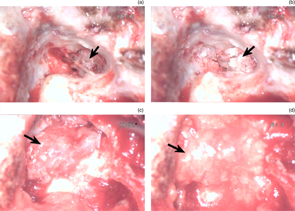
Subtotal petrosectomy was accomplished after removal of all air cells. Blind sac closure of the external auditory canal was performed after removing the external auditory canal cartilage, and was reinforced with a small anterior-based Palva flap. The middle-ear mucosa, tympanic membrane, ossicles and external auditory canal skin were removed. Abdominal fat was used to obliterate the cavity. A superiorly based temporalis muscle flap pedicled on the deep temporal artery was fashioned for sealing the cavity. This technique was adopted for all patients who had a major defect, or for those in whom middle-ear preservation was not desirable because of poor residual hearing (Figure 2).
Subtotal petrosectomy with middle-ear obliteration in a patient with a congenital defect on the promontory: (a) sealing of the bony defect on the promontory using an ossicle (arrow); (b) bone dust used to augment the sealed promontory defect (arrow); (c) superiorly based temporalis muscle pedicle rotated in the middle ear and mastoid cavity (arrow); and (d) abdominal fat used for packing the mastoid and middle ear (arrow).

Patient 1 was initially managed with a soft tissue plug to seal the stapes footplate fistula, and the contents of the middle ear were left untouched. However, the CSF rhinorrhoea recurred after one year of follow up and he subsequently underwent subtotal petrosectomy with middle-ear obliteration and cul de sac closure of the external auditory canal.
Transmastoid approach
This approach was used in two patients (numbers 3 and 7), both of whom had minor temporal bone defects. Mastoidectomy was conducted using a standard post-aural approach. The meningoencephalic herniation was identified and amputated. The dura was freed from the surrounding bony plate. In patient 3, the defect was repaired by placing a pedicled temporalis muscle flap in an hourglass fashion. A second layer consisting of fascia was placed over the site of the defect. In patient 7, a triple-layer closure was performed by placing Scarpa's fascia and conchal cartilage in an underlay fashion; a third layer consisting of fascia was placed over the defect in an overlay fashion.
Combined middle cranial fossa and transmastoid approach
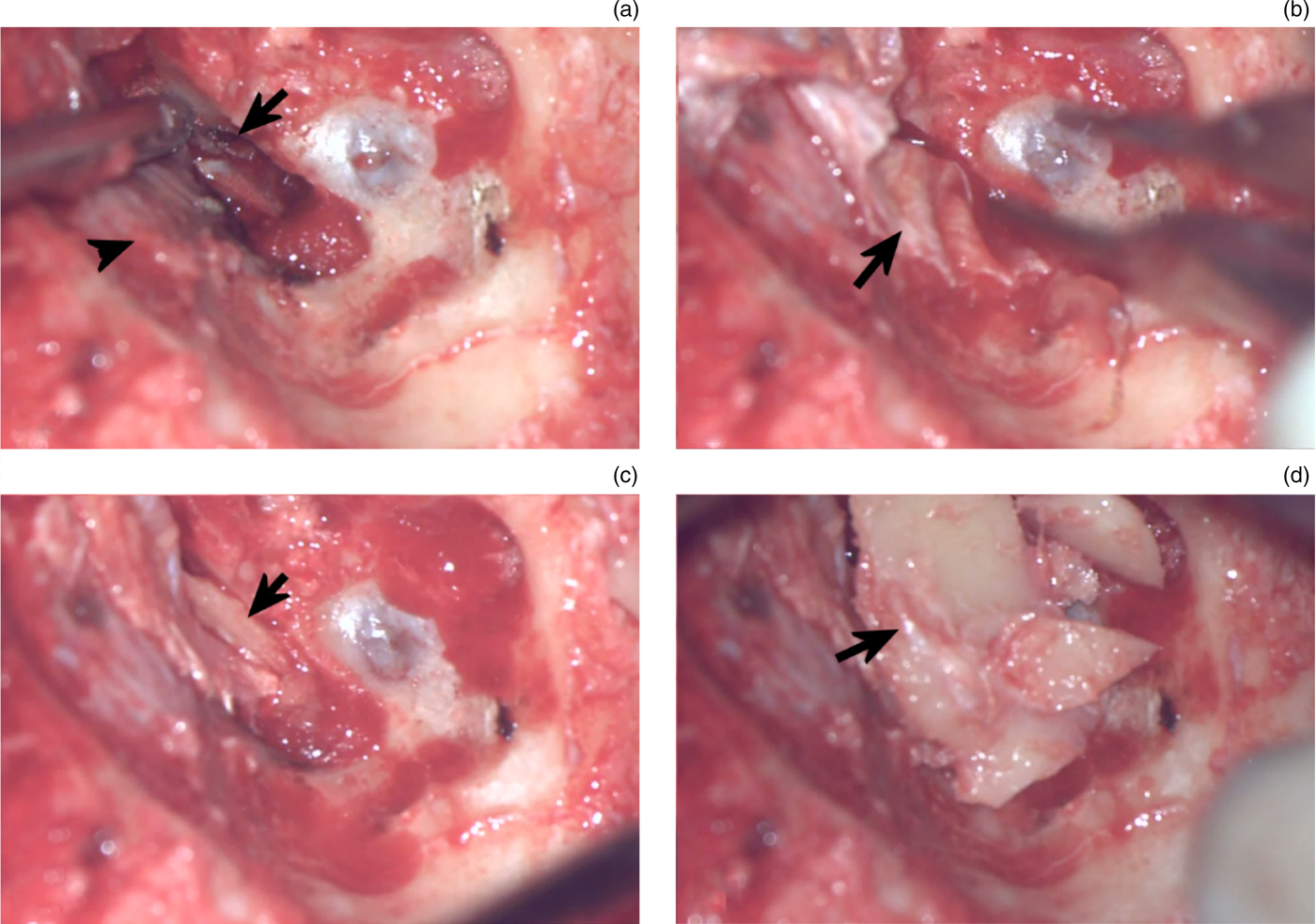
In this technique, the meningoencephalic herniation was initially managed through a transmastoid route. The post-aural incision was then extended superiorly as a reverse question mark and a posteriorly based skin flap was elevated. The incision was deepened through temporalis muscle, which was reflected anteriorly. The root of zygoma was bared. A small part of the outer table was harvested using a small cutting burr, which was later used for sealing the defect. A 3 × 3 cm craniotomy, centred on the site of the temporal bone defect, was performed, with its lower edge at the level of the zygomatic root. At this stage, intravenous (IV) mannitol was infused to decompress the brain. Extradural elevation of the temporal lobe was conducted to expose the defect site. An oversized piece of fascia was positioned extradurally, to cover the dural defect. A harvested piece of bone was then positioned carefully between the fascia and the bony defect. The bone removed during craniotomy was replaced and the wound was closed in layers.
This technique was used in three patients (numbers 12, 13 and 16), in whom the residual hearing status warranted preservation of middle-ear function (Figure 3).
Intra-operative photograph of a combined middle cranial fossa and transmastoid approach in a patient with a spontaneous-onset temporal bone defect: (a) slit-shaped defect in tegmen antri (arrow), and middle cranial fossa dura mater (arrowhead); (b) extradural placement of temporalis fascia graft to cover the bony slit-shaped defect (arrow); (c) extradural placement of bone graft at the defect site (arrow); and (d) bone plate repositioned to cover the middle cranial fossa window (arrow).

Post-operative course
All patients were administered IV antibiotics (ceftriaxone 100 mg/kg/day) for a period of 5 days, followed by oral antibiotics for another 5 days. In order to reduce the CSF pressure and allow the repair to heal adequately, injectable mannitol (1 g/kg, IV) was infused for 2 days; this was followed by oral acetazolamide for a period of 6 weeks. A lumbar drain was inserted at the time of surgery only in one patient (number 10).
Patients 2 and 6 developed post-aural CSF collection, which was managed with aspiration and a pressure dressing. The IV antibiotics had to be stepped up in patient 10, who developed meningitis on the 2nd post-operative day. Patient 6 developed delayed-onset facial nerve palsy (grade II on the 2nd post-operative day), which resolved after a course of oral steroids.
The mean follow-up time after surgery was 48.38 months (± 30.07 months). All patients were followed up clinically, except for patients 4 and 8, who had cholesteatoma at the time of surgery and required radiological follow up. Only one recurrence (in patient 1) was noted, after one year of follow up. This patient underwent subtotal petrosectomy with middle-ear obliteration, after which time the CSF leak did not recur.
Discussion
Temporal bone defects can be congenital, of spontaneous onset or consequent to: iatrogenic trauma, chronic osteitis due to chronic otitis media (both with and without cholesteatoma), or non-surgical trauma.Reference Sanna, Fois, Russo and Falcioni1, Reference Feenstra, Sanna, Zini, Gamoletti and Delogu3, Reference Glasscock, Jackson and Wiet4, Reference Neely and Kuhn7 These defects can be located in the tegmen plate or posterior fossa plate, within the otic capsule, or in the perilabyrinthine area.Reference Savva, Taylor and Beatty8, Reference Ferguson, Wilkins, Hudson and Farmer9 The defects associated with congenital malformations of the inner ear are located in the otic capsule and are seen in children.Reference Lundy, Graham, Kartush and La Rouere10, Reference Wetmore, Herrmann and Fisch11 Other defects of spontaneous origin are rare and can occur through congenital dehiscences involving the tegmen plate.Reference Sanna, Fois, Russo and Falcioni1, Reference Jackson, Pappas, Manolidis, Glasscock, Von Doersten and Hampf6, Reference Kamerer and Caparosa12 Defects due to surgical trauma or osteolysis secondary to an infective process are located in the tegmen plate or posterior fossa plate, with the latter being rare. The location of temporal bone defects following head injury depends upon the fracture site.
The clinical spectrum of temporal bone defects is varied. The initial presentation can be clinically evident as a CSF leak, recurrent meningitis or an external auditory canal mass. The herniated tissue is dysfunctional and referred to as brain fungus. It may be a potential epileptic focus, or cause visual and olfactory hallucinations.Reference Glasscock, Jackson and Wiet4 The external auditory canal mass may retain keratin flakes and cause a chronically discharging ear.Reference Feenstra, Sanna, Zini, Gamoletti and Delogu3 Temporal bone defects can be clinically silent, and discovered incidentally on radiology or at the time of surgery. Sometimes, a more perplexing clinical situation, such as recalcitrant middle-ear fluid or watery rhinorrhoea, may present, and only a high index of suspicion may direct our investigations to the site of the defect.
Various factors play a role in the aetiopathogenesis of meningoencephalic herniation. A bony defect alone is not sufficient for meningoencephalic herniation to develop, as the dura mater can support the weight of brain tissue over large bony defects without prolapsing.Reference Glasscock, Jackson and Wiet4, Reference Jackson, Pappas, Manolidis, Glasscock, Von Doersten and Hampf6 Dural weakening by infection or trauma, exposure to long-standing brain pulsations, or raised intracranial pressure due to an abscess or local cerebritis may cause it to prolapse.Reference Glasscock, Jackson and Wiet4, Reference Jackson, Pappas, Manolidis, Glasscock, Von Doersten and Hampf6, Reference Mosnier, Fiky, Shahidi and Sterkers13 In the pre-antibiotic era, blind trephination through an infected field for drainage of otogenic brain abscess was the commonest cause of meningoencephalic herniation,Reference Glasscock, Jackson and Wiet4 while in recent times the most common aetiology is surgical trauma.Reference Sanna, Fois, Russo and Falcioni1, Reference Feenstra, Sanna, Zini, Gamoletti and Delogu3, Reference Iurato, Ettore and Selvini5
Congenital temporal bone defects have been classified as: type I, which involve the otic capsule; type II, which occur adjacent to the otic capsule; and type III, which are distant from the otic capsule.Reference Neely and Kuhn7 These defects can present with spontaneous meningoencephalic herniation, and CSF otorrhoea or otorhinorrhoea, or recurrent meningitis.Reference Ferguson, Wilkins, Hudson and Farmer9, Reference Wetmore, Herrmann and Fisch11, Reference Rao, Merenda and Wetmore14–Reference Nahas, Tatlipinar, Limb and Francis16 Meningoencephalic herniation is seen with type III defects only. Type I defects are mostly associated with cochleovestibular anomalies and profound hearing loss, and present as either meningitis or a CSF leak. An abnormal communication between the subarachnoid space and vestibule through a deficient lateral wall of the internal auditory canal is present in these defects. Communication into the middle ear is usually from the oval window area, stapes footplate, round window and promontory.Reference Shetty, Shroff, Kirtane and Karmarker17, Reference da Cruz, Ahmed and Moffat18 Type II defects are rare; common sites include the fallopian canal, Hyrtl's fissure, cochlear aqueduct or roof of the Eustachian tube.Reference Wetmore, Herrmann and Fisch11, Reference Rao, Merenda and Wetmore14 These defects manifest as conductive hearing loss, meningitis, middle-ear fluid or CSF otorhinorrhoea. Type I and II defects are commonly seen in children.Reference Wetmore, Herrmann and Fisch11 Type III defects occur in adults. All cases of congenital temporal bone defects in our series, present in three patients, were type I (Table 1).
The adult-onset type of spontaneous CSF leak or meningoencephalic herniation was in the past considered uncommon,Reference Wetmore, Herrmann and Fisch11 though more recent literature suggests otherwise.Reference Rao, Merenda and Wetmore14, Reference Kutz, Husain, Isaacson and Roland19, Reference Brown, Grundfast, Jabre, Megerian, O'Malley and Rosenberg20 It can occur through isolated or multiple congenital dehiscences in the temporal bone, with a reported incidence of around 20 per cent in cadaveric studies.Reference Ahren and Thulen21, Reference Kapur and Bangash22 Not all cases of natural dehiscences develop meningoencephalic herniation or CSF leak, and factors previously discussed are important in their genesis. Other causes include bone erosion due to aberrant arachnoid granulations,Reference Gacek, Gacek and Tart23 and benign intracranial hypertension.Reference Rao, Merenda and Wetmore14 The diagnosis is seldom suspected, because of non-specific manifestations such as middle-ear fluid, recurrent acute otitis media episodes, conductive hearing loss and subtle otomicroscopic findings. More often, these patients may present with persistent ear discharge following myringotomy and grommet insertion.Reference Lundy, Graham, Kartush and La Rouere10, Reference Rao, Merenda and Wetmore14, Reference Kutz, Husain, Isaacson and Roland19, Reference Brown, Grundfast, Jabre, Megerian, O'Malley and Rosenberg20 Only one adult patient in our series (number 16) had a spontaneous-onset CSF leak secondary to benign intracranial hypertension.
Although the diagnosis may be clinically evident, a thorough radiological assessment is necessary. Both high-resolution CT and MRI can corroborate the clinical diagnosis of temporal bone defect. High-resolution CT is useful in establishing the diagnosis, and identifying the size, location and number of defects.Reference Aristegui, Falcioni, Saleh, Taibah, Russo and Landolfi24 In addition, associated cochlear anomalies and the presence of meningoencephalic herniation can be identified. When high-resolution CT shows a bony defect in the tegmen plate or posterior fossa plate, and a soft tissue density in the mastoid or middle ear contiguous with brain tissue, MRI should be conducted. Magnetic resonance imaging helps to characterise the soft tissue in the middle-ear cleft as either cholesteatoma, fluid or meningoencephalic herniation. In addition, the contents of dural outpouching can be identified pre-operatively as CSF or brain tissue. Multiple defects, if small, can be missed on high-resolution CT and should be searched for carefully.Reference Merchant and McKenna25 Brain fungus can be identified on MRI as a non-enhancing soft tissue density contiguous with the neural tissue of the temporal lobe and cerebellum. Magnetic resonance imaging can also identify any co-existing cholesteatoma. When the defect is in the petrous apex or otic capsule, the exact site may remain obscured on high-resolution CT, and ancillary investigations such as intrathecal contrast CT, radionuclide cisternography and intrathecal fluorescein injection may be required for identification.Reference Gubbels, Selden, Delashaw and McMenomey26, Reference Stone, Castillo, Neelon and Mukherji27 Both radionuclide and CT contrast can be instilled in the intrathecal space in the same sitting, as smaller defects may have only one positive study finding. While radionuclide cisternography will only confirm the presence of a CSF leak, CT cisternography can indicate the anatomical localisation of the defect.Reference Stone, Castillo, Neelon and Mukherji27
All temporal bone defects, other than those caused by non-surgical trauma, require surgical management for closure.Reference Aristegui, Falcioni, Saleh, Taibah, Russo and Landolfi24 Most temporal bone defects following head injury respond to conservative management, and only a few cases require surgical intervention.Reference Savva, Taylor and Beatty8 Given the fibrous nature of healing in the temporal bone, such defects remain a potential route for ascending infection. Development of meningoencephalic herniation or meningitis at a later stage may necessitate surgical intervention.Reference Lundy, Graham, Kartush and La Rouere10, Reference Kamerer and Caparosa12
The choice of surgical technique is dictated by the size and location of the bony defect, residual hearing status, and co-existent disease in the ear. The size of herniated tissue is inconsequential.Reference Glasscock, Jackson and Wiet4 Surgical approaches for management include transmastoid, middle cranial fossa or a combined middle cranial fossa and transmastoid route. The ideal technique is one that allows good exposure of the defect and facilitates the repair.
The middle cranial fossa approach, either alone or combined with a transmastoid route, provides the best exposure for graft placement and meningoencephalic herniation management.Reference Glasscock, Jackson and Wiet4, Reference Jackson, Pappas, Manolidis, Glasscock, Von Doersten and Hampf6, Reference Lundy, Graham, Kartush and La Rouere10, Reference Brown, Grundfast, Jabre, Megerian, O'Malley and Rosenberg20 The middle cranial fossa approach is used exclusively for tegmen plate defects. While the hernia is better managed from below, the graft is securely placed from the middle cranial fossa side.Reference Jackson, Pappas, Manolidis, Glasscock, Von Doersten and Hampf6, Reference Lundy, Graham, Kartush and La Rouere10 Both intradural and extradural graft placement has been described. Intradural graft placement prevents graft migration and achieves a watertight seal.Reference Wootten, Kaylie, Warren and Jackson2, Reference Glasscock, Jackson and Wiet4, Reference Jackson, Pappas, Manolidis, Glasscock, Von Doersten and Hampf6 Anteriorly placed defects can be managed without manipulation of the ossicular chain using a middle cranial fossa approach.Reference Sanna, Fois, Russo and Falcioni1 Similarly, congenital tegmen plate defects, which may be multiple, are better sealed using a middle cranial fossa approach.Reference Lundy, Graham, Kartush and La Rouere10 Savva et al. have advocated a middle cranial fossa approach for defects larger than 2 cm.Reference Savva, Taylor and Beatty8 Middle cranial fossa exposure, however, increases the operative time, hospital stay and cost. The approach is technically demanding and may require the availability of a neurosurgeon. Complications such as extradural haematoma, meningitis and seizures antecedent to temporal lobe retraction are known to occur.
The transmastoid approach can be used for small and single defects placed posteriorly in the tegmen plate, and for congenital defects involving the otic capsule. In addition, posterior fossa plate defects are accessible only through this route. Any co-existing ear disease can be managed simultaneously. Anteriorly placed defects can be managed using this approach after disruption of the ossicular chain.Reference Sanna, Fois, Russo and Falcioni1 Originally described for CSF leaks following vestibular schwannoma, subtotal petrosectomy with middle-ear obliteration using fat, along with cul de sac closure of the external auditory canal, is a simple, safe and a definitive closure techniqueReference Sanna, Fois, Russo and Falcioni1, Reference Jackson, Pappas, Manolidis, Glasscock, Von Doersten and Hampf6, Reference Montgomery28 using a transmastoid approach. However, this technique results in loss of the sound conduction system of the outer and middle ear. Furthermore, clinical monitoring in cases with co-existing cholesteatoma is not possible with this technique.Reference Sanna, Fois, Russo and Falcioni1 It necessitates radiological follow up in the form of high-resolution CT or diffusion-weighted MRI for cholesteatoma surveillance. This approach is also favoured for managing recurrences.Reference Sanna, Fois, Russo and Falcioni1 Subtotal petrosectomy with middle-ear obliteration was a preferred technique in our series, and none of the patients managed with it had recurrence.
A mini-craniotomy through which the graft material can be manipulated into place from above without temporal lobe retraction has also been described.Reference Feenstra, Sanna, Zini, Gamoletti and Delogu3, Reference Adkins, Osguthorpe and Charleston29 It is not a preferred technique, as manipulation of bone chip or cartilage placed through the restricted space can cause dural shearing with resultant bleeding.Reference Sanna, Fois, Russo and Falcioni1
The repair involves amputation or reduction of herniated contents with placement of a graft. As the herniated tissue is infected and non-viable, amputation is preferred.Reference Glasscock, Jackson and Wiet4, Reference Jackson, Pappas, Manolidis, Glasscock, Von Doersten and Hampf6 Only very rarely, in previously non-operated ears, can herniated brain tissue be re-cranialised.Reference Feenstra, Sanna, Zini, Gamoletti and Delogu3, Reference Glasscock, Jackson and Wiet4 Successful outcomes have been reported with multilayer closure of the defect using materials such as conchal cartilage, calvarial bone, fascia and perichondrium.Reference Savva, Taylor and Beatty8 Fascia, which is compliant enough to be juxtaposed to dura, and calvarial bone or cartilage, which are sturdy enough to sustain the weight of brain tissue, can be used in combination. Smaller defects can be secured by inserting fascia in a dumbbell-shaped fashion, followed by another layer of fascia over it. Hydroxyapatite cement, which is an easily sculpted biomaterial, can be used for sealing tegmen plate, posterior fossa plate and otic capsule defects.Reference Gubbels, Selden, Delashaw and McMenomey26, Reference Kveton and Goravalingappa30, Reference Pelosi, Bederson and Smouha31 The closure of defects can be augmented by using a pedicled temporalis flap to isolate the cranial and mastoid cavities. Synthetic materials such as titanium mesh and synthetic dura are not preferred in the presence of infection or when placement has to be from above.Reference Glasscock, Jackson and Wiet4, Reference Jackson, Pappas, Manolidis, Glasscock, Von Doersten and Hampf6, Reference Manolidis32 The reported recurrence rate using various approaches is 3–30 per cent.Reference Sanna, Fois, Russo and Falcioni1, Reference Wootten, Kaylie, Warren and Jackson2, Reference Jackson, Pappas, Manolidis, Glasscock, Von Doersten and Hampf6, Reference Savva, Taylor and Beatty8
Congenital temporal bone defects involving the otic capsule can be managed with stapedectomy followed by obliteration of the vestibule when there is profound deafness. If attempting to preserve inner-ear function, subtotal petrosectomy with middle-ear obliteration and blind sac closure of the external auditory canal is preferred. Congenital leaks are usually high volume leaks, and concomitant use of a lumbar drain may be required.Reference Jackson, Pappas, Manolidis, Glasscock, Von Doersten and Hampf6 We considered its use only in one patient.
• Dural injury or exposure in mastoidectomy may result in cerebrospinal fluid (CSF) otorrhoea or meningoencephalic herniation several years later
• Congenital, spontaneous and post-traumatic temporal bone defects may present as recurrent meningitis, middle-ear fluid or paradoxical CSF rhinorrhoea
• High-resolution computed tomography (CT) is useful for diagnosis, and identification of defect size, location and number; CT cisternography is useful when exact site remains elusive
• Gadolinium-enhanced magnetic resonance imaging can confirm meningoencephalic herniation in external auditory canal mass patients
• Temporal bone defect surgical management includes middle cranial fossa dural repair, transmastoid multilayer defect closure and subtotal petrosectomy with middle-ear obliteration
• Subtotal petrosectomy with middle-ear obliteration has advantages over middle cranial fossa dural repair alone
In our series, the most common site of temporal bone defect was the tegmen plate, and a transmastoid approach was our preferred method. The repair technique consisted of subtotal petrosectomy with middle-ear obliteration and cul de sac closure of the external auditory canal in 11 patients, including all cases of congenital defects. Only in three patients with good middle-ear function was middle-ear obliteration not desirable; in these cases, we used a combined transmastoid and middle cranial fossa approach for secure graft placement. A transmastoid route without middle-ear obliteration was employed in two patients who had minor defects. Though a middle cranial fossa approach is most favoured in the literature, we prefer a transmastoid route with subtotal petrosectomy, with middle-ear obliteration, for permanent closure of temporal bone defects.
Conclusion
Dural injury or exposure in mastoidectomy may occasionally result in CSF otorrhoea or meningoencephalic herniation several years after the initial operation. Congenital, spontaneous and traumatic temporal bone defects may present in a similar fashion. These defects can also manifest as episodes of recurrent meningitis. Though rare, these defects need prompt and effective surgical intervention.
Middle cranial fossa dural repair, transmastoid multilayer closure of the defect and subtotal petrosectomy with middle-ear obliteration were found to be successful procedures for surgical closure of these defects in our series. Subtotal petrosectomy with middle-ear obliteration offers conceptual advantages over middle cranial fossa dural repair alone, as the soft tissue closure is more robust and is preferred in situations where hearing preservation in the affected ear is not a priority. This technique is also preferred when the defect site remains obscured. The middle cranial fossa approach is excellent for anteriorly placed defects and congenital tegmen plate dehiscences, which may be multiple and slit-like, and it also preserves both middle- and inner-ear function. Posterior fossa plate defects, and congenital defects involving the labyrinth, Hyrtl's fissure, fallopian canal or cochlear aqueduct, require a transmastoid approach for closure. The decision making is guided by residual hearing status, co-existing ear disease, defect size and location, and surgical expertise for an intracranial intervention.
Competing interests
None declared