Introduction
Cyanotic congenital heart disease has a poor prognosis and is associated with reduced exercise capacity and increased risk for vascular events, such as cerebrovascular accidents and pulmonary thromboemboli.Reference Engelfriet, Boersma and Oechslin 1 Although hyperviscosity is a likely contributor, the role of impaired vascular reactivity in disease pathogenesis has been relatively poorly studied.
The ability of vessels to dilate normally in response to a variety of physical and chemical stimuli depends on normal endothelial function. A large body of literature has demonstrated that an important relationship exists between endothelial dysfunction and predisposition to vascular events in various disease states,Reference Deanfield, Halcox and Rabelink 2 as well as contributing to functional impairment, morbidity, and mortality in heart failure.Reference Katz, Hryniewicz and Hriljac 3 We hypothesised that adults with cyanotic congenital heart disease might have a widespread and significant impairment of endothelial function. In this regard, we conducted vascular and biochemical studies to characterise endothelial function in such patients.
Endothelial progenitor cells are central to endothelial repair,Reference Schmidt-Lucke, Rossig and Fichtlscherer 4 and reduced circulating numbers have been shown to be associated with cardiovascular risk.Reference Hill, Zalos and Halcox 5 In a subset of cyanotic congenital heart disease patients with Eisenmenger syndrome, circulating endothelial progenitor cell numbers have been shown to be reduced and levels correlate with functional status,Reference Diller, van Eijl and Okonko 6 although data do not exist for the broader group of adults with cyanosis. Thus, we also studied endothelial progenitor cell number and function in adults with cyanosis, including those with Eisenmenger syndrome and a variety of other cardiac lesions.
Materials and methods
Subjects
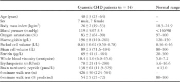
A total number of 14 adults with cyanotic congenital heart disease (seven female, seven male) were recruited from the Congenital Heart Disease Database at Royal Prince Alfred Hospital (RPAH), Sydney, Australia. All these patients had resting transcutaneous oxygen saturations ⩽90%. Exclusion criteria included major physical or intellectual impairment. Cyanotic congenital heart disease patient characteristics are shown in Table 1. Age- and sex-matched subjects were recruited from the general community to act as controls for endothelial function assessment and plasma assays. They were required to be healthy, normotensive, non-smokers not performing more than two regular exercise sessions per week, and not on any regular medications.
Cyanotic CHD subject characteristics.

CHD=congenital heart disease
Values are expressed as mean±SEM (range)
Simplified cardiac anatomical characteristics for cyanotic congenital heart disease patients included atrioventricular septal defect with Eisenmenger physiology (n=3), ventricular septal defect with Eisenmenger physiology (n=2), ventricular septal defect with severe pulmonary stenosis (n=2, both with Blalock–Taussig shunt, one with Glenn shunt), pulmonary atresia with ventricular septal defect and major aorta to pulmonary artery collaterals (n=3, one palliated with “lay open” right ventricular outflow tract), pulmonary atresia with intact septum (n=1, with Glenn shunt), pulmonary atresia with single ventricle (n=1, with Potts shunt), and pulmonary stenosis with essentially single ventricle (n=2, one with Blalock–Taussig shunt, one with Glenn shunt).
All patients provided informed written consent and the Sydney Local Health District Ethics Review Committee (RPAH Zone) approved the study.
Study design
For cyanotic congenital heart disease patients, assessment comprised endothelial function testing including brachial artery flow-mediated dilatation, peripheral artery tonometry, retinal flicker testing together with plasma assays for asymmetric dimethylarginine, endothelin-1, 3-nitrotyrosine, and high-sensitivity C-reactive protein. Endothelial progenitor cells were characterised using flow cytometry and day 7 harvest-cell culture. Functional capacity was assessed with a 6-minute walk test (method outlined in Supplementary material). Endothelial assessment and plasma assays were carried out in age- and sex-matched controls. All patients were free from infection, inflammatory illness, and episodes of clinical decompensation in the month preceding testing. Female patients were tested in the first 7 days of their menstrual cycle. For cyanotic congenital heart disease patients who had amenorrhoea (n=2), matched controls had to undergo endothelial testing during the menstrual phase of their cycle. Cyanotic congenital heart disease patients also underwent haematological and biochemical assessment that included a full blood count, NT pro-brain natriuretic peptide, erythropoietin, creatinine, iron studies, and whole blood viscosity (Brookfield Digital Viscometer, rotational speed of 12 rpm, shear rate 90/second).
Owing to functional limitation and work-related time commitments, three cyanotic congenital heart disease study subjects chose to undergo only some aspects of testing.
Brachial artery flow-mediated dilatation
Brachial artery flow-mediated dilatation was assessed using our previously described method summarised in online Supplementary Methods. The right brachial artery was used for testing except in patients who previously had a pulmonary to subclavian artery shunt procedure on that side; in those patients, the left brachial artery was tested instead. One patient’s data could not be analysed because of technical issues; a second patient was on sildenafil and therefore nitrate-mediated dilatation was not assessed in that patient.
Retinal vascular response to flickering light
Pupils were fully dilated with 1% tropicamide (Alcon Laboratories, Frenchs Forrest, Australia). Digital fundus images and reactivity parameters of retinal arterioles and veins were obtained using a Retinal Vessel Analyzer (Imedos Systems (UG), Jena, Germany).Reference Polak, Dorner and Kiss 7 Retinal diameters were measured at a sampling rate of 25 Hz. A 500-µm segment of the superior temporal artery and vein was chosen 1.5–2 disc diameters from the margin of the optic disc. Once the image had been optimised, 50 seconds of baseline measurements were taken followed by three cycles of flickering light (20 seconds each, 12.5 Hz, 80 seconds recovery time). The baseline diameters of the vessels were recorded and used to calculate values for maximal dilatation, a response mediated via nitric oxide-dependent pathways.Reference Dorner, Garhofer and Kiss 8
Peripheral artery tonometry
Endothelial function of the distal arterial bed of the fingertip was measured using an Endo-PAT device (Itamar Medical, Caesarea, Israel) to assess the change in digital pulse wave amplitude in response to reactive hyperaemia, expressed as the reactive hyperaemia PAT index. This method has been described in detail elsewhereReference Celermajer 9 and is summarised in online Supplementary Methods.
Plasma assays of vascular biomarkers
To understand contributing factors related to endothelial function such as endothelium-derived vasodilators, vasocontrictors, oxidative stress, and inflammation, we chose four circulating markers for measurement. Asymmetric dimethylarginine is a potent nitric oxide synthase inhibitor,Reference Vallance, Leone, Calver, Collier and Moncada 10 endothelin-1 is a potent endothelium-derived vasocontrictor,Reference Hasdai, Holmes, Garratt, Edwards and Lerman 11 3-nitrotyrosine reflects both nitric oxide bioavailability and oxidative stress,Reference Halliwell, Zhao and Whiteman 12 and high-sensitivity C-reactive protein is an important acute phase reactant.Reference Anand, Latini and Florea 13 Methods are described in online Supplementary material.
One patient had insufficient plasma for testing owing to extreme erythrocytosis; he and his healthy matched control were not included in this aspect of testing.
Circulating endothelial progenitor cell analysis
A volume of 10 ml of venous blood was collected from patients in the morning after an overnight fast into K3EDTA-anticoagulated vacutainer tubes (BD Biosciences, Oxford, United Kingdom).
Flow cytometric assessment of circulating endothelial progenitor cells
Flow cytometry was used to quantify endothelial progenitor cells and their subsets in both cyanotic and acyanotic healthy controls after staining with the fluorochrome-conjugated monoclonal antibodies CD45-PECy7, CD34-PECy5, KDR-CFS, and CD133-PE.
Using various combinations of markers, we analysed the different subsets, focusing on CD34+CD45− and CD34+CD45dim-expressing cells, further immunophenotyping this population as also KDR+ only, CD133+ only, or coexpressing KDR+/CD133+. The number of progenitor cells and subsets were then expressed per million white blood cells.
See Supplementary material for more detailed methods and sequential gating strategy.
Endothelial progenitor cell culture
Early endothelial progenitor cells (15) were isolated from peripheral blood using previously described methodsReference Lin, Weisdorf, Solovey and Hebbel 14 adjusted to compensate for marked increased packed cell volume in cyanosed patients as detailed in Supplementary material.
Cultured cells were assessed for the ability to ingest AcLDL (4 μg/ml) (Invitrogen, Carlsbad, United States of America) and to bind fluorescein isothiocyanate – Ulex europaeus agglutinin lectin (10 μg/ml, Sigma, St. Louis, United States of America). A patient and one matched control were removed from the analysis owing to technical issues.
Statistical analysis
All variables are expressed as mean±SEM. Data were tested for normality using a Shapiro–Wilk test. As data were normally distributed, unpaired Student’s t-tests were used for between-group analysis. We prospectively defined a primary parameter of interest for each category of physiological variable studied. For vascular function testing, this was arterial flow or flicker-mediated dilatation; for biochemical testing this was endothelin-1; for endothelial progenitor cell characterisation, this was circulating numbers of CD34+CD45dimKDR+CD133+ cells as a proportion of total white cells. All the other t-tests were adjusted for the multiple comparisons made by the Hochberg modification of the Bonferroni procedure.Reference Hochberg 15 Primary variables together with 6-minute walk distance and oxygen saturations were compared using bivariate analysis to characterise potential relationships; Spearman’s rank correlation was used because of the small sample size and non-linear distribution of data (p<0.1 are reported for correlations). SPSS statistics Data Editor (IBM Corporation, New York, United States of America) was used for statistical calculations. A two-tailed p-value ⩽0.05 was considered statistically significant.
Results
Study group characteristics
Patient characteristics together with relevant haematological and biochemical results are summarised in Table 1. All patients were NYHA Class II or III (n=8 were Class II and n=6 were Class III).
From the cyanotic congenital heart disease patients, seven male and five female had flow-mediated dilatation, peripheral artery tonometry, and endothelial progenitor cell analysis. Mean age was 41±3 years compared with 43±3 years in healthy controls (p=0.95). Blood pressure was similar between groups (systolic: 119±3 versus 118±3 mmHg, p=0.76 and diastolic: 67±3 versus 66±5 mmHg, p=0.99). Total white cell count did not differ significantly between the groups (5.71±0.44 in cyanosis versus 5.75±0.55×10⁹/L, p=0.95). There were significant differences in resting heart rate (71±3 in cyanosis versus 57±2 bpm in controls, p=0.001), body mass index (23.6±0.9 in cyanosis versus 26.4±1.2 kg/m2 in controls, p=0.03), red cell count, and platelets (6.98±0.34 in cyanosis versus 4.75±0.19×10¹²/L in controls and 148±12 versus 238±13×109/L, respectively, p<0.0001 for both).
The seven male and six female cyanotic congenital heart disease patients who had retinal flicker testing were 40±3 years compared with 38±3 years in matched controls (p=0.77). Blood pressure did not differ significantly between these groups (systolic: 119±3 versus 123±4 mmHg, p=0.42 and diastolic: 68±3 versus 74±4 mmHg, p=0.24). Body mass index was 22.5±0.8kg/m2 in the cyanosed group and 25.8 kg/m2 in controls (p=0.22).
Four of the cyanotic congenital heart disease patients who underwent endothelial function testing and endothelial progenitor cell analysis were on potentially vasoactive medications (bosentan (n=2), bosentan combined with sildenafil (n=1), and sitaxentan (n=1)); there were no significant differences in the endothelial function measures assessed between these patients and those not on vasoactive medications (key data demonstrated in Supplementary material). None of the subjects took angiotensin-converting enzyme inhibitors or statins. Two patients took aspirin, one was prescribed aspirin and persantin, and four were on warfarin.
Brachial artery flow-mediated dilatation
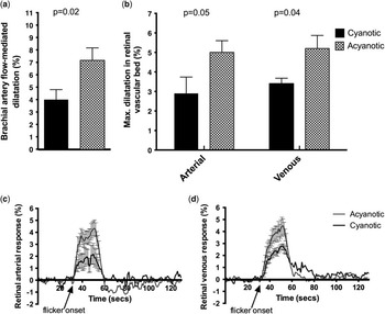
Flow-mediated dilatation was significantly impaired in cyanotic congenital heart disease compared with controls as shown in Figure 1a (4.0±0.8 versus 7.2±1.0%, p=0.019, n=11). Nitrate-mediated dilatation, a nitric oxide-independent response, was not significantly different between groups (15.8±1.9 versus 17.2±2.0%, p=1.0, n=10). Baseline brachial artery flow and diameter did not differ significantly between cyanosed patients and controls (177.0±36.9 versus 118.0±19.1 ml/second, p=0.51 and 3.86±0.26 versus 3.81±0.19 mm, p=0.89).
Retinal vascular response to flickering light
Patients with cyanotic congenital heart disease had significantly reduced maximal dilatory responses to flickering light as shown in Figures 1b–d (2.9±0.8 versus 5.0±0.6%, p=0.05 for arterial and 3.4±0.3 versus 5.2±0.7%, p=0.04 for venous, n=13 for both).
Endothelial function testing in cyanosed patients versus acyanotic healthy controls. Figure 1a demonstrates brachial artery flow-mediated response (n=11), Figure 1b represents maximal flicker-induced dilatory response in the retinal vessels (n=13) and Figures 1c and 1d demonstrate mean change in retinal vessel diameter in response to 20 seconds of flickering light. Error bars shown are SEM.

Peripheral artery tonometry
There was a tendency towards reduced PAT index detected in cyanosed adults compared with controls (1.68±0.12 versus 2.00±0.16, p=0.12, n=12).
Plasma assays of vascular biomarkers
Plasma levels of endothelin-1 and asymmetric dimethylarginine were higher in cyanosed adults compared with healthy controls (3.0±0.6 versus 1.1±0.1 pg/ml, p=0.004 and 0.68±0.05 versus 0.52±0.02 μmol/, p=0.03, respectively, n=11 for both). Although levels of 3-nitrotyrosine were higher in cyanosed patients, these differences were not statistically significant (415.4±196.3 versus 155.2±32.4 pg/ml, p=0.4, n=11). High-sensitivity C-reactive protein did not differ significantly between the two groups (3.3±1.1 versus 2.9±0.9 μg/ml, p=0.81, n=11).
Circulating endothelial progenitor cells
Flow cytometric assessment of circulating endothelial progenitor cells
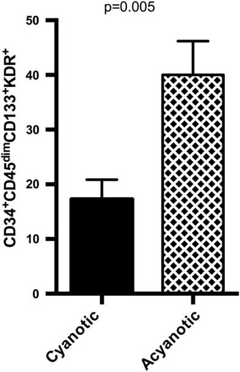
The number of circulating CD34+CD45dimKDR+CD133+ cells were lower in cyanosed patients than in controls (17±4 versus 40±6 per million white blood cells, p=0.005, n=11; see Figure 2), whereas counts for CD34+CD45dimCD133+ and CD34+CD45dimKDR+ cells did not differ significantly between groups (292±30 versus 372±24 per million white blood cells, p=0.196 and 303±35 versus 324±36 per million white blood cells for cyanotic and acyanotic, p=0.68, n=11 for both). The number of CD34+ cells did not differ significantly between cyanosed patients and controls (590±29 versus 550±57 per million white blood cells, p=1.0, n=11).
Flow cytometric assessment of circulating endothelial progenitor cells in cyanosed patients versus acyanotic healthy controls. Figure 2 demonstrates the number of circulating CD34+CD45dimCD133+KDR+ cells per million white blood cells in cyanosed patients and healthy controls (n=11). Error bars shown are SEM.

Endothelial progenitor cell culture
The number of early endothelial progenitor cells collected from culture at day 7 tended to be lower in patients with cyanotic congenital heart disease compared with healthy controls (85±13 versus 118±11 cells per 10× microscopy field, p=0.18, n=11).
Bivariate correlation
The number of circulating CD34+CD45dimKDR+CD133+ cells tended to correlate with oxygen saturations (R=0.58, p=0.06) and flow-mediated dilatation (R=0.5, p=0.09) and endothelin-1 negatively correlated with 6-minute walk distance (R=−0.87, p=0.001). Results of clinical endothelial function testing did not correlate significantly with 6-minute walk distance or oxygen saturations.
Discussion
Our investigation into the effects of chronic cyanosis on endothelial function suggests that a widespread endotheliopathy exists in this setting. Cyanosed adults had impaired flow-mediated dilatation in the brachial artery, a large conduit vessel; an attenuated flicker response in the small arteries and veins of the retina; and a tendency towards abnormal endothelial function in the distal arterial bed of the fingertip. Plasma assays showed increased levels of asymmetric dimethylarginine and endothelin-1 consistent with an imbalance between vasodilator and vasoconstrictor-related endothelial factors. Levels of circulating endothelial progenitor cells were significantly reduced in the setting of chronic cyanosis and this may have contributed, at least partly, to the abnormal vascular responses observed.
Adults with chronic cyanosis generally have a poor prognosis.Reference Engelfriet, Boersma and Oechslin 1 The prevalence of a documented cerebrovascular event has been reported to be around 14%;Reference Engelfriet, Boersma and Oechslin 1 , Reference Ammash and Warnes 16 a proportion of these are the result of venous sinus thrombosis and lacunar infarction that could potentially be related to endothelial dysfunction. Similarly, radiological series have demonstrated that 29% of adults with Eisenmenger syndrome have moderate to massive pulmonary artery thrombosesReference Perloff, Hart, Greaves, Miner and Child 17 that might also have important clinical implications. A large amount of evidence has accumulated emphasising the important role of endothelial dysfunction in the pathogenesis of vascular events in other diseases,Reference Lerman and Zeiher 18 as well as a significant association with functional status, morbidity, and mortality in chronic heart failure.Reference Fischer, Rossa and Landmesser 19 Increased understanding of this relationship has led to the investigation of therapies that might interrupt or attenuate this pathophysiology.Reference Andrews, Prasad and Quyyumi 20 , Reference Rector, Bank and Mullen 21 The clinical implications of endothelial dysfunction in the setting of cyanotic congenital heart disease have, however, been largely neglected until now.Reference Diller, van Eijl and Okonko 6 , Reference Oechslin, Kiowski, Schindler, Bernheim, Julius and Brunner-La Rocca 22
The ability of vessels to respond to a stimulus known to act via nitric oxide-dependent pathways reflects endothelial function. Our research demonstrated a markedly impaired flow-mediated dilatory response in cyanosed congenital heart disease patients similar to our other assessments of nitric oxide-dependent vascular responsiveness. To the best of our knowledge, in this setting, only single aspects of endothelial function have been investigated previously. Oechslin et alReference Oechslin, Kiowski, Schindler, Bernheim, Julius and Brunner-La Rocca 22 assessed endothelial function in 11 patients using venous occlusion plethysmography, reflecting microvascular reactivity. Response to acetylcholine, which acts via a nitric oxide-dependent mechanism, was markedly reduced in the cyanotic congenital heart disease group. Furthermore, blocking endothelial nitric oxide synthase with l-NG-monomethyl arginine produced less change in the cyanosed group, supporting the notion of reduced bioavailability of nitric oxide. The degree of hypoxaemia correlated with the degree of endothelial dysfunction. Another study on 13 patients with cyanotic congenital heart disease did not find any impairment of brachial flow-mediated dilatation compared with controls either at baseline or after an infusion of vitamin C to evaluate the role of reactive oxygen species.Reference Pedersen, Schmidt and Mortensen 23 This study did not match female patients for day of menstrual cycle but tested out of the menstrual phase.
Dynamic assessment of the retinal vessels has not been reported previously in the cyanotic congenital heart disease cohort but has been shown to reflect cardiovascular risk factors in other populations.Reference Nguyen, Kawasaki and Wang 24 , Reference Schiel, Vilser, Kovar, Kramer, Braun and Stein 25 Given the ophthalmic vessels’ close anatomical relationship with the cerebral circulation and the increased risk of stroke in our study population, our findings of attenuated retinal endothelial function may be of clinical relevance.
We did not demonstrate a statistically significant impairment of endothelial function in the distal arterial bed in the fingertip, as assessed with peripheral artery tonometry, in cyanotic patients, although our results were suggestive of blunted responsiveness. This may be related to the small sample size. Alternatively, among the patients we studied, the pulse-related volume changes in the distal arterial bed of the fingertip may be relatively less affected compared with our other diameter-based measurements (flow-mediated dilatation and retinal flicker testing), because of a differing run-off arrangement or increased arteriolar density in the setting of chronic cyanosis. We did not find a significant correlation between these measures of endothelial function and functional status; however, given our sample size, an important association should not be discounted. Long-term data are needed to characterise the relationship, if any, between endothelial dysfunction and morbidity and mortality in this group.
We have shown for the first time that numbers of circulating endothelial progenitor cells are reduced, and proliferative capacity in cell culture tends to be impaired in a broad group of cyanotic congenital heart disease patients. Consistent with our results, Diller et alReference Diller, van Eijl and Okonko 6 demonstrated that the number of circulating endothelial progenitor cells were low in 41 patients with Eisenmenger syndrome compared with healthy controls, and reduced numbers correlated with NYHA class, 6-minute walk distance, and plasma brain natriuretic peptide levels. Cultured cells also demonstrated a reduced capacity to form tube-like structures. It is likely that the subset of CD34+45dim133+KDR+ cells that we identified specifically reflect circulating putative endothelial progenitor cell numbers.Reference Fadini, Baesso, Albiero, Sartore, Agostini and Avogaro 26 In our series, we found that oxygen saturations tended to correlate with circulating endothelial progenitor cell numbers. It is possible that worsening hypoxia inhibits endothelial progenitor cell production either directly or via associated haemorrheological changes, further affecting the endothelium in the setting of cyanosis. We also found that a relationship generally existed between the numbers of circulating endothelial progenitor cells and several aspects of functional endothelial assessment; this may reflect abnormal endothelial repair capabilities related to low endothelial progenitor cell numbers.
Our results demonstrated increased levels of asymmetric dimethylarginine and endothelin-1 in a broad group of patients with cyanotic congenital heart disease that might reflect and/or contribute to impaired endothelial function. Asymmetric dimethylarginine is a cellular by-product and potent nitric oxide synthase inhibitor and has been previously found to be elevated in patients with Eisenmenger syndrome.Reference Diller, van Eijl and Okonko 6 Endothelin-1 is a powerful vasoconstrictor and is released in response to elevated vascular shear stressReference Hasdai, Holmes, Garratt, Edwards and Lerman 11 that may underlie our findings of raised levels in the setting of cyanosis. Others have previously shown that nitric oxide metabolites are elevated in adults with cyanotic congenital heart disease.Reference Diller, van Eijl and Okonko 6 , Reference Han, Perloff and Liao 27 This probably reflects a compensatory increase in nitric oxide production despite impaired endothelial function, suggesting reduced bioavailability, possibly the result of nitric oxide scavenging related to increased levels of haemoglobin and reactive oxygen species. In our study, the levels of 3-nitrotyrosine were higher in the setting of cyanosis but not significantly so.
The pathophysiology underlying the dysfunctional endothelium in the setting of cyanotic congenital heart disease is complex. As a group, our patients were heterogeneous with regard to cardiac anatomy and thus their reason for cyanosis; Eisenmenger physiology, low pulmonary blood flow, or right to left shunt without Eisenmenger physiology may all result in cyanosis. Some degree of abnormal cardiac function is universal, and pulmonary arterial hypertension exists in many, both conditions known to contribute to abnormal vascular function.Reference Katz, Hryniewicz and Hriljac 3 , Reference Wolff, Lodziewski, Bollmann, Opitz and Ewert 28 Oxygen is well recognised as a local regulator of vascular tone; acute hypoxia leads to compensatory vasodilatation,Reference Armstead 29 whereas prolonged hypoxia (>1 hour) leads to vasoconstriction probably via a nitric oxide-dependent mechanism, most likely downregulation of endothelial nitric oxide synthase.Reference Pape, Beuchard, Guillo, Allain and Bellissant 30 , Reference Toporsian, Govindaraju, Nagi, Eidelman, Thibault and Ward 31
In addition, in the setting of chronic cyanosis, compensatory elevation in haematocrit causes increased whole blood viscosity that imposes marked shear stress on vessel walls.Reference Tsai, Friesenecker, McCarthy, Sakai and Intaglietta 32 The effect of viscosity on blood vessels is, however, varied and alters, depending on the flow rate and diameter;Reference Pries, Neuhaus and Gaehtgens 33 therefore, it is likely that the effects of chronic erythrocytosis differ at the capillary level compared with that within larger calibre vessels. Furthermore, whereas an acute increase in wall shear stress leads to vasodilatation, chronic elevation leads to compensatory mechanisms that attenuate the load on the vessel wall, such as vascular remodelling to increase diameterReference Ogunshola, Djonov, Staudt, Vogel and Gassmann 34 and a blunted response to mechanical stimuli.Reference Belhassen, Pelle and Sediame 35
Our findings are of potential clinical relevance, given the known link between endothelial dysfunction and vascular events in a wide group of other conditions and the marked predisposition to serious vascular events in chronic cyanosis. Many of the factors that likely contribute to the dysfunctional endothelium – hypoxia, haemorrheological alterations, etc. – are not modifiable in the setting of cyanotic congenital heart disease, but it may be possible to optimise endothelial function in other ways. Agents exist that increase the bioavailability of nitric oxide, such as l-arginine and antioxidants – vitamin C, flavonoids, etc. Numerous pharmacologic agents have been shown to improve endothelial function in other diseases; blockade of endothelin receptors, for example, may hold promise. In addition, progenitor cell therapy to facilitate endothelial repair is currently an area of vigorous research.
Limitations
This current report was limited by small subject numbers; however, significant differences in numerous types of endothelial assessment suggest that extensive derangement exists and make a type I error unlikely. Owing to the multitude of factors that likely contribute to endothelial dysfunction in cyanotic congenital heart disease, isolating a primary or most significant mechanism is difficult. Furthermore, the group that we studied was heterogeneous with differing underlying cardiac anomalies and vascular arrangements; nevertheless, we have demonstrated significant vascular dysfunction is present, generally, in this group of cyanosed adults.
Although other functional assays for endothelial progenitor cells exist, such techniques require the collection of larger volumes of blood than was clinically appropriate in our study population; thus, we were limited to the assessment of early endothelial progenitor cell culture.
Conclusions
Endothelial function is impaired in the systemic arteries and retinal vessels in adults with cyanotic congenital heart disease, suggesting a widespread endotheliopathy that likely results from a complex interplay of factors. In addition to deranged haemorrheology and hypoxaemia, increased asymmetric dimethylarginine, endothelin-1, and oxidative stress are all possible contributors. Circulating numbers of endothelial progenitor cells are reduced that may also be significant for disease pathogenesis.
Acknowledgements
The authors thank Dr Lissa Spencer, Physiotherapy Department, Royal Prince Alfred Hospital, for her assistance with 6-minute walk tests and Dr Phil Munoz, Respiratory Department, Royal Prince Alfred Hospital, for his assistance with lung function testing.
Conflicts of Interest
None.
Financial Support
Professor Stuart Graham has received financial support from the Hillcrest Foundation and Novartis Australia.
Supplementary Material
To view supplementary material for this article, please visit http://dx.doi.org/10.1017/S1047951114000262.