In diurnal primates, the short-wavelength-sensitive (S) cones constitute only 5–10% of the cone mosaic (DeMonasterio et al., Reference DeMonasterio, Schein and McCrane1981; Wikler & Rakic, Reference Wikler and Rakic1990; Martin & Grunert, Reference Martin and Grunert1999). Their peak spectral sensitivity is well separated from those of the middle-wavelength-sensitive (M) and the long-wavelength-sensitive (L) cones (Nathans, Reference Nathans1999). Due to chromatic aberrations, light of short wavelength carries little information at high spatial frequencies. Based on these features, it has been proposed that the primary function of S-cones is coding color information (De Valois et al., Reference De Valois, Cottaris, Elfar, Mahon and Wilson2000; Hashemi-Nezhad et al., Reference Hashemi-Nezhad, Blessing, Dreher and Martin2008). However, psychophysical and brain imaging studies have shown that the S-cone signals may also contribute to the coding of other visual features, such as motion, luminance, and orientation (Stockman et al., Reference Stockman, MacLeod and DePriest1991; Gegenfurtner & Hawken, Reference Gegenfurtner and Hawken1995; Sumner et al., Reference Sumner, Anderson, Sylvester, Haynes and Rees2008). Therefore, understanding the cortical processing of the S-cone signals is critical for understanding how color and other features are represented in the cortex.
The majority of the cortical projections from the lateral geniculate nucleus (LGN) terminate in the striate cortex (V1), although a minority of them reach extrastriate areas (e.g., Benevento & Yoshida, Reference Benevento and Yoshida1981; Bullier & Kennedy, Reference Bullier and Kennedy1983; Lysakowski et al., Reference Lysakowski, Standage and Benevento1988; Weller et al., Reference Weller, Steele and Kaas2002; Sincich et al., Reference Sincich, Park, Wohlgemuth and Horton2004). It is therefore reasonable to assume that the main portion of the S-cone signals is processed in V1 before they reach other areas, and only a small portion of these signals may bypass V1. This review focuses on the processing of the S-cone signals in areas V1 and V2.
The S-cone inputs from the LGN to V1
The S-cone signals are mostly carried by the koniocellular (K) layers in the LGN, with little or minor presence in the parvocellular (P) or magnocelluar (M) layers (see the review by Hendry & Reid, Reference Hendry and Reid2000; Chatterjee & Callaway, Reference Chatterjee and Callaway2002; Sun et al., Reference Sun, Smithson, Zaidi and Lee2006; Hashemi-Nezhad et al., Reference Hashemi-Nezhad, Blessing, Dreher and Martin2008). The projections from the K layers to V1 terminate in layer 4A and in the cytochrome oxidase (CO) blobs of layers 2/3 (see the reviews by Casagrande, Reference Casagrande1994; Hendry & Reid, Reference Hendry and Reid2000). However, the K layers contain several types of cells, not all of them carrying the S-cone signals (see the reviews by Casagrande, Reference Casagrande1994; Hendry & Reid, Reference Hendry and Reid2000). So the precise distribution of the LGN terminals in V1 that carry the S-cone signals remained unknown until it was described by Chatterjee and Callaway (Reference Chatterjee and Callaway2003). In that study, the distribution of the LGN terminals in V1 that carry various cone-specific signals was mapped after the neuronal activity within V1 was silenced by a GABA agonist. Their results suggested that the terminals that carry S-on signals innervate layer 4A and lower layer 3, whereas the terminals that carry S-off signals innervate lower layer 4A (Fig. 1A). The terminals that carry the nonopponent and opponent L- and M-cone signals were found in layers 4Cα and 4Cβ, respectively. Based on their results from a small number of recording sites, Chatterjee and Callaway (Reference Chatterjee and Callaway2003) hypothesized that the S-on terminals in layer 4 are uniformly distributed, whereas the S-on terminals in lower layer 3 and S-off terminals in lower layer 4A are confined to small patches (Fig. 1A). Further studies with more recording sites are needed to test the patchiness or lack of it associated with these terminals. Given that the middle and upper parts of the CO blobs also receive direct projections from the LGN (Fitzpatrick et al., Reference Fitzpatrick, Itoh and Diamond1983) and that these projections are largely from the koniocellular layers (Hendry & Yoshioka, Reference Hendry and Yoshioka1994), it is possible that S-on and/or S-off terminals also innervate these locations but were not encountered by Chatterjee and Callaway (Reference Chatterjee and Callaway2003) due to their small number of recording sites in layers 2 and 3.
Anatomical pathways associated with the S-cone signals in V1. (A) A schematic diagram of the cone-specific projections from the LGN to V1. Terminals that carry S-off signals innervate layer 4A, whereas those carrying S-on signals innervate both layers 4A and 3. Terminals that carry opponent and nonopponent L/M-cone signals innervate layers 4Cβ and 4Cα, respectively. It was hypothesized that the S-on terminals in layer 3 and the S-off ones in layer 4 are concentrated in small patches. M, P, and K denote projections from the magnocellular, parvocellular, and koniocellular layers of the LGN, respectively. Reprinted by permission from Macmillan Publishers Ltd: Nature (Chatterjee and Callaway, Reference Chatterjee and Callaway2003), copyright 2003. (B) A schematic diagram of the distribution of axonal terminals originating from cells in layer 4A. Most of these terminals in layer 3 are clustered in the CO blobs, although they are missing in a minority of the blobs (marked with *). There are also extensive connections between cells within layer 4A. From Yoshioka et al. (Reference Yoshioka, Levitt and Lund1994). Copyright 1994, Cambridge University Press.

Since layer 4A is the major recipient of the LGN terminals that carry the S-cone signals, the efferent pattern of the cells in this layer may determine the spatial distribution of the next stage of the processing of the S-cone signals. One study based on microinjection of an anatomical tracer suggested that cells in layer 4A project mainly to the blobs in layers 2/3, although a minority of the blobs do not receive this projection (Fig. 1B) (Yoshioka et al., Reference Yoshioka, Levitt and Lund1994). This projection pattern, as well as the direct projections from the K layers to the blobs, suggests that the blobs play a prominent role in the cortical processing of the S-cone signals immediately after layer 4A. Cells in the interblob regions of layers 2/3 may receive weaker S-cone signals via their dendrites in blobs (Hubener & Bolz, Reference Hubener and Bolz1992; Malach, Reference Malach1992). However, the pattern of connectivity depicted in Fig. 1B is inconsistent with the results of two other studies (Lachica et al., Reference Lachica, Beck and Casagrande1992; Sawatari & Callaway, Reference Sawatari and Callaway2000). Thus, the question of whether the blobs receive more S-cone signals from layer 4A than the interblob regions of layers 2/3 remains controversial.
Strength of the S-cone inputs to cells in V1
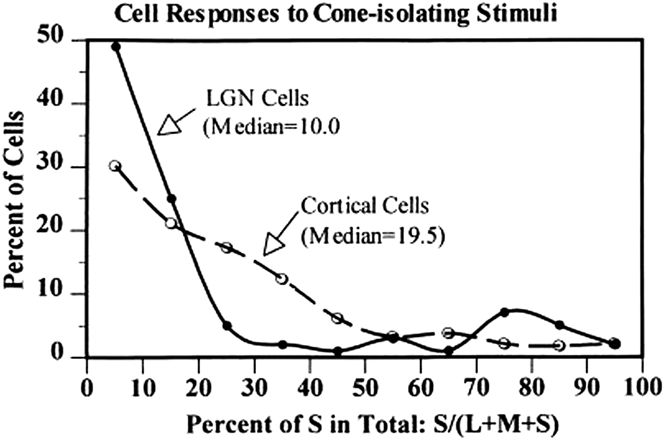
Although the S-cone signals are largely confined to the K layers in the LGN, they are more widespread in V1. De Valois et al. (Reference De Valois, Cottaris, Elfar, Mahon and Wilson2000) compared the strength of the S-cone inputs across cells in V1 and LGN (Fig. 2). They measured the responses of each cell to gratings that stimulated L-, M-, or S-cones only. In Fig. 2, the abscissa represents the S-cone weight, which is the response magnitude elicited by an S-cone-isolating grating, divided by the sum of response magnitudes that were elicited by all cone-isolating gratings. The ordinate represents the percentage of cells with a given S-cone weight. The solid line represents the distribution across cells in the LGN, and the dashed line shows the distribution in V1. The bimodal distribution in the LGN reflects the existence of two distinct populations of cells in terms of the S-cone weights. The vast majority of cells in the M and P layers had small S-cone weights, whereas many cells in the K layers had large S-cone weights. The distribution in V1 suggests that the relative separation between S- and L/M-cone inputs was lost in V1, and most cells were driven robustly by all three cone inputs. The median S-cone weight in V1 was twice as large as that in the LGN. In addition, the continuous distribution of S-cone weights in V1 suggests that V1 contains color-selective cells that are tuned to various color directions, as suggested by many other studies (reviewed by Gegenfurtner & Kiper, Reference Gegenfurtner and Kiper2003; Lennie & Movshon, Reference Lennie and Movshon2005). The activities of neurons that combine S- and L/M-cone inputs were also detected in human V1 in an fMRI study that differentiated patterns of response elicited by different combinations of the two opponent cone inputs (Goddard et al., Reference Goddard, Mannion, McDonald, Solomon and Clifford2010). Therefore, the results of many studies are consistent with the hypothesis of De Valois et al. (Reference De Valois, Cottaris, Elfar, Mahon and Wilson2000) that the S-cone signals are amplified in V1 relative to the LGN. This hypothesis is further supported by fMRI studies that directly compared the activity in human LGN and V1 in response to cone-specific stimuli (Mullen et al., Reference Mullen, Thompson and Hess2010; D’Souza et al., Reference D’Souza, Auer, Strasburger, Frahm and Lee2011). It is also consistent with imaging studies on marmosets (Buzas et al., Reference Buzas, Szmajda, Hashemi-Nezhad, Dreher and Martin2008) and tree shrews (Johnson et al., Reference Johnson, Van Hooser and Fitzpatrick2010) that found widespread activation in V1 in response to S-cone stimulation. Finally, an EEG study on human V1 found strong S-cone inputs to the mechanism underlying surround suppression (Xiao & Wade, Reference Xiao and Wade2010), although this mechanism was found to receive predominantly achromatic inputs in a study of single neurons in monkey V1 (Solomon et al., Reference Solomon, Peirce and Lennie2004).
Distributions of the relative S-cone weights across LGN cells and V1 cells. The LGN contains two distinct groups of cells, one with small S-cone weights and the other with large S-cone weights. There are few cells with intermediate weights. The first group corresponds to cells in the parvocellular and magnocellular layers of the LGN, which constitute the majority of LGN cells. The second group corresponds to cells in the koniocellular layers. V1 cells cannot be divided into distinct groups according to their S-cone weights, and the median weight is twice as large as that found in LGN cells. From De Valois et al. (Reference De Valois, Cottaris, Elfar, Mahon and Wilson2000). Copyright 2000, National Academy of Sciences, USA.
In the study by De Valois et al. (Reference De Valois, Cottaris, Elfar, Mahon and Wilson2000), the cone contrast for the S-cone-isolating grating was much higher than those for the M- and L-cone-isolating gratings. Because of this difference in cone contrast, the abscissa value in Fig. 2 may not reflect the true contribution of the S-cone inputs relative to other cone inputs to a given cell. To make the comparison more meaningful, some studies used comparable cone contrasts for different cone-isolating stimuli (Johnson et al., Reference Johnson, Hawken and Shapley2004) or normalized the cone weights with the respective contrasts (Lennie et al., Reference Lennie, Krauskopf and Sclar1990; Solomon & Lennie, Reference Solomon and Lennie2005; Hashemi-Nezhad et al., Reference Hashemi-Nezhad, Blessing, Dreher and Martin2008). These studies reported much smaller S-cone contribution to V1 cells than did the studies that did not take into account the difference in cone contrast (De Valois et al., Reference De Valois, Cottaris, Elfar, Mahon and Wilson2000; Conway & Livingstone, Reference Conway and Livingstone2006; Horwitz et al., Reference Horwitz, Chichilnisky and Albright2007). However, to determine the contribution of various cones to V1 activity in the natural visual environment, the distributions of various cone contrasts elicited by natural scenes also need to be taken into account. Such distributions are largely unknown as previous studies on natural scenes did not analyze the cone contrasts between a given surface and the adaptation color (Ruderman et al., Reference Ruderman, Cronin and Chiao1998; Nascimento et al., Reference Nascimento, Ferreira and Foster2002), which are what physiological studies usually use to normalize the responses.
Interactions between various cone inputs to individual cells in V1
In the LGN, the majority of S-cone sensitive cells are S-on cells that receive excitatory S-cone inputs and inhibitory L- and M-cone inputs. The rare S-off cells receive excitatory L-cone inputs opposed by inhibitory M- and S-cone inputs (Solomon & Lennie, Reference Solomon and Lennie2005; Tailby et al., Reference Tailby, Solomon and Lennie2008b). In V1, however, for both S-on and S-off cells that receive robust S-cone inputs and opponent L/M-cone inputs, the S-cone inputs usually take the sign of the M-cone inputs according to some studies (Conway, Reference Conway2001; Solomon & Lennie, Reference Solomon and Lennie2005; Conway & Livingstone, Reference Conway and Livingstone2006; Horwitz et al., Reference Horwitz, Chichilnisky and Albright2007). In addition, there seems to be a comparable number of S-on and S-off cells in V1 (De Valois et al., Reference De Valois, Cottaris, Elfar, Mahon and Wilson2000). These results suggest that the S-off signals from the LGN are amplified in V1 more than the S-on signals (Solomon & Lennie, Reference Solomon and Lennie2005) or that about half of the S-on signals from the LGN are inverted after amplification in V1 (De Valois et al., Reference De Valois, Cottaris, Elfar, Mahon and Wilson2000). However, while studies from one group found that the S-cone inputs had the same sign as the M-cone inputs in nearly all cells that received opponent L/M-cone inputs (Conway, Reference Conway2001; Conway & Livingstone, Reference Conway and Livingstone2006), other studies found either more exceptions to this tendency (Solomon & Lennie, Reference Solomon and Lennie2005; Horwitz et al., Reference Horwitz, Chichilnisky and Albright2007) or no such tendency at all (Cottaris & De Valois, Reference Cottaris and De Valois1998; De Valois et al., Reference De Valois, Cottaris, Elfar, Mahon and Wilson2000). The latter results were more consistent with a continuous distribution of the preferred colors across V1 cells (reviewed by Gegenfurtner & Kiper, Reference Gegenfurtner and Kiper2003; Lennie & Movshon, Reference Lennie and Movshon2005). As Conway (Reference Conway2001) and Conway and Livingstone (Reference Conway and Livingstone2006) focused on a subset of color-selective cells that responded well to a small cone-isolating spot, it is possible that the S- and M-cone inputs to this subset of cells are always aligned.
A V1 cell that receives opponent L/M-cone inputs has either a single-opponent or a double-opponent receptive field (Conway, Reference Conway2001; Conway & Livingstone, Reference Conway and Livingstone2006; Johnson et al., Reference Johnson, Hawken and Shapley2008; reviewed by; Shapley & Hawken, Reference Shapley and Hawken2011). In a single-opponent receptive field, the center receives opponent inputs from L- and M-cones and the surround receives inputs from only one cone type that provides the weaker drive to the center. A double-opponent receptive field contains two parts. Each part receives opponent inputs from L- and M-cones, and the signs of the cone inputs are opposite between the two parts. However, the precise spatial structure of a double-opponent receptive field is still hotly debated (reviewed by Conway et al., Reference Conway, Chatterjee, Field, Horwitz, Johnson, Koida and Mancuso2010; Shapley & Hawken, Reference Shapley and Hawken2011). Some of the originally identified double-opponent receptive fields might have a cone opponent center and a suppressive surround that are not cone opponent (Ts’o & Gilbert, Reference Ts’o and Gilbert1988; Solomon et al., Reference Solomon, Peirce and Lennie2004).
The S-cone signals are present in both single-opponent and double-opponent receptive fields, including various configurations of the latter (Johnson et al., Reference Johnson, Hawken and Shapley2004; Conway & Livingstone, Reference Conway and Livingstone2006). Therefore, both single-opponent and double-opponent receptive fields are potentially capable of representing the full gamut of colors, but the specific role that each type of receptive field plays in representing color remains unclear. One possibility is that a single-opponent receptive field encodes wavelength composition of the light reflected from a surface, whereas a double-opponent receptive field encodes chromatic contrast at the border between surfaces and may play an important role in color constancy (Gegenfurtner, Reference Gegenfurtner2003). The single-opponent receptive fields are unselective for orientation and are larger than other types of receptive fields that are orientation selective (Solomon et al., Reference Solomon, Peirce and Lennie2004). These characteristics could be the result of the cortical adaptation during evolution to the statistical structure of the visual world (Cecchi et al., Reference Cecchi, Rao, Xiao and Kaplan2010).
Many V1 cells that receive nonopponent L- and M-cone inputs also receive the S-cone inputs (Johnson et al., Reference Johnson, Hawken and Shapley2004; Solomon & Lennie, Reference Solomon and Lennie2005; Conway & Livingstone, Reference Conway and Livingstone2006; Horwitz et al., Reference Horwitz, Chichilnisky and Albright2007). In these cells, the S-cone inputs were either opposed to or aligned with those from L- and M-cones. The opponent inputs may originate from S-on cells in the LGN, which also receive opponent S- and (L + M)-cone inputs. V1 cells that receive opponent S- and (L + M)-cone inputs may contribute primarily to color vision.
The alignment of the inputs from all cone types may result from the merging between the S-cone signals and luminance signals in some cells. A previous study found that some cells in the magnocellular layers of the LGN also received aligned inputs from all cone types (Chatterjee & Callaway, Reference Chatterjee and Callaway2002), although the conclusion of that study was challenged later on methodological grounds (Sun et al., Reference Sun, Smithson, Zaidi and Lee2006; Hashemi-Nezhad et al., Reference Hashemi-Nezhad, Blessing, Dreher and Martin2008). In any case, V1 cells that receive aligned inputs from all cone types might be part of the pathway that sends the S-cone signals to area MT (Seidemann et al., Reference Seidemann, Poirson, Wandell and Newsome1999) and could mediate the S-cone’s contributions to luminance and motion-sensitive mechanisms (Stockman et al., Reference Stockman, MacLeod and DePriest1991; Gegenfurtner & Hawken, Reference Gegenfurtner and Hawken1995).
Dynamics of the S-cone signals in V1
In a study based on reverse correlation between spikes and uniform stimuli of various colors, it was found that among the responsive cells in V1, the majority received either opponent L/M-cone signals with a short latency (<96 ms) or S-cone signals with a long latency (>96 ms), or a combination of these two signals (Cottaris & De Valois, Reference Cottaris and De Valois1998). The latency was measured as the interval between stimulus presentation and the peak response. The longer latency of the S-cone signals relative to other cone signals was confirmed in another study (Conway & Livingstone, Reference Conway and Livingstone2006). This difference in latency might contribute to the difference in latency at the behavioral level between the responses to different cone-isolating stimuli (Smithson & Mollon, Reference Smithson and Mollon2004). Since this latency difference was not detected in the LGN (Tailby et al., Reference Tailby, Solomon and Lennie2008b), it is likely caused by processing at the input layers of V1.
In addition to the difference in latency, cells with or without the S-cone inputs also differed in the dynamics of their color tuning (Cottaris & De Valois, Reference Cottaris and De Valois1998). Cells without any S-cone input had a more stable color tuning over time than did those with the S-cone inputs. Taken together, the results of Cottaris and De Valois (Reference Cottaris and De Valois1998) suggest that relative to other cone signals, the S-cone signals go through a longer and more active stage of processing before exerting maximal impact on V1 cells. This processing stage might be responsible for the amplification of the S-cone signals in V1 (De Valois et al., Reference De Valois, Cottaris, Elfar, Mahon and Wilson2000). The neural substrate of this postulated processing stage remains unknown, but a possible candidate is the network of neurons in layer 4A that is formed through extensive intralaminar connections (Fig. 1B) (Yoshioka et al., Reference Yoshioka, Levitt and Lund1994).
In the study by Cottaris and De Valois (Reference Cottaris and De Valois1998), a minority of the cells received the S-cone inputs with very short latencies (<70 ms). Most of these cells were found in layer 6 and were assumed to mediate the contribution of the S-cone signals to the luminance and motion-sensitive mechanisms (Stockman et al., Reference Stockman, MacLeod and DePriest1991; Gegenfurtner & Hawken, Reference Gegenfurtner and Hawken1995). In contrast, cells that only received slower S-cone inputs were found mainly in the supragranular layers, whereas those receiving both the S-cone and L/M-cone inputs were found across all layers.
Several fMRI studies on humans have found that the V1 responses to S-cone-specific stimuli were tuned to a narrower bandwidth of temporal frequencies than were the responses to other cone-specific stimuli (Liu & Wandell, Reference Liu and Wandell2005; D’Souza et al., Reference D’Souza, Auer, Strasburger, Frahm and Lee2011). For instance, D’Souza et al. (Reference D’Souza, Auer, Strasburger, Frahm and Lee2011) found that while the V1 response to opponent L/M-cone signals remained relatively constant for temporal frequencies up to 12 Hz, the response to the S-cone signals declined sharply as the temporal frequency increased in the same range. In contrast, similar differences in temporal frequency tuning were not found in the LGN (D’Souza et al., Reference D’Souza, Auer, Strasburger, Frahm and Lee2011). These results are consistent with the finding that the latency of the S-cone signals in V1 cells is longer than that of other cone signals and support the hypothesis that the delay is caused by the S-cone processing mechanism in V1. However, another fMRI study did not find significant difference in temporal frequency tuning between V1 responses to the S-cone compared with the L/M-cone signals (Mullen et al., Reference Mullen, Thompson and Hess2010).
The relationship between cone signals and color perception
When the S-cone opponent cells that difference the S-cone signals from the sum of the L- and M-cone signals were first found in the LGN, they were thought to mediate our color perception along the blue-yellow axis, and the LGN cells that difference the L- and M-cone signals were thought to mediate our perception along the red-green axis (De Valois, Reference De Valois1965; De Valois et al., Reference De Valois, Abramov and Jacobs1966). However, a more recent study showed that these opponent-cell color axes do not precisely correspond to the perceptual red-green and blue-yellow axes (De Valois et al., Reference De Valois, De Valois, Switkes and Mahon1997). Instead, they correspond to the two “cardinal directions” along which psychophysically measured chromatic sensitivity can be adapted independently (Krauskopf et al., Reference Krauskopf, Williams and Heeley1982). The specificity of adaptation along the cardinal directions was taken as evidence suggesting that color is represented by two discrete postreceptoral mechanisms (Krauskopf et al., Reference Krauskopf, Williams and Heeley1982). However, later psychophysical studies with various measurements, including an reanalysis of the data of Krauskopf et al. (Reference Krauskopf, Williams and Heeley1982), suggested that there are many “higher order” color mechanisms that are tuned to various color directions that include both the cardinal and intermediate ones (reviewed by Eskew, Reference Eskew2009). This hypothesis is consistent with physiological findings that the majority of color selective cells in V1 prefer intermediate colors between the cardinal axes (Lennie et al., Reference Lennie, Krauskopf and Sclar1990; De Valois et al., Reference De Valois, Cottaris, Elfar, Mahon and Wilson2000; Wachtler et al., Reference Wachtler, Sejnowski and Albright2003; Hass & Horwitz, Reference Hass and Horwitz2013). More recent studies suggested that the independent adaptation along the cardinal directions occurs at the geniculate-input layers of V1 where the S/(L + M) and L/M opponent signals are separated (Tailby et al., Reference Tailby, Solomon, Dhruv and Lennie2008a; Stoughton et al., Reference Stoughton, Lafer-Sousa, Gagin and Conway2012).
The studies reviewed above are consistent with a multistage model that contains a cortical stage in which the S-cone opponent signals are amplified and combined with the opponent L/M signals (Fig. 3) (De Valois & De Valois, Reference De Valois and De Valois1993). According to this model, the S-cone signals modulate the cortical activity in V1 that is predominantly determined by the opponent L/M signals in trichromatic primates. This hypothesis is consistent with a recent study on trichromatic marmosets that showed a predominant role of the opponent L/M signals in the response of the color patches in V1 (Valverde Salzmann et al., Reference Valverde Salzmann, Bartels, Logothetis and Schuz2012). It is also consistent with our recent study of response patterns in V1 to various uniform colors (Xiao et al., Reference Xiao, Kavanau, Bertin and Kaplan2011). When the response patterns are divided into two groups according to their similarity, one group is usually associated with hues that have a positive L–M cone contrast, whereas the other group is associated with hues that have a negative L–M cone contrast. This bipartite division is not correlated with the value or sign of the S-cone contrast. The S-cone contrast seems to fine-tune the response pattern within each group, as predicted by the multistage model. Interestingly, a similar bipartite analysis of color names across many languages (Lindsey & Brown, Reference Lindsey and Brown2006) found a universal division between “warm” and “cool” colors, suggesting a fundamental difference between these two groups of colors. These two groups of colors also have opposite signs of L–M cone contrasts (Xiao et al., Reference Xiao, Kavanau, Bertin and Kaplan2011), supporting the view that the S-cone signals play a largely modulatory role on the opponent L/M-cone signals in producing color perception of trichromatic subjects (De Valois & De Valois, Reference De Valois and De Valois1993; De Valois et al., Reference De Valois, Cottaris, Elfar, Mahon and Wilson2000).
The transformation from cone-opponent signals in the LGN to a continuous representation of color in visual cortex. The S-cone signals from the LGN are amplified in V1 relative to the L/M-cone signals, as symbolized by the change in the line width that denotes the signal strength. According to the multistage model by De Valois and De Valois (Reference De Valois and De Valois1993), the S-cone signals play a modulatory role on the opponent L/M-cone signals in V1. As a result, chromatic preferences of V1 neurons cover the full gamut of color.
Since CO blobs in V1 receive direct projections from the koniocellular layers of the LGN, which carry the S-cone signals, CO blobs may play a particularly important role in processing color information (Livingstone & Hubel, Reference Livingstone and Hubel1984). This hypothesis is supported by studies on macaque monkeys using imaging of intrinsic optical signals. In one study, CO blobs were found to be co-localized with color-preferring regions that were preferentially activated by equiluminant chromatic stimuli (Lu & Roe, Reference Lu and Roe2008). In another study, these color-preferring regions were found to contain hue maps in which perceptually similar hues elicited peak responses at nearby locations (Xiao et al., Reference Xiao, Casti, Xiao and Kaplan2007). The finding of hue maps in color-preferring regions suggests clustering of neurons according to their cone inputs in these regions, a suggestion that was confirmed by a preliminary study using two-photon calcium imaging (Chatterjee et al., Reference Chatterjee, Ohki and Reid2008; Conway et al., Reference Conway, Chatterjee, Field, Horwitz, Johnson, Koida and Mancuso2010).
A few fMRI studies suggest that human V1 also contains an inhomogeneous, spatial distribution of hue preferences, as stimulus hue can be decoded from the spatial pattern of response in V1 (Brouwer & Heeger, Reference Brouwer and Heeger2009; Parkes et al., Reference Parkes, Marsman, Oxley, Goulermas and Wuerger2009; Goddard et al., Reference Goddard, Mannion, McDonald, Solomon and Clifford2010). This inhomogeneity was taken as an evidence for the existence of hue maps in human V1 (Parkes et al., Reference Parkes, Marsman, Oxley, Goulermas and Wuerger2009), although the low spatial resolution of fMRI prevented it from excluding other possible spatial arrangement of hue preferences. One of these fMRI studies reported that the similarity between the response patterns in human V1 did not follow that between the stimulus hues, which seems to contradict the existence of hue maps in V1 (Brouwer & Heeger, Reference Brouwer and Heeger2009). One possible explanation for the apparent discrepancy between Brouwer and Heeger (Reference Brouwer and Heeger2009) and Xiao et al. (Reference Xiao, Casti, Xiao and Kaplan2007) is that while Xiao et al. (Reference Xiao, Casti, Xiao and Kaplan2007) found hue maps in the color-preferring regions of supragranular layers, the results of Brouwer and Heeger (Reference Brouwer and Heeger2009) were based on responses that were averaged across various regions and layers due to the large size of each voxel.
Hue maps or spatial clustering of hue preferences was also found in color-preferring regions of higher visual areas in macaque monkeys (Zeki, Reference Zeki1980; Xiao et al., Reference Xiao, Wang and Felleman2003; Conway et al., Reference Conway, Moeller and Tsao2007; Tanigawa et al., Reference Tanigawa, Lu and Roe2010). A study on human subject suggested the existence of a similar hue map in the temporal cortex, where an electrical stimulation elicited the percept of blue even when no visual stimulus was presented (Murphey et al., Reference Murphey, Yoshor and Beauchamp2008). The presumed hue map in human temporal cortex was also located in a color-preferring region as identified by fMRI, suggesting that it originates from the hue maps in V1.
In summary, many experimental and theoretical studies reviewed here suggest that V1 is the first stage of the process that combines various cone-specific signals from the LGN to form our color perception. In addition, by mixing the S-cone signals with luminance signals, V1 is likely part of the pathway through which S-cone signals contribute to the coding of other features, such as luminance and motion.
The S-cone signals in the visual area V2
Like V1 neurons, the majority of V2 neurons also receive S-cone inputs and have chromatic preference intermediate to the cardinal directions (Kiper et al., Reference Kiper, Fenstemaker and Gegenfurtner1997; Solomon et al., Reference Solomon, Peirce and Lennie2004). However, unlike V1 where S-off signals were more frequently recorded than S-on signals (De Valois et al., Reference De Valois, Cottaris, Elfar, Mahon and Wilson2000; Wachtler et al., Reference Wachtler, Sejnowski and Albright2003), V2 was found to contain more neurons with S-on inputs than those with S-off inputs (Kiper et al., Reference Kiper, Fenstemaker and Gegenfurtner1997). It is unclear whether this difference was caused by a skewed sampling in each area or by the processing of the S-cone signals in V2. In addition, it is possible that V2 receives the S-cone signals directly from the LGN since one study found direct projections from the koniocellular layers of the LGN to V2 (Bullier & Kennedy, Reference Bullier and Kennedy1983).
A few fMRI studies on humans reported robust responses to the S-cone-isolating stimuli in V2 (Mullen et al., Reference Mullen, Dumoulin, McMahon, de Zubicaray and Hess2007; Sumner et al., Reference Sumner, Anderson, Sylvester, Haynes and Rees2008; Mullen et al., Reference Mullen, Thompson and Hess2010; D’Souza et al., Reference D’Souza, Auer, Strasburger, Frahm and Lee2011). D’Souza reported that the V2 response to the S-cone inputs declined steeply as the temporal frequency increased from 2 to 12 Hz. But Mullen et al. (Reference Mullen, Thompson and Hess2010) reported that those responses were flat up to 8 Hz. In both studies, the temporal frequency tuning of the S-cone signals was the same in V1 and V2, suggesting that there is no additional temporal filtering of the S-cone signals in V2. The relative strengths of the S-cone signals versus the L/M-cone signals are the same in V2 as in V1 (Mullen et al., Reference Mullen, Dumoulin, McMahon, de Zubicaray and Hess2007), suggesting that there is no additional amplification of the S-cone signals in V2.
V2 contains CO-rich thin and thick stripes and CO-poor interstripes (Tootell et al., Reference Tootell, Silverman, De Valois and Jacobs1983; Horton, Reference Horton1984; Livingstone & Hubel, Reference Livingstone and Hubel1984). Most electrophysiological studies have found that the thin stripes contain more color selective neurons than the other compartments, although the precise percentage of color selective neurons in each compartment varies considerably among studies (for a thorough study and review, see Shipp & Zeki, Reference Shipp and Zeki2002). The thin stripes also represent the only compartment in V2 that receives the projections from the V1 blobs (Livingstone & Hubel, Reference Livingstone and Hubel1984; Sincich & Horton, Reference Sincich and Horton2002; Xiao & Felleman, Reference Xiao and Felleman2004). Consistent with the electrophysiological and anatomical results, imaging studies found color-preferring regions centered at the thin stripes (Roe & Ts’o, Reference Roe and Ts’o1995; Xiao et al., Reference Xiao, Wang and Felleman2003; Tootell et al., Reference Tootell, Nelissen, Vanduffel and Orban2004; Valverde Salzmann et al., Reference Valverde Salzmann, Bartels, Logothetis and Schuz2012). In addition, these color-preferring regions were found to contain hue maps (Xiao et al., Reference Xiao, Wang and Felleman2003). All these results suggest a particularly important role of the thin stripes in processing color information including the S-cone signals. A relevant question is whether the aforementioned direct projections from the LGN terminate in the thin stripes since these projections are likely to carry the S-cone signals (Bullier & Kennedy, Reference Bullier and Kennedy1983).
Acknowledgment
The preparation of this review was supported by a grant from the US National Eye Institute, EY021575. The author thanks Dr. Ehud Kaplan for helpful comments.