Introduction
Exposure to short duration, high level noise can cause either temporary or permanent hearing loss depending on the level, duration and spectral content of the traumatising stimulus.Reference Eddins, Zuskov and Salvi1 Temporal bone drilling is an integral part of otological surgery and the drills used have been shown to produce noise in excess of 100 dB. Therefore, it is reasonable to assume that the drilling carried out during such surgery may have detrimental effects on the cochlea in terms of acoustic and possible vibrational trauma.
The impact of instrument-generated noise on cochlear damage during ear surgery has received relatively little attention.Reference Da Cruz, Fagan, Atlas and McNeill2 The effect of drill-generated noise on the non-operated ear has been discussed even less. Although there is only a 5–10 dB reduction in noise intensity on the contralateral side,Reference Tos, Lau and Plate3 the issue of cochlear damage to the contralateral ear due to temporal bone drilling has received inadequate attention. Vibration of the temporal bone induced by drilling may be an important contributing factor in cochlear damage,Reference Zou, Bretlau and Pyykkö4 and movement of cochlear compartments in the presence of stimulation appears to cause more severe damage to the cochlea than that induced by noise alone. Pyykkö et al. stated that there is only a slight reduction in contralateral temporal bone oscillation produced by vibration.Reference Pyykkö, Färkkilä, Toivanen, Korhonen and Hyvärinen5 In addition, in animal experiments, Zou et al. found little or no reduction in the contralateral threshold shift after exposure to vibration.Reference Zou, Bretlau and Pyykkö4
Most studies demonstrate a temporary hearing loss or threshold shift in the normal contralateral ear due to drilling in the opposite ear. However, this can have potentially significant consequences, especially in ‘susceptible cochleas’ (e.g. patients on ototoxic drugs) and in patients where the contralateral ear is the only hearing ear. The prospect of an inherent genetic susceptibility to such noise and vibrational cochlear trauma needs to be investigated as persistent or prolonged hearing loss may develop under these circumstances. The study of an effect of drilling on hearing in the normal contralateral ear assumes importance as contralateral hearing is often thought to be unaffected during surgery on the other ear. In addition, there are confounding factors when investigating the effect of drilling on the operated ear, such as actual contact of the ossicular chain with the high speed drill and resultant sensorineural hearing loss. We therefore decided to test the non-operated ear to evaluate the noise- and vibration-induced hearing loss leading to cochlear damage during ear surgery.
Variables such as drill speed, burr size and type, and site of drilling have been systematically investigated on isolated temporal bones, cadavers and an animal model.Reference Kylen, Stjernvall and Arlinger6 The characteristics of the drill variables and duration of skull vibration determine the extent of vibration exposure. The effect of drill-induced trauma on the cochlea in ear surgery has been investigated previously using pure tone audiometry,Reference Urquhart, McIntosh and Bodenstein7 high frequency audiometry,Reference Hegewald, Heitman and Weiderhold8 electrocochleographyReference Kylen, Arlinger and Bergholtz9 and otoacoustic emissions (OAEs).Reference Da Cruz, Fagan, Atlas and McNeill2, Reference Karatas, Miman, Ozturan, Erdem and Kalciglu10 Pye and Ulehlova reported that intense noise caused drastic changes, initially in the first row of outer hair cells, followed by inner hair cells, and then the change spread to all rows in the affected area.Reference Pye and Ulehlova11
As outer hair cells are the initial target of noise-induced cochlear damage,Reference Stockwell, Ades and Engstrom12 it seems logical to assess hearing in the normal contralateral ear using OAEs and audiometry. This study aimed to evaluate the effect of mastoid drilling on hearing in the normal contralateral ear. We wanted to determine whether hearing loss, if any, was temporary or persistent, and to examine the association between hearing loss and certain drill variables.
Materials and methods
This study was conducted over a period of 1.5 years, from 2008 to 2009. Thirty patients with unilateral cholesteatomatous chronic otitis media, who required drilling during surgery, and with (otoscopically and audiologically) normal contralateral ears, were selected for the study. All of the patients were aged between 10 and 50 years. Patients not conforming to the above criteria, and revision cases, were excluded from the study. The study design was approved by the college ethics committee, and informed consent was received from all patients included in the study. We performed pre-operative and post-operative evaluations of the normal contralateral ear using: pure tone high frequency audiometry (with a Grason Stadler clinical audiometer; Grason Stadler Inc. MN, USA), transient evoked OAEs (TEOAEs) and distortion product OAEs (with a Vivosonic Integrity V500 System; Vivosonic Inc. ON, Canada).
Otoacoustic emissions testing
Transient evoked otoacoustic emissions
The probe was fitted successfully using the auto-adjust function. The ears were then stimulated using click stimuli: click amplitude was maintained at 80 dB SPL, with a duration of 80 µs, a click interval of 21 ms and a recording window of 2.8–12.0 ms. We recorded TEOAEs at frequencies of 1.0, 2.0, 3.0 and 4.0 kHz.
Distortion product otoacoustic emissions
These were elicited with primary tone levels of L1 = 65 dB SPL and L2 = 55 dB SPL, in ascending frequency order and with a frequency ratio (F2/F1) of 1.22. Distortion product OAE 2F1–F2 data were recorded and plotted as distortion product grams as a function of F2 evaluated at seven frequencies of F2, i.e. 0.5, 1.0, 2.0, 3.0, 4.0, 6.0 and 8.0 kHz. We performed pure tone audiometry and OAE testing as per the above criteria for each patient in the pre-operative period.
Surgical procedure
All of the patients underwent mastoid exploration. Two drill types were used during surgery, the cutting burr and the diamond burr, with speeds of 60 000 revolutions per minute (rpm) and 35 000 rpm. The surgical procedures were performed by different surgeons. We noted total intra-operative drilling times, and a dedicated assistant recorded the individual drilling times for each type of drill.
Post-operative testing
We carried out the first post-operative OAE testing in the recovery room in the immediate post-operative period. The testing was carried out after due elimination of interfering sounds, including those of monitors and of other personnel. The next two readings were taken on consecutive days thereafter. Audiometric readings, however, could only be taken beginning on the day immediately following surgery. The time interval between successive readings was kept constant.
Analysis
Exploratory analysis showed that the data for all parameters were non-normally distributed (the sample size for each parameter was less than 30). Hence, non-parametric statistical analyses were carried out. We used the Mann–Whitney U test to compare the drilling times between patients who had developed changes in OAEs post-operatively and those who had not. For the purpose of analysis, all patients found to have changes in OAEs at any of the three post-operative evaluations were used for the association with drill variables. Repeated measures analyses (using Cochran's Q test) were carried out to evaluate changes in pure tone averages and (the presence or absence of) OAEs at various time points.
Results and analysis
In the present study, the mean age of patients was 23 years (standard deviation (SD) 8.7). The mean total intra-operative drilling time was 53.93 min (SD 24.576), and the mean drilling times using the cutting burr and diamond burr were 28.63 min (SD 13.687) and 24.63 min (SD 15.897) respectively.
Pure tone audiometry
For convenience of analysis, we divided pure tone averages into: bone conduction (calculated as the average of thresholds at 0.5, 1.0, 2.0 and 4.0 kHz), air conduction at low frequencies (the average of thresholds at 0.5, 1.0 and 2.0 kHz) and air conduction at high frequencies (the average of thresholds at 4.0, 8.0 and 12.0 kHz). No statistical significance was observed when comparing pre- and post-operative bone conduction and air conduction averages at low frequencies, and pre- and post-operative air conduction thresholds at high frequencies.
Otoacoustic emissions
For the purpose of analysis, we divided the distortion product OAE (DPOAE) amplitudes into two groups: low frequency DPOAEs (0.5, 1.0 and 2.0 kHz) and high frequency DPOAEs (3.0, 4.0, 6.0 and 8.0 kHz). Similarly, transient evoked OAEs (TEOAEs) were also divided into two groups: low frequency TEOAEs (1.0 and 2.0 kHz) and high frequency TEOAEs (3.0 and 4.0 kHz).
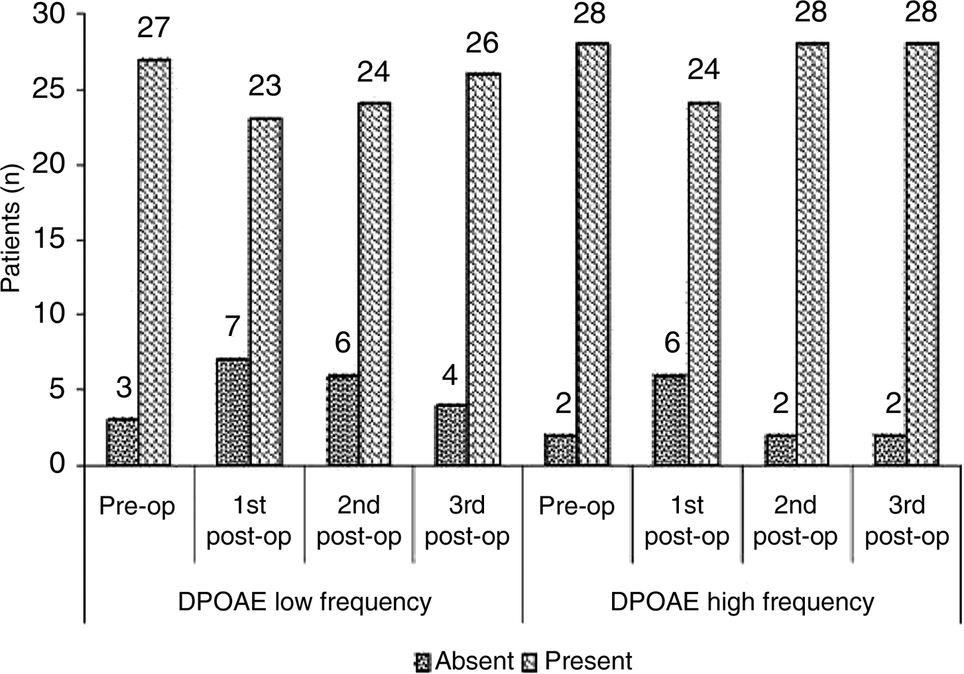
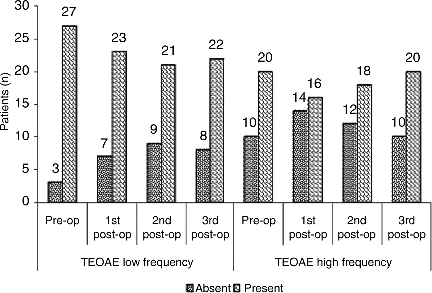
The data showing the presence or absence of OAEs, pre-operatively and on serial post-operative testing, are shown in Figures 1 and 2. For some patients, OAEs were absent over certain frequency ranges in the pre-operative period and these patients' data were excluded from the statistical analysis. Repeated measures analysis (using Cochran's Q test), conducted to examine changes in low and high frequency DPOAEs, and in low and high frequency TEOAEs, revealed respective p values of 0.182, 0.016, 0.035 and 0.021. As the value for low frequency DPOAEs was not significant, we did not examine the association between this parameter and drill variables.
Presence or absence of distortion product otoacoustic emissions at various time points. Pre-op = pre-operation; post-op = post-operation; DPOAE = distortion product otoacoustic emissions

Presence or absence of transient evoked otoacoustic emissions at various time points. Pre-op = pre-operation; post-op = post-operation; TEOAE = transient evoked otoacoustic emissions
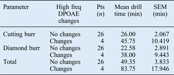
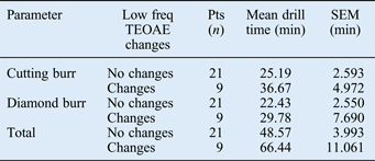
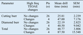
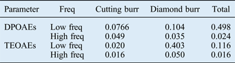
Changes in TEOAEs and DPOAEs were associated with various drilling times; the results are shown in Tables I–III. We used the Mann–Whitney U test to determine the exact significance of associations between the changes in OAEs and drilling times (Table IV).
High frequency distortion product otoacoustic emission data
Freq = frequency; DPOAE = distortion product otoacoustic emissions; pts = patients; min = minutes; SEM = standard error of the mean
Low frequency transient evoked otoacoustic emission data
Freq = frequency; TEOAE = transient evoked otoacoustic emissions; pts = patients; min = minutes; SEM = standard error of the mean
High frequency transient evoked otoacoustic emission data
Freq = frequency; TEOAE = transient evoked otoacoustic emissions; pts = patients; min = minutes; SEM = standard error of the mean
Associations between otoacoustic emission data and drill times
Data represent p values calculated using the Mann–Whitney U test. Freq = frequency; DPOAEs = distortion product otoacoustic emissions; TEOAEs = transient evoked otoacoustic emissions
No statistically significant associations were observed between changes in DPOAEs and TEOAEs and drill speed (rpm).
Discussion
Bone drilling is an important component of otological surgery, but it exposes both cochleas to a significant level of noise and vibration. Transcranial vibration represents a complex interaction between transmission, and damping effects of the skull, cranial content and surrounding soft tissue.
Various studies have shown that hearing in either the operated or contralateral ear may be temporarily affected by mastoid drilling.Reference Zou, Bretlau and Pyykkö4, Reference Kylen, Arlinger and Bergholtz9, Reference Karatas, Miman, Ozturan, Erdem and Kalciglu10 In a study using isolated temporal bones and cadavers, Kylen and Arlinger demonstrated that, during drilling, the exposed cochlea was subjected to noise levels of about 100 dB, while in the contralateral cochlea, the levels were 5–10 dB lower.Reference Kylen and Arlinger13
We could not find any study in the literature that discussed the effect of drilling on the contralateral ear using high frequency audiometry. However, Hegewald et al. reported the use of high frequency electrostimulation audiometry for hearing assessment in the post-operative period in the operated ear, and found statistically significant changes.Reference Hegewald, Heitman and Weiderhold8 In the present study, no statistically significant changes were observed when comparing pre- and post-operative pure tone threshold averages at high frequencies. Several investigators have emphasised that hearing over higher frequencies is most sensitive to noise-induced damage;Reference Flottorp14 hair cell degeneration may begin in the basal portion of the cochlea, which is the region of high frequency reception.Reference Johnsson and Hawkins15
Whereas previous studies have demonstrated a detrimental effect of mastoid drilling on hearing in the normal contralateral ear using OAEs,Reference Da Cruz, Fagan, Atlas and McNeill2, Reference Karatas, Miman, Ozturan, Erdem and Kalciglu10 no attempt has previously been made to associate these changes with drill variables as reported in the present study.
Karatas et al. observed statistically significant changes in pre- and post-operative transient evoked OAEs (TEOAEs) at all frequencies tested, and distortion product OAEs (DPOAEs) at frequencies of 2319, 2759, 3284 and 5164 Hz.Reference Karatas, Miman, Ozturan, Erdem and Kalciglu10 There were no statistically significant changes for DPOAEs at frequencies of 818, 977, 1160, 1379, 1636, 1953 and 4638 Hz. In our study, statistically significant changes were revealed for the comparison of pre- and post-operative TEOAEs over both low and high frequency ranges (with respective p values of 0.035 and 0.021), and DPOAEs over high frequency ranges (p = 0.016). There were no statistically significant changes in DPOAEs over low frequency ranges (p = 0.182).
Similarly, Da Cruz et al. reported reversible drill-related DPOAE changes in the contralateral ear in 2 out of 12 cases.Reference Da Cruz, Fagan, Atlas and McNeill2 This seems reasonable, as loud noise induces hearing loss by changing the way that outer hair cells function within the cochlea. In the case of temporary hearing loss, the outer hair cells are physically intact, but there is a reversible physiological disruption of their function.Reference Hamernik, Turrentine, Roberto, Salvi and Henderson16
Tos et al. reported no significant change in post-operative hearing status in the normal contralateral ear in 50 cases of translabyrinthine removal of acoustic neuromas.Reference Tos, Trojaborg and Thomsen17 There are several possible reasons for this. First, except for the cortical mastoidectomy, the predominant part of the surgery was carried out using a small (1–4 mm) diamond burr. As shown in the study by Kylen et al., which investigated variables affecting drill-generated noise during ear surgery,Reference Kylen, Stjernvall and Arlinger6 the noise produced by the diamond burrs differs considerably from that of the cutting burrs. The mean noise levels of the diamond burrs are 5–11 dB lower than those of the cutting ones. Of all of the variables, the size of the burr has the most pronounced effect on noise levels. The smaller the burr, the lower the noise; a 2 mm diamond burr results in a 20 dB reduction in noise at 8 kHz. In comparison with the size of the burr, all of the other variables have little effect on the noise levels produced. In the study by Tos et al., surgery was predominantly carried out using a small diamond burr; this is not likely to have generated a noise sufficient to damage the contralateral cochlea. Second, Tos et al. evaluated hearing using pure tone and speech audiometry up to only 8000 Hz, and this was conducted three months after surgery. Thus, the detection of a temporary threshold shift was unlikely, and as frequencies above 8000 Hz were not used, a change in higher frequencies could not be revealed.
Da Cruz et al. used intra-operative DPOAE recordingsReference Da Cruz, Fagan, Atlas and McNeill2 and Karatas et al. used immediate post-operative recordings,Reference Karatas, Miman, Ozturan, Erdem and Kalciglu10 and both groups reported statistically significant results. We too utilised the immediate post-operative period for hearing evaluation and found statistically significant results.
Our findings demonstrated statistically significant associations between cutting burr times and changes in: DPOAEs at high frequencies, and TEOAEs at both low and high frequencies (p values of 0.049, 0.020 and 0.016, respectively). However, the only significant association with diamond burr times was DPOAEs at high frequencies (p = 0.035). In addition, no association was revealed between drill speed (rpm) and changes in OAEs.
• Temporal bone drilling during ear surgery has the potential to cause significant acoustic trauma to the contralateral ear
• Otological surgeons must minimise drilling time during surgical management of chronic otitis media
• Surgeons should use a diamond burr over a cutting burr when feasible, particularly in an only hearing ear
• Drill revolutions per minute do not seem to affect hearing
Both Da Cruz et al. and Karatas et al. demonstrated reversible hearing loss after mastoid drilling.Reference Da Cruz, Fagan, Atlas and McNeill2, Reference Karatas, Miman, Ozturan, Erdem and Kalciglu10 Our study, however, showed complete recovery in 10 out of 15 patients where changes or reductions occurred in OAEs post-operatively, and ‘incomplete recovery’ or persistent deficit in 5 patients at the end of the post-operative follow-up period (i.e. at 72 hours). Whether this hearing loss would have recovered or become permanent after a prolonged follow-up period requires further investigation. Nevertheless, the prospect of a prolonged hearing deficit after drill-induced trauma certainly highlights the possibility of increased or differential susceptibility of certain individuals or groups (e.g. the very young or the elderly) to noise- and vibration-induced trauma to the cochlea. Studies that encompass a larger sample size (to enable multiple linear regression analysis), more drill variables and a longer follow-up time may elucidate drill-generated acoustic or vibrational trauma to the human cochlea.