The importance of the intestinal microbiota in the protection against disease has been confirmed (Tappenden & Deutsch, Reference Tappenden and Deutsch2007). The intestinal microbiota contains many useful microbes among them lactic acid bacteria (LAB) and bifidobacteria have a very important role to play. The beneficial effect of the intestinal microbiota, can be affected by diet, antibacterial drugs, stress, etc., and probiotic bacteria such as some LAB can repair these deficiencies (Verdu et al. Reference Verdu, Bercik, Verma-Gandhu, Huang, Blennerhassett, Jackson, Mao, Wang, Rochat and Collins2006; Eutamene et al. Reference Eutamene, Lamine, Chabo, Theodorou, Rochat, Bergonzelli, Corthesy-Theulaz, Fioramonti and Bueno2007; Zuccotti et al. Reference Zuccotti, Meneghin, Raimondi, Dilillo, Agostoni, Riva and Giovannini2008).
There are many beneficial claims attributed to probiotic supplementation in human health especially on the immune system. Since LAB are usually ingested as part of the normal daily diet, many studies have been done to demonstrate the effect of these bacteria on the systemic and mucosal immunity (Galdeano & Perdigon, Reference Galdeano and Perdigon2006; Galdeano et al. Reference Galdeano, de Moreno de LeBlanc, Vinderola, Bonet and Perdigon2007). Nowadays, the challenge is to establish the scientific basis for probiotic use. With this aim, experimental models in mice were used in the present study. Even though not all the results from animal models can be extrapolated to humans, they are useful in the understanding of the different process where the immune system is involved.
It has been shown that the substances that stimulate the immune response (adjuvant or immunomodulatory) can enhance the non-specific, the innate and or the adaptive immune response. Probiotics could exert their influence on the immune system by stimulating one or all of these types of responses.
The reports about the influence of the oral administration of fermented milk containing probiotic microorganisms on the immune state have been mainly focused on the alleviation or prevention of different pathologies (de Moreno de LeBlanc et al. Reference de Moreno de LeBlanc, Matar, LeBlanc and Perdigon2005b; Medici et al. Reference Medici, Vinderola, Weill and Perdigon2005).
These previous studies showed the important role of probiotic bacteria and fermented milks containing probiotic in the activation of the systemic and the mucosal immune response. The effects of the fermented milk on the gut mucosal immune system can be exerted by the probiotic microorganisms and by the biomolecules contained in the non microbial fraction.
In the present work we analyzed the effect of a probiotic fermented milk (PFM) containing the probiotic bacterium Lactobacillus (Lb.) casei DN 114001, on different parameters of the gut mucosal immunity related to the non-specific, innate and adaptive response.
The interaction of the probiotic bacterium with the epithelial and immune cells and the residence time of this bacterium in the gut were studied. For the PFM, the number of IgA+ B lymphocytes and T cells as well as the expression of receptors and markers present in the cells of the innate immunity (macrophages and dendritic cells) were analyzed. The IL-6 release from intestinal cells, the number of goblet cells associated with the epithelium and the activation of the transcriptional factor NF-AT through the phosphatase calcineurine enzyme expression were studied. A scheme to describe the immune mechanisms elicited by the fermented milk on the effectors site of the gut is also suggested.
Materials and Methods
Animals and probiotic bacterium
Six-week-old BALB/c mice were obtained from the random-bred colony kept at CERELA (Argentina). Each experimental and control group consisted of 5 mice per assay. Each assay was performed in duplicate or triplicate. All the animals were fed a conventional balanced diet ad libitum. Animal protocols were pre-approved by the Animal Protection Committee of CERELA and all experiments comply with the current laws of Argentina.
The probiotic strain was Lb. casei DN 114001. The procedure for the administration of this bacterium to the mice was as described by Galdeano & Perdigon (Reference Galdeano and Perdigon2004) for Lb. casei CRL431.
TEM assay
Mice received by gavage a suspension of Lb. casei DN 114001 (109 CFU ml−1) and were sacrificed 10 min after that. The small intestine was removed and treated according to the technique described previously (Galdeano & Perdigon, Reference Galdeano and Perdigon2004) for electron microscopy studies.
Bacteria labelling procedure
The bacterial pellets were labelled with fluorescein isothiocyanate (FITC) according to Galdeano & Perdigon (Reference Galdeano and Perdigon2004). The persistence of lactobacilli in the gut was determined in small intestine at 10 min, 24, 48 or 72 h after administration of FITC-labelled bacteria. Mice were sacrificed at these time points and the fluorescent bacteria were observed in the intestinal tissues using a fluorescence light microscope.
Fermented milk and feeding procedure
Probiotic fermented milk (PFM) containing Lb. delbrueckii subsp. bulgaricus 108 CFU/ml, Streptococcus thermophilus 108 CFU/ml and the probiotic bacterium Lb. casei DN-114001 (108 CFU/ml) was used in this study. The animals were subdivided into two groups: (a) those receiving PFM and (b) those that did not. The PFM was administered ad libitum during 5 consecutive days, optimal period as determined previously (de Moreno de LeBlanc et al. Reference de Moreno de Leblanc, Chaves, Carmuega, Weill, Antoine and Perdigon2008a). At the end of the feeding period mice were sacrificed and samples of small intestine were obtained.
Determination of goblet cells in small intestine
Histological slides from the small intestine were stained with 1% (w/v) Alcian Blue 8Gx solution (Merck, Darmstadt, F.R. Germany) in 3% (w/v) acetic acid. The slides were then incubated in eosin solution and then in 0·5% safranin solution in 0·1 m-HCl. The results were expressed as the number of goblet cells per ten intestinal villi.
Immunofluorescence assays for detection of IgA, CD4, CD8, CD206 and TLR4 positive cells in the lamina propria of the small intestine from mice feeding with PFM
The number of IgA, CD4, CD8, CD-206 and TLR-4 positive cells was determined using immunofluorescence assay on the histological slides from the small intestine for treated and untreated groups. IgA-secreting cells were determined using alpha-chain mono-specific antibody conjugated with FITC (Sigma, St Louis, MO, USA). For CD4 and CD8 positive cells, monoclonal antibodies conjugated with FITC were used (Cederlane, Ottawa, Canada).
CD-206 and TLR-4 receptors were measured according to Galdeano & Perdigon (Reference Galdeano and Perdigon2006). Mouse anti-human CD-206 monoclonal antibody (BD Biosciences Pharmingen, USA) or rabbit anti-mouse TLR-4 polyclonal antibody (eBioscience, USA) was used. FITC conjugated rabbit anti-mouse or goat anti-rabbit antibodies (Jackson Immuno Research Labs. Inc., USA) were the second antibodies.
Results were expressed as the mean of the number of positive fluorescent cells per 10 fields (magnification 1000X).
Identification of macrophages and dendritic cells in lamina propria of the small intestine
Macrophages were determined using the BM8 monoclonal antibody (eBioscience, San Diego, CA), which reacts with mouse F4/80 antigen. Dendritic cells were determined using the 33D1 monoclonal antibody (eBioscience, San Diego, CA) which recognizes a dendritic cell-specific surface marker. Both cells were determined by indirect immunofluorescence assays according de Moreno de LeBlanc et al. (Reference de Moreno de LeBlanc, Dogi, Maldonado Galdeano, Carmuega, Weill and Perdigón2008b). Results were expressed as the mean of the number of positive cells per 10 fields (magnification 1000X).
Intestinal epithelial cell isolation. IL-6 determination
The effect of the PFM on the intestinal cells (IC) was measured by IL-6 release. The intestinal cells were isolated as previously described (Vinderola et al. Reference Vinderola, Matar and Perdigon2005) from the small intestine of mice that received or not the PFM; this is a preparation enriched in IEC. The IC suspension was transferred to 96 well cell culture plates and incubated for 8 h (37°C, 5% CO2). Supernatants were recovered for IL-6 determination by a commercial ELISA kit (BD Biosciences San Diego, USA).
Determination of the expression of calcineurin
The animals received by intragastric intubation 100 μl PFM. They were sacrificed 15 min after PFM administration and the small intestine was removed.
Histological slices were deparaffinized, rehydrated and incubated for 16 h at 4°C with monoclonal anti-calcineurin (BD Biosciences Pharmingen, San Diego, CA, USA). The sections were then treated with a FITC conjugated goat anti-mouse antibody (Jackson Immuno Research). The results were expressed as the number of positive cells per 10 fields (magnification 1000X).
Statistical analysis
For each trial, the test and control groups contained 10–15 animals. Five mice for each group were sacrificed in each sample taken (n=5). Unless otherwise indicated, all values (n=15) were the means of 3 independent trials (no significant differences were observed between individual replicates) ± standard deviation (sd). The Student's T-test was used to asses the statistical significance (P<0·05) of the differences between the test and the control group.
Results and Discussion
Interaction of Lb. casei DN-114001 with the gut by TEM and its persistence in the intestine
Two important features used in the selection of a probiotic bacterium are the ability to persist in the gut and the ability to adhere to the mucosal surfaces (Bezkorovainy, Reference Bezkorovainy2001).
Adhesion to the intestinal epithelial cells is the first step in the modulation of the immune system by probiotic bacteria. Good adhesion properties should enhance the possibility of long-term survival of the organism in the intestinal tract by countering the peristaltic action of the intestine.
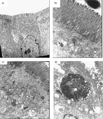
The intestinal epithelium is known to possess a number of specialized protective adaptations such as tight junctions which restrict the passage of molecules and micro-organisms. Since the interaction with the epithelial intestinal cells is the first step in the signal transduction to stimulate the gut immune system, it was studied using electron microscopy. The probiotic bacterium (Lb. casei DN-114001) was observed interacting with the microvilli of the enterocytes (Fig. 1b). This interaction induced an activation of the epithelial cells evidenced by the increase of multivesicular bodies in the cytoplasm of these cells (Fig. 1c & d).
Interaction of Lb. casei with the intestinal epithelial cells.

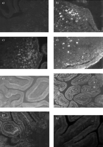
To induce a gut immune response, the probiotic bacteria must also make contact with the immune cells, thus the interaction of Lb. casei DN 114001 with the immune cells associated with the gut was studied. Figures 2b, c & d show the interaction of the whole bacteria or their fragments and the sequence of the persistence of the probiotic bacterium in the Peyer's patches (PP).
Time of persistence of FITC labelled Lb. casei DN 114-001 in Peyer′s patches (inductor site) and in lamina propria (effector site) of the small intestine.

It was observed that after 10 min, fluorescent cells in the PP and the number of fluorescent cells increased reaching a peak at 24 h post administration (Fig. 2c). A few fluorescent cells were observed in the sample taken at 72 h (Fig. 2d). In the LP of small intestine (Fig. 2f, g & h), fluorescence followed the same sequence as in the PP with the highest number of fluorescent cells in the sample taken after 24 h. Considering previous results obtained with Lb. casei CRL 431 (Galdeano & Perdigon, Reference Galdeano and Perdigon2004), it is possible to suggest that the fluorescence observed in the lamina propria could be from bacterial fragment. However, the whole bacteria could also have translocated to the LP and this fact is possible through M cells present in the gut villous (Jang et al. Reference Jang, Kweon, Iwatani, Yamamoto, Terahara, Sasakawa, Suzuki, Nochi, Yokota, Rennert, Hiroi, Tamagawa, Iijima, Kunisawa, Yuki and Kiyono2004) or by internalization through dendritic cells, which express proteins of the tight junctions and penetrate between the intestinal epithelial cells (Rescigno et al. Reference Rescigno, Urbano, Valzasina, Francolini, Rotta, Bonasio, Granucci, Kraehenbuhl and Ricciardi-Castagnoli2001). In the present work the fluorescence observed in the LP can be due to either bacterial fragments or intact bacteria (Fig. 2). Micrographs in Fig. 1 also showed that the epithelial barrier was preserved in the mice fed with the probiotic bacterium in contrast to pathogens, which are able to invade the gut epithelium. The fluorescence observed in the immune cells diminished significantly at 72 h meaning that the whole bacterium or the bacterial fraction suffered a normal clearance (Fig. 2). This last observation is important because these bacteria are introduced in probiotic products that are consumed on a regular basis.
Effect of PFM on cells of the nonspecific barrier and on the gut immune cells
The gastrointestinal epithelium is covered by a protective mucus containing predominantly mucin glycoproteins that are synthesized and secreted by goblet cells. The concept of the mucus layer functioning as a dynamic defensive barrier is suggested by studies showing altered mucus-related indexes in germ-free animals (Gaskins, Reference Gaskins, Wiseman and Varley1998) and from consistent evidence of enhanced mucus secretion in response to intestinal microbes (Mack et al. Reference Mack, Michail, Wei, McDougall and Hollingsworth1999).
In our study (Table 1), the goblet cells increased significantly only in mice that received the PFM (95±15) versus (65±12). This observation agrees with previous reports where the administration of this PFM, to mothers during the suckling period or to their offspring after weaning, increased the number of goblet cells in the small intestine of the offspring (de Moreno de LeBlanc et al. Reference de Moreno de LeBlanc, Dogi, Maldonado Galdeano, Carmuega, Weill and Perdigón2008b).
Effect of the PFM on Goblet, IgA+, CD4+ and CD8+ cells

Animals were given PFM during 5 consecutive days. Histological slices were performed from small intestine of control and treated animal. Goblet cells were determined by Alcian blue stained. For IgA, CD4+ and CD8+ cells direct immunofluorescence assay was performed. Values are the mean of 9 animals (three independent trials)±SD. Significant values P<0·05
T and B cells associated with the gut were analyzed and it was observed that PFM administration significantly increased IgA+ B cells (185±12) in the LP of the small intestine compared with the control mice (119±14, Table 1). These results are desirable in the stimulation of the mucosal immune system by probiotic bacteria or a fermented milk (de Moreno de LeBlanc et al. Reference de Moreno de LeBlanc, Maldonado Galdeano, Chaves and Perdigón2005a). Enhanced intestinal IgA production, provides defence for the mucosal surface (Lamm, Reference Lamm1998), and it is one of the beneficial effects associated with probiotic consumption.
For T cells, it was observed that PFM significantly increased (P<0·05) CD8+ and CD4+ cells compared with control mice (Table 1). These results were different from those obtained using the probiotic bacterium Lb. casei CRL 431 alone where CD4+ or CD8+ T lymphocytes did not increase (Galdeano et al. Reference Galdeano, de Moreno de LeBlanc, Vinderola, Bonet and Perdigon2007). Our results show that the effects and the mechanisms are different for each probiotic strain but also that it is important to consider for a PFM the total product because of the multiple immunogenic components produced during the fermentation process, in addition to the probiotic bacterium.
We analyzed the effects of this PFM on the antigen presenting cells: macrophages and dendritic cells. Macrophages play an important role in the innate immune response and in the regulation of acquired immune responses (Wijburg et al. Reference Wijburg, Heemskerk, Boog and Van Rooijen1997). The F4/80 marker is present on the surface of cells of the mononuclear phagocyte system of mice. The expression of this antigen is higher in mature macrophages and its expression is required for regulatory T cell development (Lin et al. Reference Lin, Faunce, Stacey, Terajewicz, Nakamura, Zhang-Hoover, Kerley, Mucenski, Gordon and Stein-Streilein2005).
Dendritic cells are known to be essential in the innate immunity and in the initiation of adaptive immunity. These cells capture, migrate and transfer information from the lumen to the cells of the adaptive immune system. It is known that the shaping of adaptive immunity by innate immunity is dependent on dendritic cells and effector molecules derived from them such as cytokines and chemokines (Wen et al. Reference Wen, Schaller, Dou, Hogaboam and Kunkel2007).
In our study, the number of macrophages (F4/80+ cells) increased significantly in the LP of intestine from mice that received the PFM, compared with control animals (Table 2). The increase in the number of dendritic cells was not significant in relation to the control. However, in a previous paper it was observed that dendritic cell populations were increased in mice receiving the PFM from weaning until the adulthood (45 days) (de Moreno de LeBlanc et al. Reference de Moreno de LeBlanc, Dogi, Maldonado Galdeano, Carmuega, Weill and Perdigón2008b). Perhaps the period of PFM administration used in the present work was not enough to observe an increase in the number of dendritic cells, or maybe the low expression of the maker recognized by 33D1 antibody in adult, is necessary to maintain the intestinal homeostasis. These findings allowed us to study other receptors that are present in the cells of the innate immune response such as TLR-4 and CD206 (mannose receptor). The mannose receptor is implicated in the homeostatic system for the clearance of endogenous molecules (Allavena et al. Reference Allavena, Chieppa, Monti and Piemonti2004). This receptor facilitates the uptake of the mannosylated antigens by dendritic cells for presentation to T cells. TLR-4 was studied because, this receptor recognizes the LPS present in the cell wall of the Gram(-) bacteria, but some authors have related this receptor as a link in the stimulation induced by the main component in Gram(+) bacteria, such as LAB, the lipoteichoic acid (LTA) (Cox et al. Reference Cox, Ofek and Hasty2007). The intestinal epithelium shows a marked upregulation of TLR4 in active inflammatory bowel diseases in a murine model of colitis and the persistence of colitis could be prevented by suppression of TLR4 expression in IEC (Isono et al. Reference Isono, Katsuno, Sato, Nakagawa, Kato, Sato, Seo, Suzuki and Saito2007). Little is known about the effects of probiotics on the expression of this receptor. The results obtained for both receptors (TLR4 and CD206) did not show increases for them (Table 2).
Determination of macrophages and dendritic cells and the expression of CD-206 and TLR-4 receptors and calcineurine+cells in lamina propria of the small intestine of mice given PFM. Levels of IL-6 released by IC
Values are means for n=15±sd mice

* Significant differences between test and control groups (P<0·05)
The number of macrophages and dendritic cells (F4/80 positive cells and cells positive for 33D1 antibody which recognizes a mouse dendritic cell-specific surface marker, respectively), CD-206+ and TLR-4+ cells were determined by indirect immunofluorescence on the small intestine of mice that received PFM during 5 days. Calcineurine+ cells were also determined by immunofluorescence after 15 min of PFM administration. Results are expressed as number of positive cells recognized for the respective primary antibody, counted in 10 fields of vision at 1000X of magnification. The IL-6 release was measured by ex vivo assay in the IC purified from mice after 5 days of PFM administration by ELISA test. Results are expressed as concentration (pg/ml)
Calcineurine (CN) is a calcium- and calmodulin-dependent phosphatase required for diverse biological processes such as T cell activation, development, hypertrophy and gene regulation in skeletal and cardiac muscle (Olson & Williams, Reference Olson and Williams2000; Rusnak & Mertz, Reference Rusnak and Mertz2000; Crabtree, Reference Crabtree2001). Tsuboi et al. (Reference Tsuboi, Masuda, Naito, Tokumitsu, Arai and Arai1994) demonstrated that in T lymphocytes, CN is involved in the coordinated induction of the GM-CSF and IL-2 genes mediated by the transcriptional factor NF-AT. We demonstrated that the administration of the PFM to mice increased the number of CN+ cells in the LP of the small intestine (Table 2). This could be related to the activation of the macrophages since this enzyme has been described in these cells (Tsuboi et al. Reference Tsuboi, Masuda, Naito, Tokumitsu, Arai and Arai1994), and with the activation of T lymphocytes. The stimulation of both cell populations was observed in a previous study where the PFM administration increased the number of IL-2 and TNFα positive cells (de Moreno de LeBlanc et al. Reference de Moreno de Leblanc, Chaves, Carmuega, Weill, Antoine and Perdigon2008a). Conboy et al. (Reference Conboy, Manoli, Mhaiskar and Jones1999) demonstrated that CN constitutively act in normal macrophages to suppress expression of inflammatory cytokines in the absence of specific activation and modulate effector gene expression at the mRNA level by inhibiting transcription factor NF-κB.
This negative regulation by CN is opposite to its crucial positive role in T cells, where it activates NFAT transcription factor(s) leading to expression of IL-2, TNFα and other cytokine genes (Kaminuma et al. Reference Kaminuma, Kitamura, Kitamura, Hiroi, Miyoshi, Miyawaki and Miyatake2008).
The IEC (intestinal epithelial cells) are in close proximity to adjacent immune cells on the basolateral side. These cells can express different toll like receptors and are able to secrete cytokines such as IL-6, IL-1 and IL-8 (Haller et al. Reference Haller, Bode, Hammes, Pfeifer, Schiffrin and Blum2000). The interaction of probiotic bacteria or PFM with the preparation enriched in IEC can activate these cells to induce cytokine release (Dogi et al. Reference Dogi, Galdeano and Perdigon2008). In the present work, PFM administration showed a significant increase in the IL-6 produced by the IC (Table 2). The physiological role for IL-6 would be to initiate or maintain the cross-talk between the epithelial and immune cells associated with the gut and to induce the IgA B cell expansion (Beagley et al. Reference Beagley, Eldridge, Aicher, Mestecky, Di Fabio, Kiyono and McGhee1991), as was observed in the present work.
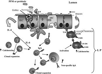
Results obtained in this study showed that the probiotic bacteria administered to the mice make contact with the IC and activate these cells. The whole bacterium or their fragments interact with the gut associated immune cells. The fermented milk containing this probiotic bacterium stimulated the IC as well as the immune cells associated with the gut. This was observed through the increase in the number of different immune cell populations: T and IgA+ B lymphocytes, cells related with both innate and adaptive response (macrophages), and cells from the nonspecific barrier (goblet cells). PFM stimulated the IC with IL-6 release. This cytokine is related to the IgA B cell expansion. The PFM studied was also able to activate the enzyme calcineurine responsible for the activation of the transcriptional factor NFAT. Figure 3 is a schematic of these results, showing that the PFM is able to exert its influence at different level of the gut immune response.
Proposed model about the effect of PFM on the effectors sites of the gut.

This work had been supported by a grant 2007 DANONE Argentina S.A.-CONICET (Consejo Nacional de Investigaciones Científicas y Técnicas), Argentina and grant for CONICET-PIP 5445. The authors grateful Dr. Jean Guy LeBlanc for the English correction.