Fundamental aspects
Introducing large, thermally labile compounds into the gas-phase environment of mass spectrometers without changing their structures has always been a challenge. To address this issue, various soft desorption/ionization techniques have been developed, such as matrix-assisted laser desorption ionizationReference Tanaka, Waki, Ido, Akita, Yoshida and Yoshida1 (MALDI), electrospray ionizationReference Fenn, Mann, Meng, Wong and Whitehouse2 (ESI), desorption electrospray ionizationReference Tak´ats, Wiseman, Gologan and Cooks3 (DESI), and laser-induced acoustic desorptionReference Lindner and Seydel4 (LIAD) coupled with various ionization methods.Reference Pérez, Petzold, Watkins, Vaughn and Kenttämaa5,Reference Shea, Petzold, Liu and Kenttämaa6 Among these techniques, LIAD is unique in that it desorbs neutral molecules instead of ions.
A typical LIAD device focuses a high-energy laser beam on the backside of a thin metal foil (typically ∼10 μm). A thin layer of a solid sample is deposited on the front side of the foil. The laser irradiation causes desorption of intact, neutral sample molecules into the gas phase from the metal surface (if the conditions are such that thermal degradation of the sample does not take place). Many different ionization methods and mass analyzers have been utilized to ionize and analyze the gaseous molecules.
LIAD exhibits several major advantages over MALDI, ESI, and DESI, including the following:Reference Shea, Petzold, Liu and Kenttämaa6,Reference Sezer, Wörner, Horak, Felix, Tüxen, Götz, Vaziri, Mayor and Arndt7 (1) LIAD can be used to desorb large saturated hydrocarbons with no polar functional groups into the mass spectrometer for ionization, while these compounds cannot be desorbed using MALDI, ESI or DESI; (2) the translational energy and average velocity of the desorbed molecules are lower than for gas-phase ions generated using MALDI, thus allowing for more efficient ion–molecule reactions; and (3) sample desorption is decoupled from ionization, making LIAD a more versatile technique than MALDI, ESI, or DESI as many different ionization methods can be coupled with LIAD. However, the plume densities (109–1010 cm3) generated upon some LIAD experiments have been reported to be less than those obtained upon MALDI, although still high enough for many experiments.Reference Freund and Surech8 Due to these and other advantages, LIAD has become a common desorption technique, with its broadening field of applications summarized later in this article.
As for the working principle of LIAD, the exact mechanism(s) of LIAD is (are) still unknown, and different mechanisms may be operational under different experimental conditions. Substantial efforts have been made thus far to understand these mechanism(s), including both computational modeling and experimental measurements. Several models have been proposed and tested; the most important ones are discussed next.
Thermal waves
Although thermal waves are generated upon laser shots on a metal and may contribute to sample desorption, this is unlikely to be the mechanism of most LIAD experiments for the following reasons. First, as the translational velocity of the molecules after desorption is independent of the laser intensity that dictates the intensity of thermal waves, it has been claimed that thermal desorption is not occurring.Reference Freund and Surech8 Second, the foil is selected to be so thick that there is no change in temperature on the sample side of the metal foil,Reference Cheng, Huang and Shiea9 thus demonstrating that the sample is not desorbed due to thermal waves. Finally, labile compounds, such as oligonucleotides, desorbed with LIAD using 12-μm-thick Ti foils, undergo minimal to no fragmentation, which invalidates the presence of strong thermal waves.Reference Lindner10,Reference Pérez, Ramírez-Arizmendi, Petzold, Guler, Nelson and Kenttämaa11
“Shake-off” mechanism
A “shake-off” model has been the most widely accepted description of the LIAD mechanism, yet it has its flaws. The presumptions are as follows: when a laser beam impacts the irradiation side of the metal foil, acoustic waves are generated and propagate through the foil, imparting energy to the sample molecules. The sample molecules obtain momentum normal to the sample surface and are thus ejected.Reference Lindner and Seydel4,Reference Dow, Wittrig and Kenttämaa12
The previously mentioned model is founded on a few well-established experiments. Pulsed laser beams with different energy densities have been demonstrated to interact with metal foils to cause different mechanical vibration responses.Reference Hutchins, Dewhurst, Palmer and Scruby13 When the irradiation side of the foil was placed against a glass surface and irradiated with a laser beam with an intensity greater than 108 W/cm,Reference Fenn, Mann, Meng, Wong and Whitehouse2,Reference Zinovev, Moore, Calaway, Pellin, Veryovkin, Kudryashov, Paxton, Ilchenko, Giesen, Nickel, Davis, Heaven and Schriempf14 a sharp mechanical wave was usually observed within 1 μs after the first laser pulse.Reference Zinovev, Veryovkin, Moore and Pellin15 Therefore, the acoustic waves were hypothesized to create a sudden displacement of the foil surface, resulting in analyte desorption.
However, some observations have been reported to be in disagreement with this model. For example, the minimum required speed of foil displacement has been calculatedReference Freund and Surech8 to be 100–500 m/s to desorb molecules with molecular weights of 200–500 Da from the surface. This range was concludedReference Cheng, Huang and Shiea9 to be in disagreement with the maximum 100 m/s speed that has been observed for LIAD experiments. Further, the observation that increasing the laser power intensity by two orders of magnitude had no effect on the measured velocities of the desorbed moleculesReference Cheng, Huang and Shiea9 indicated that although acoustic waves were generated, they were not solely responsible for LIAD. However, it should be noted that the relationship between the laser power intensity and the velocities of the desorbed molecules is not fully understood at this time. Further, while the first and largest foil displacement has been reported to occur in less than 1 μs, desorption has been found to occur over a range of tens of μs.Reference Shea, Petzold, Campbell, Li, Aaserud and Kenttämaa16 Desorption due to “shake- off” was postulated to occur during the largest displacement, not over time.Reference Shea, Petzold, Campbell, Li, Aaserud and Kenttämaa16 These observations led to the introduction of the “film strain” mechanism that is discussed next.
“Film strain” mechanism
A new mechanism for LIAD was reported in 2007.Reference Shea, Petzold, Campbell, Li, Aaserud and Kenttämaa16 The basis of the mechanism was that the sample on the foil is not a continuous film, but crystals that experience “film strain.” The molecules are liberated from the crystals due to surface deformation caused by a combination of thermal and acoustic waves. This may explain why the energy transfer to the molecules appears to be independent of laser power, as it would instead be derived from the stored energy of individual crystals.Reference Shea, Petzold, Campbell, Li, Aaserud and Kenttämaa16 However, the feasibility of this mechanism for the experiments involved in most analytical applications of LIAD is not clear because these experiments use a continuous sample layer on the foil, not crystals.
LIAD experimental setups
A typical LIAD setup consists of a laser with relevant optics, a substrate (usually metal foil) with a sample deposited on one side, and a means for positioning the system so that laser sampling can be performed on different spots of the foil. In the following section, different experimental setups for LIAD are discussed, followed by a discussion of several ionization methods that have been coupled with LIAD.
Desorption
The substrate onto which the sample is deposited is an essential part of a LIAD experiment. The best substrate is expected to absorb laser light, be capable of propagating acoustic waves, be thin enough to enable propagation of the acoustic waves to the sample side, and be thick enough to prevent thermal waves from reaching the sample during the desorption event.Reference Lindner10 Metal foils are often used because of their high thermal expansion coefficients, low thermal conductivities, and low reflectivity at certain wavelengths of light.Reference Shea, Petzold, Campbell, Li, Aaserud and Kenttämaa16 Considering these three properties, titanium excels compared to many other metals, including Ag, Al, Au, Cu, and Fe.Reference Shea, Petzold, Campbell, Li, Aaserud and Kenttämaa16 About 10-μm-thick titanium foils have been widely and successfully employed for LIAD of intact neutral molecules. Hg has even better properties, but is much more difficult to deal with experimentally.Reference Golovlev, Allman, Garrett and Chen17
More recently, tantalum has been introduced as an alternative metal due to its much higher melting point (3290 K compared to 1941 K for titanium) as it can therefore withstand higher laser intensities.Reference Sezer, Wörner, Horak, Felix, Tüxen, Götz, Vaziri, Mayor and Arndt7,Reference Calvert, Belshaw, Duffy, Kelly, King, Smyth, Kelly, Costello, Timson, Bryan, Kierspel, Rice, Turcu, Cacho, Springate, Williams and Greenwood18,Reference Huang, Ossenbrüggen, Rubinsky, Schust, Horke and Küpper19 However, consideration of the melting point may be irrelevant as no laser damage to titanium foils has been reported in the literature. Furthermore, research groups using tantalum have reported extensive fragmentation even to such simple and stable compounds as glycine. However, as these groups have employed high-energy methods (high-energy laser irradiation) to ionize the gas-phase molecules, it is difficult to determine whether the fragmentation was caused by LIAD or by ionization.Reference Sezer, Wörner, Horak, Felix, Tüxen, Götz, Vaziri, Mayor and Arndt7,Reference Zinovev, Moore, Pellin and Veryovkin20 Another disadvantage of using tantalum includes the finding that molecules desorbed from tantalum move faster compared to those desorbed from titanium foil.Reference Sezer, Wörner, Horak, Felix, Tüxen, Götz, Vaziri, Mayor and Arndt7
Other than metal foils, aluminum-based thin-layer chromatography (TLC) plates have also been employed in LIAD experiments. The samples analyzed included mixtures of dye standards and drug standards. The samples were first separated using TLC plates that were composed of a 100-μm-thick layer of aluminum and a 200-μm-thick layer of gel. After separation, the plates were glued on microscopic glass slides by using glycerol. The glass slides were set on a stage that moved linearly at a speed of 0.01–0.02 cm/s. A pulsed infrared laser with a wavelength of 1064 nm and a power density of 2.1 × 10⁹ W/cm2 was employed to perform LIAD. The desorbed molecules were ionized by ESI and analyzed using an ion trap mass spectrometer.Reference Cheng, Huang and Shiea9 Dye standards were deprotonated or doubly deprotonated under negative ion mode ESI, whereas drug standards were protonated under positive ion mode ESI. Most ions derived from TLC spots could be assigned to known mixture components. However, gel particles desorbed together with the analyte were a concern.Reference Cheng, Huang and Shiea9
Experiments carried out using copper foils with different thicknesses have demonstrated that a foil thickness of ∼10 μm is optimal for LIAD experiments.Reference Pérez, Ramírez-Arizmendi, Petzold, Guler, Nelson and Kenttämaa11 Thinner foils have been found to enable thermal waves to reach the analyte, thereby causing thermal degradation of the sample. On the other hand, thicker foils have been reported to result in less efficient desorption.Reference Cheng, Huang and Shiea9–Reference Pérez, Ramírez-Arizmendi, Petzold, Guler, Nelson and Kenttämaa11
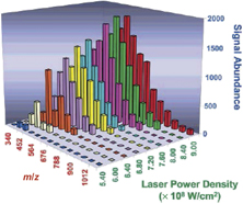
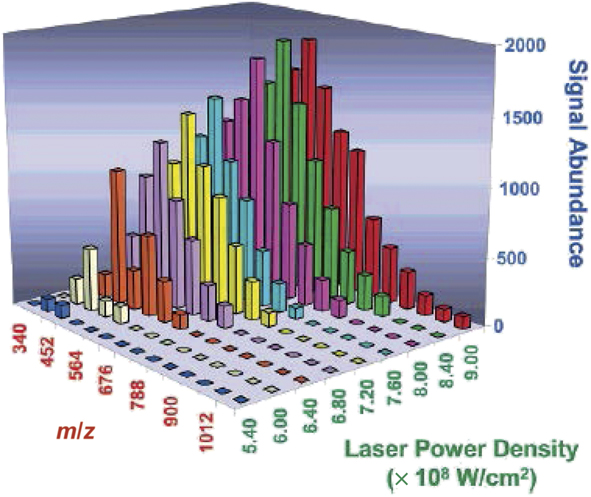
Laser power density is another key factor controlling LIAD efficiency. Increasing laser power density from 5.4 × 10⁸ to 9.0 × 10⁸ W/cm2 was foundReference Shea, Petzold, Campbell, Li, Aaserud and Kenttämaa16 to increase ion abundances. This also resulted in a shift in the distribution of the m/z (mass-to-charge ratio) values of ions derived from a polymer mixture to higher m/z values, as shown in Figure 1. These findings indicate that higher laser power densities result in more efficient desorption of larger molecules.
Relative ion abundances of polyisobutenyl succinic anhydride oligomers as a function of laser power density (different colors are used to make it easier to see which oligomers desorbed at each laser power density). Higher laser power densities result in more efficient desorption of larger molecules.Reference Shea, Petzold, Campbell, Li, Aaserud and Kenttämaa16 Note: m, mass; z, charge.
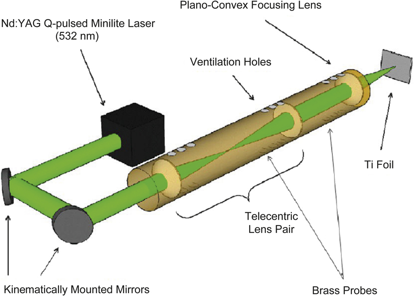
However, laser power density that is too high can be detrimental to the generation of acoustic waves via a shielding effect wherein the plume of metal ablated from the foil surface (when using high power density) attenuates the energy of the later laser pulses at the foil.Reference Vadillo, Fernández Romero, Rodríguez and Laserna21 A high-power LIAD probe with kinematically mounted high-power reflective mirrors and a telecentric lens pair, shown in Figure 2, has been developed to decrease power loss from the beam path by reducing beam divergence.Reference Borton, Amundson, Hurt, Dow, Madden, Simpson and Kenttämaa22 Power loss of 75% in earlier experiments was reduced to 2% by using this device, resulting in better sensitivity and reproducibility.
A high-throughput laser-induced acoustic desorption probe with kinematically mounted high-power reflective mirrors and a telecentric lens pair developed to increase power density by decreasing power loss from the beam path as well as reducing beam divergence.Reference Borton, Amundson, Hurt, Dow, Madden, Simpson and Kenttämaa22 Note: YAG, yttrium aluminum garnet (laser).
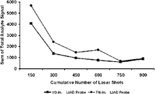
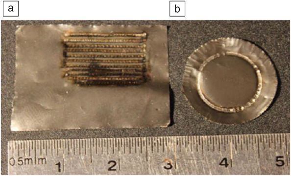
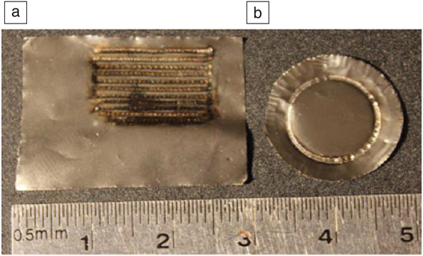
Analyte signal intensity in LIAD experiments relies heavily on the number of laser shots employed. However, each laser shot fired on a given spot of the foil depletes the sample at that spot. Therefore, the first few laser shots at any given foil location contribute the most to the amount of desorbed analyte. This phenomenon is illustrated in Figure 3.Reference Shea, Petzold, Campbell, Li, Aaserud and Kenttämaa16 Furthermore, firing many laser shots on a given spot on the foil may heat the sample. To increase the overall signal quality, a sampling mechanism can be employed that moves the laser to fresh spots on the sample foil or that moves the foil so that each laser shot is fired at a different spot. One way to do this is to rotate the sample foil while keeping the laser beam fixed, creating a circular sampling ring (Figure 4). The disadvantage is that this ring of sampled area is relatively small compared to the entire foil area and therefore most of the sample is not analyzed.Reference Shea, Petzold, Liu and Kenttämaa6,Reference Jarrell, Owen, Riedeman, Prentice, Pulliam, Max and Kenttämaa23 Improving on this method, a long rotating metal foil tape driven by a taper platform has been introduced to continuously provide new sample for a fixed laser beam, thus increasing the sampling area.Reference Huang, Ossenbrüggen, Rubinsky, Schust, Horke and Küpper19 Yet another approach involves placing the sample foil on a stage that can be controlled in the xy-plane (Figure 4). This setup utilized nearly the entire sample area. Additionally, this setup made switching between analytes or samples easier because different samples could be deposited to different areas of the same piece of foil.Reference Jin, Daiya and Kenttämaa24
Sum of total analyte signal for a protonated tetrapeptide, valyl alanyl phenylalanine, as a function of cumulative number of laser shots fired on a circular sample foil. Two different laser-induced acoustic desorption (LIAD) probes, with a 1/2 in. and 7/8 in. dia., were employed. Each data point represents the total analyte signal obtained using n × 150 laser shots (n = 1–6). The first 150 laser shots were each fired on a fresh spot on the foil, corresponding to one full rotation of the sample foil. The following set of 150 laser shots (data point at 300 total laser shots) were fired on the same spots that had already been sampled once, showing a decrease in analyte signal. After a total of about 750 laser shots (n = 5, meaning that each spot on the foil had been sampled five times), most of the analyte had been desorbed.Reference Shea, Petzold, Campbell, Li, Aaserud and Kenttämaa16

(a) A raster laser-induced acoustic desorption (LIAD) foil can be used to examine the analyte over a much larger area of the foil surface than (b) a circular LIAD foil (pictures taken after experiments).Reference Borton, Amundson, Hurt, Dow, Madden, Simpson and Kenttämaa22
Ionization
LIAD has been coupled with electron ionization (EI) and chemical ionization (CI) in Fourier transform ion cyclotron resonance (FT-ICR) mass spectrometers without external ion sources.Reference Huang, Ossenbrüggen, Rubinsky, Schust, Horke and Küpper19,Reference Jin, Daiya and Kenttämaa24,Reference Pinkston, Duan, Gallardo, Habicht, Tan, Qian, Gray, Müllen and Kenttämaa25 In these experiments, sample desorption and ionization took place in the cell (ion trap) inside the mass spectrometer under high vacuum (∼10–Reference Cheng, Huang and Shiea9 Torr, 1.33 × 10³ Pa). A LIAD probe loaded with the sample was positioned inside the vacuum chamber close to a trapping plate of the cell, and the laser was fired to desorb neutral analytes into the cell. This was followed by EI (electron kinetic energies of 60 eV or 70 eV) performed by firing an electron beam through the cell from a filament placed close to the cell. EI is a universal ionization method (all organic compounds are ionized) and highly reproducible, but can cause extensive fragmentation. The same LIAD setup was coupled with CI, a softer ionization method. This approach involved using the electron beam to ionize a reagent in order to generate CI reagent ions that were trapped in the cell, isolated, and then used to ionize the neutral analyte introduced by LIAD. A careful selection of the CI reagent ion can greatly reduce fragmentation. This approach, using protonated diethylaniline as the reagent ion, enabled LIAD and ionization of the tetrapeptide Val-Ala-Ala-Phe (VAAF, valyl alanyl alanyl phenylalanine containing the amino acids valine [Val], alanine [Ala] and phenylalanine [Phe]) with only minor fragmentation.Reference Huang, Ossenbrüggen, Rubinsky, Schust, Horke and Küpper19
The LIAD/CI experiment was later adapted to a linear quadrupole ion trap mass spectrometer.Reference Habicht, Amundson, Duan, Vinueza and Kenttämaa26 A LIAD probe was inserted into the back of the instrument (Figure 5) to desorb neutral molecules into the ion trap. The desorbed molecules were then ionized by reagent ions that had been previously generated using ESI, transferred into the trap, isolated, and trapped. This method was demonstrated to be more sensitive for analysis of VAAF than the FT-ICR analysis previously discussed.Reference Habicht, Amundson, Duan, Vinueza and Kenttämaa26
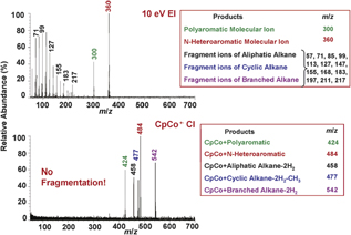
Comparison of laser-induced acoustic desorption/electron ionization (LIAD/EI) (top) and LIAD/CpCo+• (bottom) mass spectra of a five-component model hydrocarbon mixture (∼1:1:1:1:1; compounds are given in the red boxes).Reference Crawford, Campbell, Fiddler, Duan, Qian, Gorbaty and Kenttämaa40 The data demonstrate that only two components of the mixture yielded a stable molecular ion upon EI while all others fragmented extensively. However, use of the CpCo+• reagent ion resulted in formation of a stable pseudo-molecular ion for all five compounds with no fragmentation. Note: Cp, cyclopentadienyl; m, mass of the ion; z, number of charges in the ion.

LIAD has also been combined with tunable synchrotron vacuum ultraviolet photoionization (SVUVPI) in a quadrupole time-of-flight (QTOF) mass spectrometer.Reference Jia, Weng, Zhou, Qi, Guo, Zhao and Chen27 Desorption and ionization of the analyte occurred in the vacuum chamber. Fragile compounds, including ergosterol (molecular weight [MW] 397 Da), were evaporated and ionized with little to no observed fragmentation by using the lowest laser energy that enabled ionization of the analyte.Reference Jia, Weng, Zhou, Qi, Guo, Zhao and Chen27
In addition to vacuum ionization methods, LIAD has been coupled with atmospheric pressure ionization methods. One of them is ESI. This is a soft ionization method typically used to characterize large polar compounds, such as proteins.Reference Cheng, Huang and Shiea9,Reference Banerjee and Mazumdar28,Reference Cheng, Cheng, Chang and Shiea29 Different types of protein mixtures, such as protein standards, human serum, and cow’s milk, were desorbed by LIAD, mixed into an ESI plume for ionization, and transferred into a QTOF mass spectrometer for analysis. Singly or multiply charged analyte ions of proteins were detected.Reference Cheng, Cheng, Chang and Shiea29
Commercial atmospheric pressure chemical ionization (APCI) sources also have been coupled to LIAD.Reference Borton, Amundson, Hurt, Dow, Madden, Simpson and Kenttämaa22,Reference Gao, Borton, Owen, Jin, Hurt, Amundson, Madden, Qian and Kenttämaa30 APCI is beneficial compared to ESI as it can be used to ionize nonpolar as well as polar analytes. In these experiments, reagent ions (such as the radical cations of nitrogen and carbon disulfide [i.e., N2+● and CS2+●]) were generated via APCI. Desorbed neutral analyte molecules were mixed with the plume of reagent ions and subsequently ionized.Reference Borton, Amundson, Hurt, Dow, Madden, Simpson and Kenttämaa22,Reference Gao, Borton, Owen, Jin, Hurt, Amundson, Madden, Qian and Kenttämaa30 Another approach involved introducing APCI reagent gas, O2, through 16 small channels surrounding the sample foil and then mixing them with the desorbed analytes.Reference Nyadong, Quinn, Hsu, Hendrickson, Rodgers and Marshall31 The mixture was subsequently ionized by Corona discharge. This approach is referred to as atmospheric pressure laser-induced acoustic desorption chemical ionization.Reference Nyadong, Quinn, Hsu, Hendrickson, Rodgers and Marshall31 Linear and branched alkanes and cycloalkanes were ionized to produce [M—H]+ ions with minimal fragmentation.Reference Nyadong, Quinn, Hsu, Hendrickson, Rodgers and Marshall31
Atmospheric pressure photoionization (APPI) is an option for ionizing molecules with chromophores.Reference Raffaelli and Saba32 A vacuum ultraviolet lamp and toluene solvent have been utilized to ionize both aromatic and nonaromatic hydrocarbons desorbed via LIAD, including phenanthrene, perylene, and squalene.Reference Benham, Hodyss, Fernández and Orlando33 Most analytes were ionized to give molecular ions with minimal fragmentation.
Applications and prospects
Applications in petroleomics
Many methods have been tested for the mass spectrometric analysis of heavy hydrocarbons (MW range 400–1500 Da) related to petroleomics (characterization of petroleum at the molecular level). These compounds are particularly challenging as they are not volatile, do not contain ionizable functionalities, and after ionization they often fragment extensively. For example, laser desorption ionization preferentially ionizes relatively small moleculesReference Herod34 and suffers from aggregation and analyte fragmentation.Reference Hortal, Martínez-Haya, Lobato, Pedrosa and Lago35,Reference Rodgers and McKenna36 On the other hand, ESI can be used to evaporate and ionize only polar components of petroleum, while the majority of the components in petroleum are nonpolar hydrocarbons.Reference Porter, Mayer and Fingas37–Reference Crawford, Campbell, Fiddler, Duan, Qian, Gorbaty and Kenttämaa40 Field desorption/field ionization (FD/FI) is the method traditionally employed in the oil industry to desorb and ionize heavy hydrocarbon samples. This method is based on an emitter composed of needle-like whiskers onto which the sample is deposited.Reference Qian and Dechert41,Reference Gross42 High voltage applied to the whiskers causes desorption and ionization of the sample. However, FD/FI suffers from several problems, such as extensive ion fragmentation, poor reproducibility, mass bias, and variable sensitivity.Reference Klesper and Röllgen43–Reference Briker, Ring, Iacchelli, McLean, Fairbridge, Malhotra, Coggiola and Young46
LIAD/EI can be used to desorb and ionize heavy hydrocarbons in petroleum samples, but it often causes extensive fragmentation at high electron energies and has low ionization efficiency at low electron energies.Reference Duan, Qian, Habicht, Pinkston, Fu and Kenttämaa47 LIAD coupled to other ionization methods besides EI is a better option. LIAD has been coupled with CI in an FT-ICR mass spectrometer to evaporate and ionize large saturated hydrocarbonsReference Campbell, Crawford and Kenttämaa48 and petroleum distillates with minor fragmentation.Reference Crawford, Campbell, Fiddler, Duan, Qian, Gorbaty and Kenttämaa40 In these studies, the cyclopentadienyl cobalt radical cation, CpCo+•, was utilized as the CI reagent ion. For the model compounds (R) and mixtures tested, the reagent ion formed an adduct ion [R + CpCo]+• or fragment ions [R + CpCo—H2]+• or [R + CpCo—2H2]+• for each analyte, but in most cases, no further fragmentation was induced by this reagent ion. This method was tested for different types of compounds, including alkanes, alkenes, aromatic compounds, and heterocyclic compounds. Only limited fragmentation was observed. Therefore, this approach enabled the determination of the MW distributions for the analyzed mixtures.
This method also showed minimal ionization bias for different types of hydrocarbons (Figure 5). However, distinction of alkenes from analogous alkanes was challenging if the alkane produced [R + CpCo—2H2]+• as the major ions (Figure 5). Therefore, a better LIAD/CI approach was developed later.Reference Duan, Qian, Habicht, Pinkston, Fu and Kenttämaa47,Reference Duan, Fu, Pinkston, Habicht and Kenttämaa49 This method employed the ClMn(H2O)+ reagent ion that only generated one product ion for each model compound (R) studied, the [R + ClMn]+ ion, which is a benefit compared to the CpCo+• reagent ion. The method showed minimal ionization bias toward different types of compounds in mixtures such as base oil samples. Further, analysis of saturated hydrocarbon polymers, such as polyethylene, was also successfully accomplished with this method and yielded results that agreed well with other techniques.Reference Campbell, Fiddler, Crawford, Gqamana and Kenttämaa50
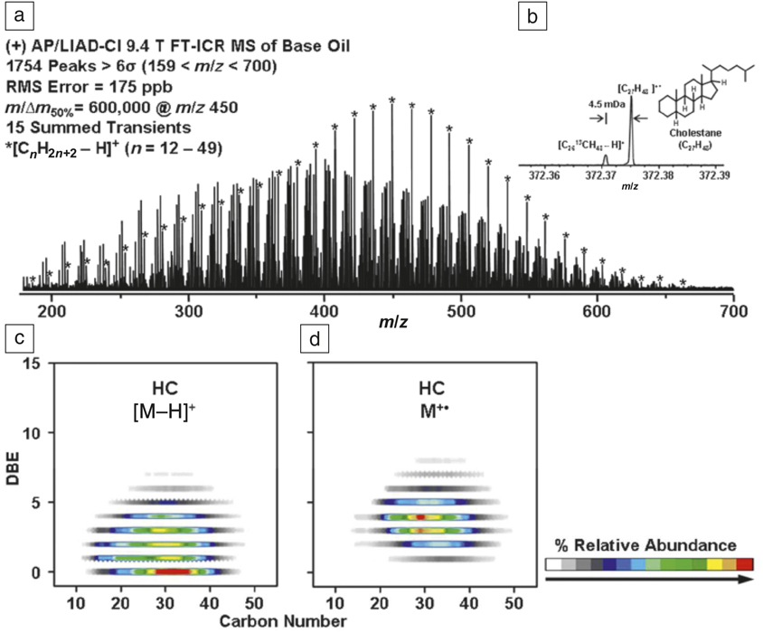
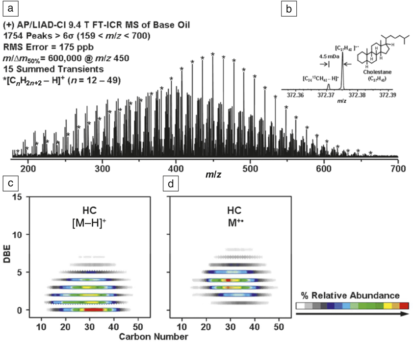
LIAD coupled with APCI and using O2 reagent gas (atmospheric pressure laser-induced acoustic desorption chemical ionization) in an FT-ICR mass spectrometer has been recommended for the analysis of large hydrocarbons, based on studies of model compound mixtures and a base oil sample.Reference Nyadong, Quinn, Hsu, Hendrickson, Rodgers and Marshall31 The effects of N2 and O2 reagent gases on the ionization of the analytes were examined. When N2 was used, molecular ions M+• were formed for linear alkanes while [M—H]+ ions were formed when O2 was used. No fragmentation was observed in either case. Analogous ions were generated for branched alkanes. However, some model compounds, such as squalene (2,6,10,15,19,23-hexamethyltetracosane), experienced extensive fragmentation after ionization when using N2 gas. Therefore, O2 was concluded to be a better reagent gas. However, the use of O2 resulted in the production of either M+• or [M–H]+ ions for cycloalkanes, depending on the temperature of the heated metal capillary. The MW distribution of the base oil sample as well as the elemental composition of each component in the sample were determined (Figure 6). These results were comparable to those obtained previously using LIAD/CI methods in an FT-ICR,Reference Duan, Qian, Habicht, Pinkston, Fu and Kenttämaa47,Reference Campbell, Crawford and Kenttämaa48,Reference Campbell, Fiddler, Crawford, Gqamana and Kenttämaa50 but the throughput of the experiment was higher (less than 3 min/sample versus ∼30 min/sample). The analysis of the paraffin series in the base oil sample was in good agreement with the results obtained previously using FI/FD.Reference Hsu51
(a) Laser-induced acoustic desorption/atmospheric pressure chemical ionization (LIAD/APCI) 9.4 T Fourier-transform ion cyclotron resonance (FT-ICR) mass spectrum for a base oil obtained using O2 reagent gas. Reprinted with permission from Reference Nyadong, Quinn, Hsu, Hendrickson, Rodgers and MarshallReference 31. © 2012 American Chemical Society. The most abundant peaks (highlighted by asterisks) correspond to a saturated hydrocarbon series. The mass-scale-expanded inset (b) highlights the requirement for ultrahigh resolution to separate ions that differ in elemental composition by 13C versus CH (4.5 mDa difference in exact mass). (c, d) Isoabundance-contoured plot of double-bond equivalent (the number of double bonds and rings) versus the number of carbons for ions containing only carbons (C) and hydrogens (H). (c) The saturated hydrocarbon series (DBE = 0) corresponding to alkanes has the highest relative abundance for ions by hydride abstraction ([M–H]+). The figure also shows higher DBE series corresponding to cycloalkanes with increasing number of rings (from one up to eight rings; DBE = 1–8). (d) When the heated metal capillary temperature was raised to ∼250°C, cycloalkanes also generated molecular ions (M+•). Note: MS, mass spectrometry; RMS, root mean square; m, mass of the ion; Δm 50%, mass spectral peak width at half maximum peak height; z, number of charges in the ion; DBE, double-bond equivalence; σ, standard deviation.
Applications in studies of aromatic compounds
Gas-phase reactivity of the radical cations of polycyclic aromatic hydrocarbons (PAHs), such as naphthalene, anthracene, and phenanthrene, and their nitrogen-containing analogs (PANHs), such as quinoline, isoquinoline, and acridine, toward hydrogen atoms has been explored by using LIAD. These reactions are of interest for improving the understanding of the chemistry occurring for PAHs and PANHs in diffuse regions of interstellar medium, where both PAH and PANH radical cations and hydrogen atoms are abundant. In order to examine these reactions, LIAD was coupled with a flowing afterglow-selected ion flow tube instrument, a special type of a mass spectrometer designed for studies of gas-phase ion-molecule reactions at higher pressures than in most mass spectrometers.Reference Demarais, Yang, Snow and Bierbaum52 LIAD enabled the introduction of the relatively large, nonvolatile PAH and PANH molecules into the flowing afterglow in sufficient concentrations for reactivity studies. LIAD/He+ CI or Penning ionization using metastable Ar atoms was used to generate molecular ions from the gaseous molecules. Hydrogen atoms were generated by thermal dissociation of H2 on a hot W filament. Addition of a hydrogen atom to all cations studied was observed. Interestingly, the reaction efficiencies of PAH and PANH cations with the same size and geometry (e.g., ionized anthracene versus acridine) were found to be identical.
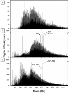
LIAD coupled to single-photon ionization ([SPI]; photon energy 7.9 eV) time-of-flight (TOF) mass spectrometer has been tested for the analysis of several organic dyes with low ionization energies.Reference Zinovev, Veryovkin, Moore and Pellin15 Dominant molecular ions were generated for all of the compounds studied, though increased laser intensities led to increased fragmentation in some of the experiments. Later, LIAD was coupled to a tunable SVUVPI source and a QTOF mass spectrometer. The SVUVPI source provided photons with energies ranging from 7.8 to 24.0 eV, thus enabling ionization of a wider range of analytes. The technique was examined for the analysis of petroleum related aromatic compounds by using several different photon energies.Reference Chen, Jia, Zhao, Lu, Guo, Weng and Qi53 The extent of fragmentation was successfully controlled by selecting the lowest photon energy capable of ionizing the analytes. Interestingly, the mass distribution measured for one of the mixtures (Figure 7), composed of aromatic compounds and separated directly from Lungu atmospheric residue and not subjected any further treatment, was bimodal, peaking at 300 and 450 Da. The relative areas of these two peaks were found to change significantly with the photon energy (9.0, 11.0, or 14.0 eV), suggesting the presence of at least two categories of compounds with different ionization energies (i.e., polycyclic aromatic hydrocarbons with a molecular weight range of 400−900 Da and aliphatic and alicyclic compounds with a mass range of 200−400 Da).Reference Chen, Jia, Zhao, Lu, Guo, Weng and Qi53
Laser-induced acoustic desorption/synchrotron vacuum ultraviolet photoionization mass spectra of a mixture of aromatic compounds separated from a Lungu atmospheric residue resin and measured using synchrotron vacuum ultraviolet photon energies of (a) 9.0 eV, (b) 11.0 eV, and (c) 14.0 eV. The relative areas of the major two peaks change significantly with the photon energy, suggesting the presence of at least two categories of compounds with different ionization energies. Adapted with permission from Reference Chen, Jia, Zhao, Lu, Guo, Weng and QiReference 53. © 2013 American Chemical Society.

Applications in life sciences
Mass spectrometric analysis of biopolymers, such as polypeptides and polynucleotides, is usually performed using MALDI or ESI to evaporate and gently ionize the molecules nearly simultaneously. However, in some instances, it is preferable to separate the evaporation and ionization steps. This is the case, for example, when ionization via reactions other than proton or metal cation attachment is desired. Furthermore, the ability to examine gas-phase reactions of net-neutral biopolymers with radical cations is sometimes desirable.Reference Petzold, Ramírez-Arizmendi, Heidbrink, Pérez and Kenttämaa54 LIAD can be used to address both of these issues by vaporizing the intact neutral biomolecules into a mass spectrometer, followed by ionization using desirable reagent ions other than Brønsted acids, (i.e., reagents that do not protonate the biomolecules) or by exploring their gas-phase reactivity toward ionic reagents of interest.
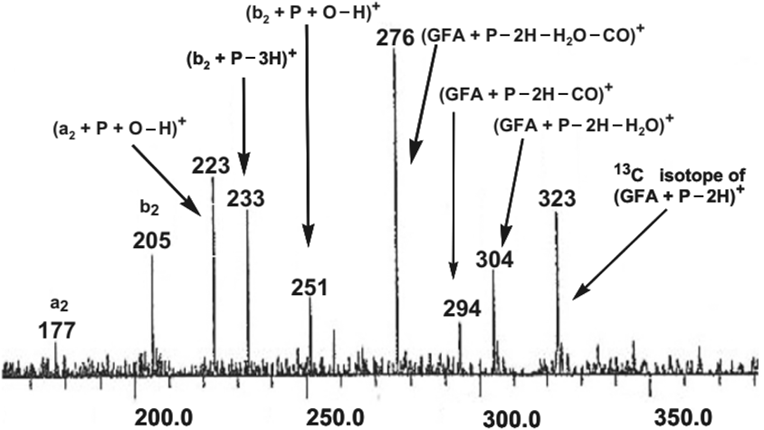
Examination of LIAD/EI mass spectra of several amino acids revealed that this method generates similar mass spectra to those obtained using EI coupled with thermal heating to evaporate the amino acids.Reference Jarrell, Owen, Riedeman, Prentice, Pulliam, Max and Kenttämaa23 Therefore, LIAD can be used to measure EI mass spectra for peptides and other compounds that cannot be evaporated using thermal heating. Indeed, this approach has been employed to measure EI mass spectra for several small peptides as well as their hydrochloride salts.Reference Jarrell, Owen, Riedeman, Prentice, Pulliam, Max and Kenttämaa23 Further, LIAD/EI has been demonstrated to enable the differentiation of peptides containing leucine versus isoleucine.Reference Jarrell, Owen, Riedeman, Prentice, Pulliam, Max and Kenttämaa23 A LIAD/CI method has been introduced for peptide sequencing.Reference Somuramasami and Kenttämaa55 Utilization of P(OCH3)2+ as a CI reagent ion to ionize peptides desorbed by LIAD generated stable [peptide + P–2H]+ ions. Subjecting these ions to collision-activated dissociation (CAD) yielded sequence-related product ions (Figure 8).
A collision-activated dissociation mass spectrum of [GFA + P–2H]+ ions (GFA or gly-phe-ala is a tripeptide containing the amino acids glycine, phenylalanine, and alanine) obtained to study peptide sequencing. The various sequence-related fragment ions are indicated.Reference Somuramasami and Kenttämaa55 Note: a2 and b2 are symbols for common peptide fragment ions.

Further, LIAD has been utilized to desorb net-neutral nucleotides, dinucleoside phosphates, dipeptides, and tetrapeptides into the gas phase in order to study their reactions with charged phenyl radicals generated in an FT-ICR mass spectrometer.Reference Liu, Petzold, Ramirez-Arizmendi, Perez and Kenttämaa56,Reference Li, Fu, Habicht, Pates, Nash and Kenttämaa57 The results were found to be consistent with those obtained for similar systems in aqueous solution.Reference Hazelwood, Davies, Gilbert and Packer58–Reference Yang, Nash, Yurkovich, Jin, Vinueza and Kenttämaa61 Therefore, LIAD coupled with mass spectrometry (MS) provides a convenient method to study the fundamental reactivity of aromatic biradicals toward biomolecules, which is not straightforward when using solution experiments.
On the other hand, LIAD/CI in an FT-ICR mass spectrometer has been demonstrated to enable the detection of nonpolar lipids and steroid model compoundsReference Jin, Daiya and Kenttämaa24 ionized using ClMn(H2O)+ as the CI reagent ion. Little or no fragmentation was observed. These analytes either could not be ionized using APCI and ESI because they are nonpolar, or extensive fragmentation was observed (upon APCI).
The previously discussed LIAD/SVUVPI approach has also been used for the analysis of fragile compounds, such as ergosterol (a sterol found in fungi). The extent of fragmentation was controlled by selecting the lowest photon energy capable of ionizing the compounds.Reference Jia, Weng, Zhou, Qi, Guo, Zhao and Chen27
LIAD has been utilized to study the mechanisms of DNA strand breakage via dissociative electron attachment (DEA) of model compounds (thymidine and D-ribose-5-phosphate).Reference Bald, Dąbkowska and Illenberger62 The neutral model compounds were desorbed by LIAD, then ionized and fragmented via DEA. When using electrons with near 0 eV kinetic energies, DEA caused the cleavage of the sugar–phosphate linkage in the d-ribose-5-phosphate, suggesting that DEA occurs directly in the DNA/RNA backbone.
Femtosecond pulsed lasers coupled to LIAD have been utilized to study extremely fast gas-phase dynamics of biomolecules, such as vibrational wave packet evolution.Reference Remacle and Levine63 As utilization of a femtosecond laser can provide information on interactions on time scales shorter than structural rearrangements and charge-transfer processes, it may be more suitable for peptide sequencing and for causing selective bond cleavages than the use of nanosecond lasers.Reference Calvert, Belshaw, Duffy, Kelly, King, Smyth, Kelly, Costello, Timson, Bryan, Kierspel, Rice, Turcu, Cacho, Springate, Williams and Greenwood18,Reference Calvert, Kelly, Duffy, Belshaw, King, Williams and Greenwood64 Fragmentation of the ionized analytes could be controlled by changing the pulse length of the laser.Reference Remacle and Levine63
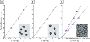
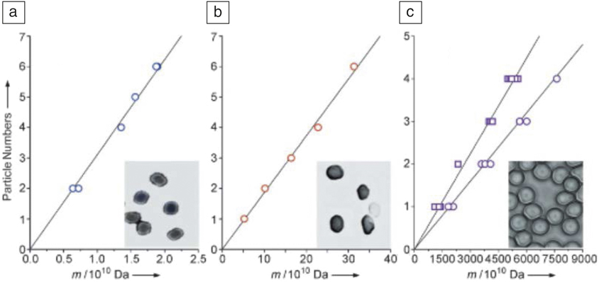
LIAD has been successfully used to desorb and detect vaccinia viruses, E. coli K-12 bacteria, red blood cells, and other bioparticles in a three-dimensional quadrupole ion trap mass spectrometer.Reference Peng, Yang, Kang, Tzeng, Nie, Chang, Chang and Chen65 These bioparticles were charged, and therefore, no external ionization was needed in this experiment (Figure 9).Reference Peng, Yang, Kang, Tzeng, Nie, Chang, Chang and Chen65 The m/z value of each single particle was determined by observing the light-scattering pattern, which is related to the trap-driving frequency (Ω/2π) and voltage amplitude (V ac), when using a charge-coupled device for collection of scattered photons. The feasibility of this method was demonstrated by measuring the sizes of polystyrene particle standards.Reference Peng, Lin, Lin, Chu, Yu, Chang and Chen66–Reference Kahler and Lloyd68
Plots of the number of particles as a function of their mass (m) measured for (a) vaccinia viruses (inset: electron microscope image), (b) E. coli K-12 bacteria (inset: electron microscope image), and (c) human red blood cells (inset: optical microscope image). The two data sets shown for (c) are those of normal (󠄀○) and anemic (□) red blood cells. As cluster particles are formed during the laser desorption process, least-squares fitting of measured masses versus particle numbers was used to derive the mass of the monomeric particles. Adapted with permission from Reference Peng, Yang, Kang, Tzeng, Nie, Chang, Chang and ChenReference 65. © 2006 Wiley.
Prospects
LIAD has thus far been demonstrated to enable the evaporation of a variety of nonvolatile and thermally fragile neutral compounds into mass spectrometers, ranging from large nonpolar compounds, such as large saturated hydrocarbons, to fragile polar biopolymers, such as oligonucleotides. LIAD has even been used to desorb charged particles, such as bacteria. The gas-phase molecules have low internal and kinetic energies when proper experimental conditions are employed. They can be ionized using a wide variety of methods (EI, CI, SPI, ESI, APCI, APPI, and Penning ionization), which sets this method apart from such commonly used evaporation/ionization methods as MALDI, ESI, and DESI. The ability to use almost any ionization method in combination with LIAD enables unprecedented control over ion energetics. LIAD can be operated in high vacuum and at atmospheric pressure and anywhere in between, and it can be coupled with most mass spectrometers, making this method quite versatile.
Future applications of LIAD/ MS will be diverse. For example, LIAD/MS is anticipated to lead to improvements in disease diagnostics; it has been used already to distinguish normal red blood cells from anemic red blood cells.Reference Peng, Yang, Kang, Tzeng, Nie, Chang, Chang and Chen65 Coupling of LIAD with new types of mass spectrometric methods, such as ion mobility MS, will enable more detailed studies of large gas-phase molecules and ions and will facilitate identification of isomers that present a challenge for other mass spectrometric methods.Reference Huang, Ossenbrüggen, Rubinsky, Schust, Horke and Küpper19 The efficiency of desorption will be increased and the extent of ion fragmentation will be decreased, thus making LIAD/MS even more powerful than it is today.
Xin Ma is a doctoral candidate in the Department of Chemistry at Purdue University. He received his BS degree in chemistry at Lanzhou University, China, in 2013. His research interests include gas-phase reactivity of organic carbon- and boron-centered bi- and polyradicals toward small organic molecules and biomolecules as well as gas-phase C–H bond activation in hydrocarbons by using halogenated closo-borate anions. His research also focuses on the synthesis of novel lignin model compounds for the exploration of the mechanisms of their fast pyrolysis reactions by using tandem mass spectrometry and the development of mass spectrometry methods for the identification of functionalities in drug metabolites via ion–molecule reactions. Ma can be reached by email at ma315@purdue.edu.

Yuyang Zhang is a doctoral candidate in the Department of Chemistry at Purdue University. He received his BS degree in chemistry at Zhejiang University, China. His research interests include the development of ionization methods for mass spectrometric analysis of large saturated hydrocarbons and mass spectrometric characterization of the structures of asphaltenes via model compound studies. His research also focuses on the development of a novel laser-induced acoustic desorption system for more efficient evaporation of large compounds. Zhang can be reached by email at yuyang@purdue.edu.

Hao-Ran Lei is a doctoral candidate in the Department of Chemistry at Purdue University. He received his BS degree in the College of Chemistry and Molecular Engineering at Peking University, China, in 2012. His research interests include the development of more efficient laser-induced acoustic desorption techniques for desorption of large biomolecules. Lei can be reached by email at lei48@purdue.edu.

Hilkka Kenttämaa is the Frank Brown Distinguished Professor of Chemistry at Purdue University. She received her PhD degree in chemistry at Helsinki University, Finland. Her research interests include fundamental understanding of organic tandem mass spectrometry, including the kinetics, mechanisms, and thermochemistry of reactions used to ionize analytes and characterize their structures, utilization of tandem mass spectrometry to study the reactivity, kinetics, and reaction mechanisms of organic and biological reaction intermediates, including organic polyradicals, delineation of mechanisms of fast pyrolysis processes of biomass via tandem mass spectrometry, and development of better chromatography/tandem mass spectrometry methods for the fast and reliable compositional characterization and quantitation of complex mixtures of organic compounds, such as drug metabolites, degraded biomass, biomass conversion products, crude oil, and aviation fuels. Kenttämaa can be reached by email at hilkka@purdue.edu.