Introduction
Foraminifers are unicellular organisms that inhabit various marine macro-environments ranging from carbonate platforms to deep-sea basins (Gooday et al., Reference Gooday, Bernhard, Levin and Suhr2000; Gooday, Reference Gooday2003; Pawlowski, Reference Pawlowski2009). They could also survive in extreme conditions, such as salt marshes or sulfidic or oxygen-depleted environments (Bernhard, Reference Bernhard1993; Kaiho, Reference Kaiho1994a; Bernhard and Alve, Reference Bernhard and Alve1996; Bernhard and Sen Gupta, Reference Bernhard and Sen Gupta2003; Koho and Piña-Ochoa, Reference Koho and Piña-Ochoa2012). Foraminifers are one of the major fossil groups for interpreting Phanerozoic geological history, especially because of their abundant fossil record. They are widely used for characterizing the biodiversity change during the end-Permian mass extinction.
The Permian–Triassic (P–T) transition was a significant interval in Earth history. Following voluminous eruptions of Siberian Traps and widespread oceanic anoxia, more than 90% of marine species and 70% of terrestrial species were wiped out in probably just a few million years (Erwin, Reference Erwin1994; Benton and Benton, Reference Benton and Benton2003). During the Early Triassic, there were large temperature perturbations and exceptional warming in the low latitudes, suggesting harsh conditions continued in the aftermath of the P–T mass extinction (Sun et al., Reference Sun, Joachimski, Wignall, Yan, Chen, Jiang, Wang and Lai2012). Foraminifers underwent a sharp decrease in diversity and abundance during the mass extinction (Tappan and Loeblich, Reference Tappan and Loeblich1988; Jin et al., Reference Jin, Zhang and Shang1994; Rampino and Adler, Reference Rampino and Adler1998; Jin et al., Reference Jin, Wang, Wang and Erwin2000; Leven and Korchagin, Reference Leven and Korchagin2001; Groves et al., Reference Groves, Altiner and Rettori2005; Mohtat-Aghaï and Vachard, Reference Mohtat-Aghaï and Vachard2005; Song et al., Reference Song, Tong and Chen2009a, Reference Song, Tong and Chen2009b). Subsequently, they slowly recovered in the late Early Triassic and diversified in the Middle–Late Triassic (Tong and Shi, Reference Tong and Shi2000; Groves and Altiner, Reference Groves and Altiner2005; Márquez, Reference Márquez2005; Mohtat-Aghaï and Vachard, Reference Mohtat-Aghaï and Vachard2005). In recent years, detailed systematic studies of latest Permian foraminifers have been widely undertaken, including faunas from South China (Wang, Reference Wang1976; Lin, Reference Lin1978; Zhao et al., Reference Zhao, Sheng, Yao, Liang, Chen, Rui and Liao1981; Lin et al., Reference Lin, Li and Sun1990; Tong and Shi, Reference Tong and Shi2000; Gu et al., Reference Gu, Pei, Yang and Gao2002; Zhang and Hong, Reference Zhang and Hong2004; Song et al., Reference Song, Tong and He2006; Gaillot and Vachard, Reference Gaillot and Vachard2007; Gaillot et al., Reference Gaillot, Vachard, Galfetti and Martini2009; Song et al., Reference Song, Tong and Chen2009b), Japan (Kobayashi, Reference Kobayashi2004, Reference Kobayashi2005, Reference Kobayashi2006, Reference Kobayashi2012), Italy (Groves et al., Reference Groves, Rettori, Payne, Boyce and Altiner2007), Turkey (Groves and Altiner, Reference Groves and Altiner2005; Groves et al., Reference Groves, Altiner and Rettori2005; Gaillot and Vachard, Reference Gaillot and Vachard2007), the Caucasus (Pronina-Nestell and Nestell, Reference Pronina-Nestell and Nestell2001), northwestern Serbia (Nestell et al., Reference Nestell, Sudar, Jovanović and Kolar-Jurkovšek2009), western Slovenia (Nestell et al., Reference Nestell, Kolar-Jurkovsek, Jurkovsek and Aljinovic2011), and southwestern Austria (Krainer and Vachard, Reference Krainer and Vachard2011). Extinction and recovery patterns of foraminifers across the P–T transition were also analyzed (Altiner et al., Reference Altiner, Groves and Özkan-Altiner2005; Groves and Altiner, Reference Groves and Altiner2005; Groves et al., Reference Groves, Altiner and Rettori2005; Márquez, Reference Márquez2005; Mohtat-Aghaï and Vachard, Reference Mohtat-Aghaï and Vachard2005; Groves et al., Reference Groves, Rettori, Payne, Boyce and Altiner2007; Song et al., Reference Song, Tong and Chen2009a; Song et al., Reference Song, Tong and Chen2009b; Angiolini et al., Reference Angiolini, Checconi, Gaetani and Rettori2010). Furthermore, foraminiferal data have provided an important basis for characterizing extinction patterns associated with the end-Permian mass extinction (Tong, Reference Tong1991; Tong and Shi, Reference Tong and Shi2000; Stanley and Yang, Reference Stanley and Yang1994; Rampino and Adler, Reference Rampino and Adler1998; Jin et al., Reference Jin, Wang, Wang and Erwin2000; Leven and Korchagin, Reference Leven and Korchagin2001; Song et al., Reference Song, Tong and Chen2011, Reference Song, Wignall, Tong and Yin2012).
Deep-water foraminifers are very important in the study of the Permian–Triassic extinction because they are less affected than the shallower representatives of the group and are mainly composed of nodosariids, which are at the origin of all the dominant hyaline modern foraminifers. Moreover, deep-water foraminiferal faunas were a major component of the end-Permian oceanic biodiversity. A reliable theory on the extinction of foraminifers in the P–T (Permian–Triassic boundary) interval cannot be established as long as both shallow- and deep-water foraminifers are not carefully studied. Most of the previous studies were focused on shallow-water faunas from carbonate platform or reef settings. Few foraminiferal faunas have been reported from the deep-water environments. Only recently, a deep-water foraminiferal fauna extracted from siliceous deposits in the Dalong Formation was systematically described from the Dongpan section in Guangxi, southwestern South China (Shang et al., Reference Shang, Vachard and Caridroit2003; Gu et al., Reference Gu, Feng and He2007). Significantly, deep-water environments would be one of the refuges during the Permian–Triassic crisis. Some foraminifers known from the Early Triassic were found to have a close affinity to latest Permian lineages based on their morphology and wall ultrastructure (Groves et al., Reference Groves, Altiner and Rettori2003; Krainer and Vachard, Reference Krainer and Vachard2011; Rigaud et al., in Reference Rigaud, Vachard, Martini and Rettoripress), suggesting that some foraminifers could have survived the end-Permian oceanic crisis.
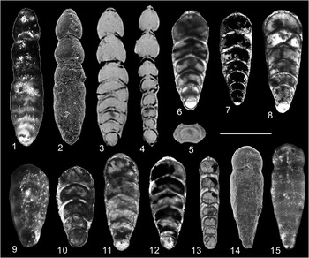
Here, we document a latest Permian foraminiferal assemblage including 19 species (belonging to 10 genera) from deep-water strata in the northern periphery of South China. Although its Changhsingian age has been confirmed, its taxonomic composition is largely different from what was previously reported from coeval shallow-water environments in South China, which are dominated by small, elongate, flattened, nodosariid foraminifers. Therefore, this study contributes to our knowledge of the evolutionary history of deep-water foraminifers during the Permian–Triassic turnover. The paleoecological significance of the fauna is also discussed.
Geological setting
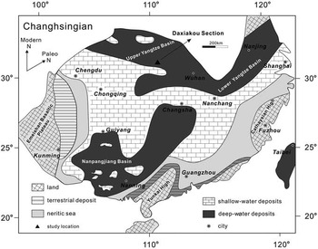
During the Permian–Triassic interval, the Daxiakou section in Xingshan County, Hubei Province, was situated along the north margin of the Yangtze Platform, which was located in the low latitudes of the eastern Paleo-Tethyan realm (Fig. 1). The latest Permian strata in this section are represented by the Dalong Formation, which is mainly composed of black siliceous mudstone and carbonaceous mudstone, overlying bafflestone and bioclastic limestone of the Wujiaping (or Wuchiaping) Formation.
Changhsingian paleogeographic map of South China showing the location of the studied section (modified from Feng and Gu, Reference Feng and Gu2002; Xie et al., Reference Xie, Pancost, Wang, Yang, Wignall, Luo, Jia and Chen2010).

The Dalong Formation, named by Zhang and Chen (Reference Zhang and Chen1938), is widely distributed in South China. It is mostly composed of chert and cherty limestone and has been extensively investigated (Sheng, Reference Sheng1962; Zhao et al., Reference Zhao, Liang and Zhen1978). Although the age of the Dalong Formation was disputed for several decades, it has been widely accepted that the Dalong Formation is contemporaneous with the Changhsing (or Changxing) Formation (Zhao et al., Reference Zhao, Liang and Zhen1978). The Dalong and Changhsing Formations were deposited during the high sea-level transgression of the Changhsingian; and they marked the uppermost strata of the Late Permian in South China. The Dalong Formation is generally recognized as deep-water facies (Jin and Li, Reference Jin and Li1987; Jin, Reference Jin1987; Yao and Zhang, Reference Yao and Zhang1996) and it accumulated on the lower part of an outer carbonate platform to the upper basin edge or at the limit of the continental slope (Yin et al., Reference Yin, Ding, Zhang, Tong, Yang and Lai1995; Niu et al., Reference Niu, Duan, Xu, Fu, Zeng and Zhu1999). Recent works on several sections situated within the studied area (western part of Hubei Province) suggest that the Dalong Formation was deposited in a deep-water, even anoxic environment (Niu et al., Reference Niu, Duan, Xu and Xu2000; Cai et al., Reference Cai, Zhang, Peng and Feng2007).
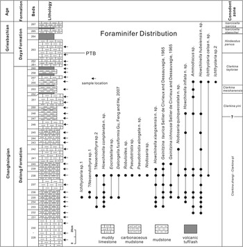
The Daxiakou section contains a continuous succession of the Permian–Triassic boundary interval conodont zones in the Dalong Formation and lower part of the Daye Formation (Wang and Xia, Reference Wang and Xia2004; Wang, Reference Wang2005; Wang et al., Reference Wang, Xia, Zhang and Dong2007; Xia et al., Reference Xia, Zhang, Wang and Yang2007), which are, in ascending order, the Clarkina (=Neogondonella) zhangi – Clarkina qii, Clarkina yini, Clarkina meishanensis, and Clarkina taylorae zones (Fig. 2) (Zhao et al., Reference Zhao, Chen, Chen and Cao2013). Based on the first occurrence of Hindeodus parvus, the Permian–Triassic boundary in the Daxiakou section is placed between beds XK262 and XK263 in the Daye Formation (Fig. 2; Xia et al., Reference Xia, Zhang, Wang and Yang2007; Zhao et al., Reference Zhao, Chen, Chen and Cao2013). The conodont zones in the Dalong Formation from the Daxiakou section correlate well with those in the Changhsing Formation in the Meishan section (GSSP of the PTB).
Stratigraphic column and foraminiferal distribution in the Daxiakou section. Conodont zones after Wang et al. (Reference Wang, Xia, Zhang and Dong2007) and Zhao et al. (Reference Zhao, Chen, Chen and Cao2013).

Methods
Sampling
Forty-three microfossil samples from the Dalong Formation in the Daxiakou section were processed and studied. The samples were collected continuously bed by bed. Each sample interval is 5 to 10 cm thick. The stratigraphic column and sampling horizons used in this study are shown in Figure 2. The sample numbers range from XK228 to XK267, in ascending order, according to the numbering system used by Wang and Xia (Reference Wang and Xia2004).
Free specimen extraction
Each sample was divided into two parts for two different disaggregation methods using hydrofluoric acid (HF) disaggregation and acetic acid (HAc) heating disaggregation methods.
Dilute HF disaggregation, a conventional method to extract radiolarians from chert or other siliceous rocks, is widely used in Paleozoic radiolarian studies with a HF concentration of 4%–5%. According to recent works on sample preparation (Gu et al., Reference Gu, Pei, Yang and Gao2002; Gu and Feng, Reference Gu and Feng2007), it is possible to retrieve many foraminiferal tests from siliceous sediments by dilute HF immersion, where HF is diluted to 2%–3% and the reaction time is shortened to less than 8 hours, which could be sufficient to extract some recrystallized or metasomatic foraminiferal tests. In this study, the samples were treated by this process, and the residues between #20 and #240 GB standard sieve meshes were retrieved to pick for fossils.
The other half of the samples was treated with the acetic acid method. In the past two decades, acetic acid has been used to separate calcareous microfossils from carbonates (Lethiers and Crasquin-Soleau, Reference Lethiers and Crasquin-Soleau1988; Costa de Moura et al., Reference Costa de Moura, de Moraes Rios-Netto, Wanderley and Pereira de Sousa1999; Lirer, Reference Lirer2000; Tur et al., Reference Tur, Smirnov and Huber2001; Kariminia, Reference Kariminia2004; Crasquin-Soleau and Kershaw, Reference Crasquin-Soleau and Kershaw2005); here we used the procedure suggested by Kariminia (Reference Kariminia2004) but slightly modified, by diluting the HAc to 90%–95%, compared to the 99.99% HAc concentration used by Kariminia (Reference Kariminia2004). The immersed samples were kept reacting in a heating sand bath with temperature 75°C–80°C. The reaction could take a few hours to a few days. When there is enough residue accumulated at the bottom of the beakers, the samples are washed and the residues between #20 and #230 meshes collected. The procedure is repeated twice or more until a sufficient amount of washed residue is retrieved from each sample for fossil examination.
Oriented thin sectioning
Well-preserved foraminiferal free tests were selected for oriented sectioning, using the method modified from Mathews (Reference Mathews1945) and Gu and Feng (Reference Gu and Feng2007). Before sectioning, the specimens were photographed under a reflected light microscope to get an overview of the tests. A drop of Canadian Balsam was put on a glass slide and heated for 10–15 minutes at 120°C until liquid. The specimen was mounted in the heated balsam and adjusted to a proper orientation, then ground using #3000 SiC grinding powder. When the desired plane is obtained, the balsam is reheated, the specimen is gently flipped over, and the other side of the specimen is ground to a desired thickness. Well-oriented foraminifer sections were observed and imaged under transmitted light and cross-polarized microscopes.
Micro-CT scanning
Selected specimens were scanned by SkyScan 1172 X-ray Micro-CT. The x-ray source was of 40 kV and 120 μA, without filters. A small camera pixel size (0.5 μm) was used, with the stage rotation step 0.10 degree. Frame averaging was four pictures per step and random movement was 20. For each scanned specimen, 1820 x-ray projection images were obtained; then, the scanned data were imported to NRecon (a SkyScan software) for reconstruction. The reconstructed images could be used by CTVox software to create 3D models, and oriented sections could be then generated.
Selected systematic paleontology
We use the classification system of higher foraminiferal taxa proposed by Cavalier-Smith (Reference Cavalier-Smith2002) and Mikhalevich (Reference Mikhalevich1998, Reference Mikhalevich2000, Reference Mikhalevich2004). Description and terminology mainly follow Gerke (Reference Gerke1967), which was partially translated into English by Karavaeva and Nestell (Reference Karavaeva and Nestell2007). Only new species are described herein. All illustrated specimens are housed in the Department of Biogeology, China University of Geosciences, Wuhan, Hubei, China.
Subphylum Foraminifera Cavalier-Smith, Reference Cavalier-Smith2002
Class Nodosariata Mikhalevich Reference Mikhalevich1992
Subclass Nodosariana Mikhalevich, Reference Mikhalevich1992 [nom. correct. Mikhalevich, Reference Mikhalevich1998]
Order Nodosariida Calkins, Reference Calkins1926 [nom. transl. Mikhalevich, Reference Mikhalevich1980 ex order Nodosalida Calkins, Reference Calkins1926=Lagenida Fursenko, Reference Fursenko1958]
Family Nodosariidae Ehrenberg, Reference Ehrenberg1838
Genus Nodosaria Lamarck, Reference Lamarck1816 emend. Marie, Reference Marie1941
Type species
Nautilus radicula Linné, Reference Linné1758.
Nodosaria quinquecostata new species
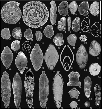
(1–3) Ammodiscus sp.: (1) XK238_10SEM_058 (SEM, frontal view, inner-mold preserved); (2) XK235_10SEM_051, (SEM, frontal view, proloculus is lost); (3) XK256_07SEM_036 (SEM, oblique lateral view). (4–12, 19) Eocristellaria sp.: (4–6) XK235_10SEM_052, (4, SEM, frontal view; 5, 6, Micro-CT reconstruction and slice showing the aperture in the terminal part of test); (7, 12) XK238_10TS_051 (7, lateral axial section; 12, hand drawing of 7); (8–11, 19) XK238_12SEM_021 (8, 9, 19, Micro-CT reconstruction showing the chambers arrangement and aperture; 10, frontal view with transmitted light; 11, hand drawing of 10). (13, 14)?Neoendothyra sp. 2: XK235_11SEM_052 (13, SEM, frontal view; 14, frontal view with reflected light); (15–18)?Neoendothyra sp. 1: (15, 16) XK236_11SEM_049 (SEM, 15, frontal views; 16, lateral apertural views); (17, 18) XK235_11SEM_052 (17, SEM, frontal view; 18, frontal view with reflected light). (20, 23–30) Dalongella fusiformis Gu, Feng, and He, Reference Gu, Feng and He2007: (20, 29) XK238_12SEM_021 (Micro-CT reconstruction, 20, transverse slice; 29, oblique apertural view showing radiate trace); (23, 24) XK238_11SEM_012 (microspheric test; 23, SEM, axial view; 24, axial view with transmitted light); (25–27) XK235_10SEM_063 (megalospheric test; (25) SEM, axial view; (26) axial slice from Micro-CT reconstruction; (27) hand drawing from 26); (28, 30) XK238_12SEM_024 (megalospheric test; axial view and transverse slice of Micro-CT reconstruction). (21, 22) Robuloides sp.: XK238_12TS_023 (21, lateral axial section; 22, hand drawing). (31–38) Nodosaria quinquecostata n. sp.: (31) XK235_11SEM_053 (paratype, megalospheric test; SEM, axial view of a broken test); (32) XK238_11SEM_207 (paratype, megalospheric test; axial view with reflected light); (33) XK238_10TS_071 (paratype, megalospheric test; frontal axial section); (34–38) XK238_10SEM_073 (megalospheric test; 34, holotype, SEM, axial view; (35, 37, 38) axial slice, transverse slice of adult stage and juvenile stage from Micro-CT reconstruction; (36) enlargement of the apertural end showing the apertural neck and radiate grooves). (39–41) Nodosaria sp.: XK238_10TS_068 (megalospheric test; 39, 40, axial and apertural views with reflected light before sectioning; 41, frontal axial section, proloculus is lost in sectioning). Scale bar is 200 μm.

Types
Holotype: Fig. 3.34 (No.XK238_10SEM_073); paratypes: Fig. 3.31 (No.XK238_11SEM_053), Fig. 3.32 (No.XK238_11SEM_207), and Fig. 3.33 (No.XK238_10TS_071). All types were recovered from the Dalong Formation, Daxiakou section, South China.
Diagnosis
Test elongate, pentagonal in transverse view, with five longitudinal costae; initial apical periphery tapering, with spiny outgrowth. Sutures horizontal, slightly depressed. Chambers high, fabiform; final chamber pear-shaped. Aperture terminal, developing external neck and lip with radial grooves.
Material
Sixteen free specimens; one was prepared for frontal axial section, three for SEM, and one for Micro-CT.
Description
Test elongate, uniserial, gently wavy in lateral outline, with pentagonal transverse section, and possessing five strong longitudinal costae. Test tapering in initial apical periphery with spiny outgrowth. Apertural end narrowly rounded, slowly expanding in width. Sutures horizontal, perpendicular to growth axis, and slightly depressed in terminal portion of test. Five to six chambers. Aperture terminal, with development of external neck and lip with apparent radial grooves. Protruding height of neck 0.018–0.026 mm. Proloculus small, spherical, with internal diameter 0.035 mm and external diameter 0.046–0.054 mm. Early chambers fabiform and become oblong cask-shaped in terminal part. Last chamber often attenuated, pear-shaped. Holotype (Fig. 3.34) measurements are height of second chamber 0.034 mm, width 0.051 mm; height of third chamber 0.062 mm, width 0.070 mm; height of fifth chamber 0.116 mm, width 0.106 mm (longest chamber in test); height of last chamber 0.068 mm, width 0.088 mm. Chambers not enveloping at initial part, but weakly enveloping in last three chambers. Attachment of chambers with sutural thickenings, and sutural facets straight, at 30° to horizontal. Wall calcareous, hyaline, radial, ortho-monolamellar, with thickness 0.013 mm in initial part of test and 0.007 mm in terminal part. Septa very thin in initial chambers with thickness 0.002–0.004 mm; in later chambers, they have same thickness as wall, with slightly mace-shaped thickenings near apertural border. Dimensions: length (L) 0.370–0.454 mm, width (W) 0.102–0.125 mm; ratio L/W 3.5–4.1; for holotype (Fig. 3.34), L 0.378 mm, W 0.106 mm, and L/W 3.6.
Etymology
From the Latin quinquecostatus, meaning with five costae.
Occurrence
Late Permian, Changhsingian, Dalong Formation, South China.
Remarks
Costate nodosariid species are common in the Mesozoic, but not well known in the Paleozoic from South China, and forms developing five costae have not yet been found in the Changhsingian. Based on the chamber shape and their growth type, Nodosaria quinquecostata is similar to Middle Permian forms such as Nodosaria opima Lin, Reference Lin1984 (p. 146, pl. 7, figs. 22–44) from the Qixia Formation in the Xintan section, west Hubei, but the new species differs by having five costae and development of an external neck. Based on the presence of costae, an apertural neck, and spiny outgrowth at the base of the test, the new species is similar to Nodosaria costata Karavaeva and Nestell, Reference Karavaeva and Nestell2007 (p. 174, pl. 1, figs. 24, 25) and Nodosaria pseudolata Karavaeva and Nestell, Reference Karavaeva and Nestell2007 (p. 171, pl. 1, figs. 7–9), two species from the Middle Permian of the Omolon Massif (northeastern Siberia, Russia), but it differs by having a smaller, elongate test with a larger number of chambers, higher chambers, thinner wall, and fewer costae (only five).
Superfamily Geinitzinacea Bozorgnia, Reference Bozorgnia1973 [nomen transl. Loeblich and Tappan, Reference Loeblich and Tappan1984, ex family Geinitzinidae Bozorgnia, Reference Bozorgnia1973]
Family Geinitzinidae Bozorgnia, Reference Bozorgnia1973 [=Lunucamminidae Haynes, Reference Haynes1981]
Genus Howchinella Palmieri, 1985 in Foster et al., Reference Foster, Palmieri and Fleming1985
Type species
Howchinella woodwardi Howchin, Reference Howchin1895.
Howchinella inflata new species
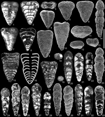
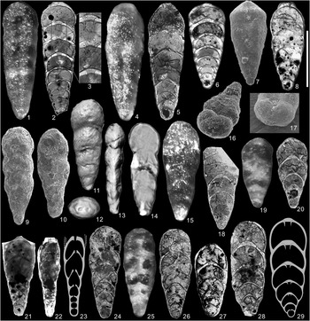
(1–8) Geinitzina ichnousa Sellier de Civrieux and Dessauvagie, Reference Sellier de Civrieux and Dessauvagie1965: (1) XK237_10SEM_214 (axial view with reflected light); (2) XK235_11TS_004 (frontal axial section); (3) XK235_10TS_073 (transverse section); (4) XK235_11SEM_043 (SEM, close to axial view, wall of proloculus and last chamber is broken); (5) XK235_11SEM_071 (SEM, axial view, wall broken); (6) XK235_11SEM_069 (SEM, axial view); (7, 8) XK240_10TS_001 (7, axial view with reflected light; 8, frontal axial section). (9–13, 15–17) Geinitzina taurica Sellier de Civrieux and Dessauvagie, Reference Sellier de Civrieux and Dessauvagie1965: (9) XK237_10SEM_067 (SEM, axial view); (10) XK235_10SEM_068 (SEM, axial view); (11) XK235_11SEM_070 (SEM, apertural view); (12) XK235_11SEM_027 (SEM, transverse view of a broken test, showing apertural view in adult test); (13) XK235_11SEM_029 (SEM, transverse view of a broken test, showing apertural view in juvenile test); (15, 16) XK235 _10TS_005 (15, frontal axial section; 16, hand drawing from 15); (17) XK237_10TS_062 (frontal axial section). (14, 18–33) Howchinella inflata n. sp.: (14) XK238_10SEM_110 (SEM, axial view); (18–20) XK238_10TS_089 (18, transverse section of adult stage; 19, transverse section of juvenile stage; 20, axial view with reflected light); (21) XK239_10TS_006 (frontal axial section); (22) XK238_10TS_078 (frontal axial section); (23) XK236_10SEM_056 (paratype, SEM, axial view); (24) XK239_10TS_086 (paratype, frontal axial section); (25) XK238_10TS_081 (frontal axial section); (26) XK236_12TS_021 (frontal axial section); (27) XK239_10TS_076 (paratype, frontal axial section); (28) XK236_12TS_002, (frontal axial section); (29–31) XK239_11SEM_006 (29, holotype, SEM, axial view; 30, oblique apertural view showing oval aperture and distinctly sutures; 31, close to axial view with reflected light, showing excavated sutures and inflated chambers); (32, 33) XK248_10TS_075 (32, axial view with reflected light; 33, frontal axial section). Scale bar is 200 μm.

Types
Holotype: Fig. 4.29 (XK239_11SEM_006); paratypes: Fig. 4.23 (No.XK236_10SEM_056), Fig. 4.24 (No.XK239_10TS_086), Fig. 4.27 (No.XK239_10TS_076). All types were recovered from the Dalong Formation, Daxiakou section, South China.
Diagnosis
Test small, elongate, laterally compressed. Sutures excavated. Chambers crescentiform in frontal axial section, inflated in adult stage. Aperture simple oval, terminal, weakly protruding.
Occurrence
Late Permian, Changhsingian, Dalong Formation, South China.
Description
Test small, lanceolate to strongly elongate, oval in transverse section. Sutures excavated, with deep longitudinal grooves in central part of test. Initial apical periphery narrowly rounded to acutely rounded. Apertural end parabolic-shaped. Surface smooth. Five to eight chambers. Aperture terminal, simple, with weakly protruding apertural face, oval. Proloculus small, spherical, with internal diameter 0.020–0.033 mm. Chambers fabiform in juvenile stage, crescentiform and always inflated in adult stage; envelopment of chambers weak in early stage, but attains to one-third height of preceding chamber in terminal stage. Attachment of chambers simple or with slightly sutural thickenings at 40° angle to sutural facets. Wall calcareous, hyaline, atelo-monolamellar, thin, with thickness 0.006 mm in initial part and 0.004 mm in terminal part. Septa of same thickness as wall, and two times thicker than wall near apertural borders of septa. Septa rounded in first chambers and arcuate in later chambers. Internal apertural tube present in most chambers, and more developed in last chamber. Dimensions: L 0.252–368 mm, W 0.088–0.108 mm, L/W 2.8–3.3; for holotype, L 0.336 mm, W 0.102 mm, and L/W 3.3.
Etymology
From the Latin inflatus, swollen, inflated.
Material
Fifty free specimens, three were prepared for SEM, eight for frontal axial section, one for transverse section.
Remarks
Based on the test shape, Howchinella inflata is similar to Spandelina kirkbyi (Richter, Reference Richter1861), which is known from northwestern Germany (Scherp, Reference Scherp1962, p. 299, pl. 10, figs. 7, 8), the Baltic area (Miklukho-Maklay and Ukharskaya, Reference Miklukho-Maklay and Ukharskaya1975, p. 74, pl. 12, figs. 5, 6, pl. 18, figs. 6, 7, 10, 11), and the Polish Lowlands (Woszczyńska, Reference Woszczyńska1987, p. 170, pl. 2, figs. 9, 10), but it differs by its smaller size. It also has a similar test shape to Spandelina kirkbyi minuta (Scherp, Reference Scherp1962) from the Zechstein Limestone in northwestern Germany (Scherp, Reference Scherp1962, p. 300, pl. 10, figs. 9, 10, 14, 15) and the Polish Lowlands (Woszczyńska, Reference Woszczyńska1987, p. 170, pl. 2, figs. 4, 5), but it differs from that form in its higher chambers. It differs from Spandelina excavata Cushman and Waters, Reference Cushman and Waters1928 (p. 364, pl. 48, figs. 3, 4) in its less compressed test and lack of a medial depression.
Howchinella hubeiensis new species
Figures 5.1–5.15, 6.1–6.7, 6.17
(1–15) Howchinella hubeiensis n. sp.: (1–5) XK238_10SEM_021 (megalospheric test; (1) axial view with reflected light; (2) holotype, SEM, axial view; (3–5) transverse, axial and lateral slices of Micro-CT reconstruction); (6) XK238_12TS_025 (paratype, megalospheric test; axial view with transmitted light); (7) XK238_10SEM_095 (microspheric test; frontal axial section, proloculus is lost in sectioning); (8) XK238_10TS_093 (megalospheric test; frontal axial section); 9, 10, XK237_10TS_091 (megalospheric test; 9, axial view with reflected light; 10, frontal axial section); (11) XK238_10TS_092 (megalospheric test, frontal axial section); (12, 13) XK238_12TS_017 (paratype, megalospheric test; 12, axial view with transmitted light; 13, lateral axial section); (14, 15) XK238_11SEM_039 (paratype, microspheric test; 14, SEM, axial view, proloculus is not well preserved; 15, axial view). Scale bar is 200 μm.

(1–7, 17) Howchinella hubeiensis: (1–3) XK238_10TS_128 (microspheric test; 1, axial view with reflected light; 2, paratype, frontal axial section; 3, enlargement of 2 to show septal thickenings and internal apertural tube); (4, 5) XK238_10TS_134 (megalospheric test; 4, axial view with reflected light; 5, frontal axial section); (6) XK239_10TS_104 (microspheric test; frontal axial section); (7, 17) XK235_11SEM_021 (microspheric test; 7, paratype SEM, axial view; 17, oblique apertural view). (8–16, 18–29) Howchinella complanata n. sp.: (8) XK238_10TS_123 (paratype, frontal axial section); (9) XK237_07SEM_027 (paratype, SEM, axial view); (10) XK237_07SEM_026 (paratype, SEM, axial view); (11–14) XK238_12SEM_023 (Micro-CT reconstruction, 11, holotype, axial view; 12, apertural view; 13, lateral view; 14, axial slice); (15, 18) XK238_10TS_106 (15, axial view with reflected light; 18, frontal axial section, proloculus and last chamber are broken in sectioning); (16) XK238_11SEM_008 (SEM, oblique apertural view); (19, 20) XK238_10TS_120 (19, axial view with reflected light; 20, frontal axial section); (21–23) XK_238_12TS_010 (21, axial view with transmitted light; 22, lateral axial section; 23, hand drawing from 22); (24) XK238_10TS_125 (frontal axial section); (25, 26) XK238_10TS_105 (25, axial view with reflected light; 26, paratype, frontal axial section); (27) XK236_12TS_031 (frontal axial section); (28, 29) XK238_10TS_106 (28, frontal axial section, part of test lost in sectioning; 29, hand drawing from 28). Scale bar is 200 μm.

2007 Howchinella? sp. A, Gu, Feng, and He, p. 317, pl. 3, fig. 9, pl. 4, fig. 13.
Types
Holotype: Figure 5.2 (No.XK238_10SEM_021); paratypes: Figure 5.6 (No.XK238_12TS_025), Figure 5.12, 5.13 (No.XK238_12TS_017), Figure 5.14, 5.15 (No.XK238_11SEM_039), Figure 6.2 (No.XK238_10TS_128), Figure 6.7 (No.XK235_11SEM_021). All types were recovered from the Dalong Formation, Daxiakou section, South China.
Diagnosis
Test elongate, lanceolate or egg-shaped, with longitudinal median depression; biconcave in transverse view. Chambers crescentiform to low pear-shaped or crescentiform in frontal axial section, slightly enveloping. Septa moderately thicken near apertural borders. Aperture oval, terminal, with apertural internal tubes.
Occurrence
Late Permian, Changhsingian, Dalong Formation, South China.
Description
Test uniserial, elongate, lanceolate or egg-shaped, lanceolate in megalospheric tests, moderately to strongly elongate lanceolate or cigar-shaped in microspheric tests. Lateral outline even, slightly convex in middle part of test, and with development of longitudinal median depression, sometimes with longitudinal costae. Transverse section rhombic biconcave, moderately depressed in both median regions. Initial apical periphery narrowly rounded. Apertural end parabolic-shaped in megalospheric tests and pointed parabolic-shaped in microspheric tests. Sutures indistinct in early stages, occasionally distinctly excavated in terminal stages, especially in microspheric forms. Five to eight chambers in megalospheric tests, and eight to ten in microspheric tests. Aperture terminal, oval, with internal apertural tubes in last few chambers. Proloculus small, spherical, with internal diameter 0.046–0.062 mm in megalospheric tests and 0.025–0.035 mm in microspheric tests. Chambers crescentiform to low pear-shaped crescentiform in frontal axial section; each chamber enveloping previous one approximately one-quarter to one-third height of chamber. Height and width of chambers gradually increasing, ratio of width to height of chambers in adult stage 2.5–3.0, and with slow increase in width; compared with earlier chambers, last few chambers become much higher but narrower, pear-shaped, with sutures distinctly excavated, that may be result of ecophenotypic variations or intra-specific divergence in adult stage of test. In paratype (Fig. 6.6) height of second chamber 0.024 mm, width 0.041 mm; height of third chamber 0.026 mm, width 0.059 mm; height of eighth and final chamber 0.048 mm, width 0.132 mm. Attachment of chambers with triangular sutural thickenings, and sutural facets at 45° to horizontal. Wall calcareous, hyaline, radial, monolamellar, with thickness 0.006–0.010 mm in initial part and 0.004–0.006 mm in terminal part. Septa curved, arcuate, thinner or of same thickness as wall in initial part of test, and gradually becoming thicker in terminal part, with thickness up to 0.015 mm, which is 1.5 times thicker than thickness of wall. Moderate thickenings near apertural borders of septa. Dimensions: for microspheric tests, L 0.350–0.482 mm, W 0.129–0.140 mm, L/W 2.3–3.5; for megalospheric tests, L 0.323–0.404 mm, rarely up to 0.544 mm, W 0.126–0.159 mm, L/W 2.2–2.9, rarely up to 4.3; thickness (T) 0.68–0.75 mm, ratio W/T 1.7–2.1; for holotype, L 0.533 mm, W 0.132 mm, L/W 4.1, T 0.072 mm, W/T 1.8.
Etymology
After the location in Hubei Province, China.
Material
One hundred free specimens showing the variation in test morphology; three were prepared for SEM, nine for frontal axial section, three for transverse section, and one for lateral axial section.
Remarks
The new species is similar to Lingulina strigata Wang, Reference Wang1976 (p. 188, pl. 1, fig. 2) from the Changhsing Formation in western Guizhou, South China, in its biconcave transverse section and longitudinal median depression. It differs, however, in its higher chambers and internal apertural tubes.
Howchinella complanata new species
Types
Holotype: Figure 6.11 (No.XK238_12SEM_023); paratypes: Figure 6.8 (No.XK238_10TS_123), Figure 6.9 (No.XK237_07SEM_027), Figure 6.10 (No.XK237_07SEM_026), Figure 6.26 (No.XK238_10TS_105). All types were recovered from the Dalong Formation, Daxiakou section, South China.
Diagnosis
Test elongate lanceolate, compressed, oval in transverse view. Sutures curved, depressed in terminal part of test. Chambers crescentiform in frontal axial section, quickly enlarged in adult stage, enveloping by one-half height of preceding chamber. Aperture terminal, oval, with internal apertural tubes.
Occurrence
Late Permian, Changhsingian, Dalong Formation, South China.
Description
Test small, oblong triangular to elongate lanceolate, lateral outline slightly waved, with narrowly rounded initial apical periphery and rounded periphery in apertural end, compressed, oval in transverse section. Sutures slightly depressed in longitudinal median, especially in terminal part of test. Surface smooth. Six to seven chambers. Aperture terminal, oval, with internal apertural tubes. Proloculus small, spherical, with internal diameter 0.028–0.040 mm. Chambers crescentiform in frontal axial section, very slowly increasing in height and width in initial part of test, and quickly enlarged in adult stage, enveloping one-third to one-half height of preceding chamber. In lateral axial section, chambers almost round in initial part and arrow-shaped crescentiform in later stages, enveloping one-half of height of preceding chamber. Height of second chamber 0.024–0.028 mm, width 0.044–0.048 mm; height of third chamber 0.020–0.026 mm, width 0.052–0.062 mm; height of sixth chamber 0.055–0.060 mm, width 0.104–0.112 mm. Attachment of chambers with sutural thickenings. Wall calcareous, hyaline, thin, monolamellar. Septa thinner than wall in initial part of test and gradually equal to thickness of wall in terminal part. Septa near apertural borders developed triangular thickenings with height up to 0.020 mm in last chamber of test. Dimensions: L 0.326–0.380 mm, W 0.120–0.136 mm, L/W 2.7–2.9; for the holotype, L 0.380 mm, W 0.136 mm, and L/W 2.8.
Etymology
From the Latin complanatus, flattened.
Material
Twelve free specimens were extracted, seven were prepared for frontal axial sections, one for lateral axial section, three for SEM, one for Micro-CT.
Remarks
Howchinella complanata is similar to Frondicularia reliqua (Gerke, Reference Gerke1961, pl. 55, fig. 5) from the Early–Middle Permian of the Nordvik region of central Siberia in overall test shape and chamber shape, but differs by its more enveloped chambers, and simple oval aperture. It differs from Howchinella hubeiensis by its higher and more arcuate chambers, especially in lateral view.
Howchinella xiangxiensis new species
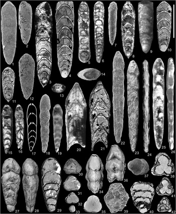
(1–14) Howchinella xiangxiensis n. sp.: (1) XK235_10SEM_062 (paratype, megalospheric test; SEM, axial view); (2) XK235_10SEM_060 (megalospheric test; SEM, axial view, last chamber lost); (3) XK239_10TS_110 (paratype, megalospheric test; frontal axial section); (4) XK235_10TS_131 (megalospheric test; frontal axial section); (5) XK238_10TS_133 (paratype, megalospheric test; lateral axial section); (6) XK238_10TS_132 (paratype, microspheric test; lateral axial section, proloculus is broken in sectioning); (7) XK237_11SEM_004 (microspheric; SEM, lateral view); (8) XK238_10TS_097 (holotype, megalospheric test; frontal axial section); (9, 10) XK235_10TS_113 (megalospheric test; 9, axial view with reflected light; 10, frontal axial section, proloculus is broken in sectioning); (11) XK238_10TS_130 (megalospheric test; juvenile test; frontal axial section); (12) XK235_10SEM_061 (megalospheric test; SEM, axial view, juvenile test); (13) XK_238_10TS_135 (transverse section); (14) XK233_12SEM_033 (SEM, transverse view of a broken test, showing oval aperture). (15–19) Ichthyolaria celsa n. sp.: (15–17) XK238_10TS_094 (microspheric test; 15, holotype, axial view with transmitted light; 16, frontal axial section, proloculus lost in sectioning; 17, hand drawing from 16); (18) XK255_07SEM_041 (megalospheric test; paratype, SEM, axial view); (19) XK238_10TS_210 (microspheric test; axial view with reflected light). (20, 21) Ichthyolaria sp. 1: XK235_10TS_096 (broken test, last four chambers preserved; 20, axial view with reflected light; 21, frontal axial section). (22–26) Ichthyolaria sp. 2: XK256_07SEM_049 (22, SEM, axial view; 23, 24, axial and lateral view of Micro-CT reconstruction; 25 axial view of the same specimen with transmitted light; 26, lateral slice of Micro-CT reconstruction). (27–37) Pseudotristix elongata n. sp.: (27–33) XK238_12SEM_028 (Micro-CT reconstruction, 27, holotype, axial view; 28, lateral view; 29, axial slice; 30, apertural view; 31–33, transverse slice in early, middle and latest stage of the same specimen); (34–36) XK238_12SEM_027 (Micro-CT reconstruction of a broken test, 34, lateral view; 35, transverse slice; 36, axial view); (37) XK238_11SEM_018 (SEM, apertural view). (38–42) Pseudotristix sp.: XK238_11SEM_032 (38, 40–42, Micro-CT reconstruction: 38, transverse slice; 40, frontal axial section; 41, transverse slice of middle stage; 42, transverse slice of early stage; 39, SEM, oblique axial view). Scale bar is 200 μm.

Types
Holotype: Figure 7.8 (No.XK238_10TS_097); paratypes: Figure 7.1 (No.XK235_10SEM_062), Figure 7.3 (No.XK239_10TS_110), Figure 7.5 (No.XK238_10TS_133), Figure 7.6 (No.XK238_10TS_132). All types were recovered from the Dalong Formation, Daxiakou section, South China.
Diagnosis
Test elongate, oval in transverse view. Sutures arcuate, slightly or not excavated. Chambers crescentiform in frontal axial section, low in initial stage and high in terminal stage, moderately enveloping. Aperture terminal, oval, with club-shaped thickenings near apertural borders.
Occurrence
Late Permian, Changhsingian, Dalong Formation, South China.
Description
Test uniserial, small to moderately large, elongate egg-shaped to strongly elongate lanceolate, gradually expanded in juvenile stage, whereas unexpanded or weakly expanded in adult stage in microspheric tests. Transverse section lens-shaped, outline of sides even. Surface smooth. Sutures arcuate, chevron-shaped, slightly excavated in terminal part of test in microspheric tests. Initial apical periphery pointed in microspheric tests and arcuate in megalospheric tests. Apertural end parabolic in microspheric tests and rounded in megalospheric tests. Always, a spiny outgrowth at apical periphery of test. Five to six chambers in microspheric tests, and seven to 11 in megalospheric tests. Proloculus spherical, with internal diameter 0.020–0.025 mm in microspheric tests and 0.045–0.055 mm in megalospheric tests; external diameter 0.045–0.050 mm in microspheric tests and 0.065–0.070 mm in megalospheric tests. In both microspheric and megalospheric forms, initial chambers low, crescentiform, weakly enveloping, embracing preceding chamber by one-third to one-half of its height, increasing gradually in height and width, and becoming arrow-shaped crescentiform in terminal stage of test; whereas in microspheric forms, height of chambers increases more quickly, chambers become very high and arrow-shaped or crescentiform in terminal stage without expanding in width. In holotype (Fig. 7.8), height of second chamber 0.018 mm, width 0.038 mm; height of third chamber 0.021 mm, width 0.045 mm; height of fourth chamber 0.022 mm, width 0.054 mm; height of fifth chamber 0.024 mm, width 0.060 mm; height of last chamber 0.074 mm, width 0.050 mm. Ratio of chamber height/width 2.1 in initial stage and 1.5 in terminal stage. Chamber shape in lateral axial section (Fig. 7.6, microspheric test; Fig. 7.5, megalospheric test) cask-shaped. Chamber weakly embracing in initial stage and increasing to one-third height in terminal stage. Attachment of chambers with slightly triangular sutural thickenings. Sutural facets at angle of 60° to horizontal. Wall calcareous, hyaline, monolamellar, radial, with thickness 0.007–0.012 mm in initial part of chamber, and 0.002–0.004 mm in penultimate chamber. Septa arcuate, with development of club-shaped thickenings near apertural borders. Height of septal thickenings in last chambers 0.016–0.020 mm. Aperture terminal, central, simple, oval; apertural canal funnel-shaped, with very short internal apertural tube. Dimensions: for microspheric tests, L 0.305–0.510 mm, W 0.090–0.110 mm, L/W 4.0–4.8; for megalospheric tests, L 0.264–0.375 mm, W 0.088–0.094 mm, L/W=3.0–3.8; for holotype, L 0.366 mm, W 0.092 mm, L/W 3.9.
Etymology
After the Xiangxi River, located in the western part of Hubei Province, China.
Material
Twenty free specimens; five were prepared for SEM, eight for frontal axial section, and three for lateral axial section.
Remarks
Howchinella xiangxiensis resembles some Ichthyolaria in frontal axial section, but differs by its higher and crescentiform chambers. The frontal axial section is also similar to that of Spandelina longissima Miklukho-Maklay, Reference Miklukho-Maklay1960 from the Kazanian of the Russian Platform (p. 158-159, pl. 27, fig. 9, 10, pl. 28, fig. 3), but this latter form possesses a radiate aperture, higher chambers, and less chamber embracement.
Family Ichthyolariidae Loeblich and Tappan, Reference Loeblich and Tappan1986
Genus Ichthyolaria Wedekind, Reference Wedekind1937
Type species
Frondicularia bicostata d’Orbigny, Reference d’Orbigny1850.
Ichthyolaria celsa new species
Types
Holotype: Figure 7.15 (No.XK238_10TS_094); paratype: Figure 7.18 (No.XK255_07SEM_041). All types were recovered from the Dalong Formation, Daxiakou section, South China.
Diagnosis
Test elongate, compressed, tapering in initial end. Chambers chevron-shaped, high, slightly enveloping, five to seven in number. Sutures arcuate, not excavated. Aperture terminal, oval.
Occurrence
Late Permian, Changhsingian, Dalong Formation, South China.
Description
Test small, cigar-shaped, tapered in initial and apertural end, oval in transverse section. Sutures slightly curved and excavated in terminal part of test. Seven to eight chambers. Aperture terminal, simple, and oval. Proloculus small, oval, 0.014 mm in width and 0.022 mm in height. Chambers arrow-shaped or crescentiform, gradually expanding in width and rapidly increasing in height, enveloping one-third height of previous chamber. For holotype (Fig. 7.15), height of second chamber 0.020 mm, width 0.028 mm; height of third chamber 0.027 mm, width 0.038 mm; height of seventh chamber 0.045 mm, width 0.052 mm. Attachment of chambers simple. Sutures slightly excavated in last two chambers. Wall calcareous, radial, atelo-monolamellar, thin, with thickness of 0.002 mm in initial stage and 0.003 mm in terminal stage. Septa arched, with club-shaped thickenings in apertural border in terminal stage. Dimensions: L 0.295–0.370 mm, W 0.066–0.068 mm, L/W 4.5–5.5; for holotype, L 0.295 mm, W 0.066 mm, L/W 4.47.
Etymology
From the Latin celsus, high (reflecting higher chambers compared to other ichthyolariids).
Material
Five free specimens were recovered, one was prepared for frontal axial section and one for SEM.
Remarks
The new species differs from Ichthyolaria primitiva from the Upper Permian Bellerophon Limestone of southwestern Turkey (Sellier de Civrieux and Dessauvagie, Reference Sellier de Civrieux and Dessauvagie1965, p. 74, pl. 5, figs. 30, 31, pl. 9, fig. 1) by its much higher but less embracing chambers, with height/width ratio up to 1.1, and its sutures which are slightly excavated in the last few chambers with distinct apertural thickenings.
Genus Pseudotristix Miklukho–Maklay, 1960 [nomen transl. Loeblich and Tappan, Reference Loeblich and Tappan1964]
Type species
Tristix (Pseudotristix) tcherdynzevi Miklukho-Maklay, Reference Miklukho-Maklay1960.
Pseudotristix elongata new species
Types
Holotype: Figure 7.27 (No. XK238_12SEM_028), recovered from the Dalong Formation, Daxiakou section, South China.
Diagnosis
Test uniserial, elongate, triangular in transverse section. Sutures slightly excavated. Chambers crescentiform, high, four to six in number, slightly enveloping. Aperture terminal, round.
Occurrence
Late Permian, Changhsingian, Dalong Formation, South China.
Description
Test small, uniserial, elongate, roundly triangular in transverse view, narrowly rounded in initial periphery, and parabola-shaped in apertural end. Transverse section round-triangular. Sutures weakly excavated, slightly arcuate in terminal part of test. Chambers number four to six. Aperture terminal, round, slightly developed. Proloculus spherical, 0.050–0.080 mm in external diameter. Following chambers trilobate in transverse section, crescentiform in axial section, very weakly enveloping, slowly increasing in width and height in initial part of test, and becoming higher crescentiform; chamber width does not increase in terminal part of test. For holotype (Fig. 7.27), height of second chamber 0.038 mm, width 0.062 mm; height of third chamber 0.046 mm, width 0.080 mm; height of fifth chamber 0.058 mm, width 0.115 mm; height of last chamber 0.060 mm, width 0.115 mm. Wall calcareous, hyaline. Septa slightly arched in initial two or three chambers of test and arched in last three chambers. Dimensions: for holotype, test L 0.322 mm, W 0.118 mm, L/W=2.7.
Etymology
From the Latin elongatus, meaning elongate.
Material
Four free specimens were recovered; one was prepared for SEM, and two for the Micro-CT.
Remarks
The typical Permian nodosariid genus with trilobate chambers was first reported from the Kazanian (Middle Permian) of the Russian Platform by Miklukho-Maklay (Reference Miklukho-Maklay1960) as a subgenus Pseudotristix of the Jurassic–Recent genus Tristix Macfadyen, Reference MacFadyen1941. Pseudotristix was proposed for forms differing from typical Tristix in their flat apertural end, straight sutures, and un-enveloped chambers. Based on these differences, Loeblich and Tappan (Reference Loeblich and Tappan1964, p. C524) raised Pseudotristix to generic rank. We concur with Loeblich and Tappan’s separation of Pseudotristix from Tristix based on chamber shape and envelopment of chambers. Pseudotristix has higher chambers, less or no chamber envelopment, and a much earlier Permian range than Tristix. The earliest appearance of Pseudotristix is in the Sakmarian–Artinskian (Lower Permian) (Palmieri, Reference Palmieri1983, Reference Palmieri1994; Palmieri, in Foster, 1985) where it is often associated with Howchinella, Ichthyolaria, and Geinitzina. This co-occurrence suggests that Pseudotristix may have derived from Howchinella, Ichthyolaria, or Geinitzina during the Lower Permian. Our species has trilobate and weakly enveloping chambers which conform to the genus Pseudotristix. The new species differs from the type species Pseudotristix tcherdynzevi by its smaller test, slightly arched sutures, crescentiform chambers, and parabolic-shaped apertural end. Based on the shape of its test, it is similar to Tristix mariusi Woszczyńska (Reference Woszczyńska1987, p. 186, pl. 3, fig. 14), but it differs in its larger test and weakly enveloping chambers.
Discussion on age
The foraminiferal fauna described in this paper includes 19 species of 10 genera. They are Ammodiscus sp., Nodosaria quinquecostata n. sp., Nodosaria sp., Dalongella fusiformis Gu, Feng, and He, Reference Gu, Feng and He2007, Robuloides sp., Neoendothyra sp. 1, Neoendothyra sp. 2, Eocristellaria sp., Geinitzina taurica Sellier de Civrieux and Dessauvagie, Reference Sellier de Civrieux and Dessauvagie1965, Geinitzina ichnousa Sellier de Civrieux and Dessauvagie, Reference Sellier de Civrieux and Dessauvagie1965, Howchinella inflata n. sp., Howchinella hubeiensis n. sp., Howchinella complanata n. sp., Howchinella xiangxiensis n. sp., Ichthyolaria celsa n. sp., Ichthyolaria sp. 1, Ichthyolaria sp. 2, Pseudotristix elongata n. sp., and Pseudotristix sp.. None of the typical Changhsingian foraminiferal genera of South China, such as Palaeofusulina Deprat, 1912, Colaniella Likharev, Reference Likharev1939, and Paraglobivalvulinoides Zaninetti and Jenny-Deshusses, Reference Zaninetti and Jenny-Deshusses1985, etc., were found in the Daxiakou section. Although most of the species are new and therefore could not provide detailed age data, the age could be confined to Late Permian by the discovery of Geinitzina ichnousa and Geinitzina taurica. Both are widely distributed in the Late Permian of the Tethyan realm, including southwestern Turkey (Sellier de Civrieux and Dessauvagie, Reference Sellier de Civrieux and Dessauvagie1965, Köylüoglu and Altiner, Reference Köylüoglu and Altiner1989, Gaillot and Vachard, Reference Gaillot and Vachard2007), Cambodia (Nguyen, Reference Nguyen1986), Transcaucasia (Pronina, Reference Pronina1989), northwestern Caucasus (Pronina-Nestell and Nestell, Reference Pronina-Nestell and Nestell2001), northwestern Serbia (Nestell et al., Reference Nestell, Sudar, Jovanović and Kolar-Jurkovšek2009) western Slovenia (Nestell et al., Reference Nestell, Kolar-Jurkovsek, Jurkovsek and Aljinovic2011), and the Southern Alps (Groves et al., Reference Groves, Rettori, Payne, Boyce and Altiner2007). In South China, they are restricted to the Changhsingian (Gaillot and Vachard, Reference Gaillot and Vachard2007). Their occurrence in the Daxiakou section indicates a latest Permian age. Furthermore, Dalongella fusiformis, which was only discovered in the end-Permian deep-water environments from the Dongpan section in southwestern South China (Gu et al., Reference Gu, Feng and He2007) confirms the Changhsingian age.
The late Changhsingian age of the Daxiakou foraminiferal fauna is also supported by conodont data reported by Zhao et al. (Reference Zhao, Chen, Chen and Cao2013), where the top of the Dalong Formation, beds 247–262 in the Daxiakou section, is subdivided into three conodont zones: Clarkina yini, Clarkina meishanensis, and Clarkina taylorae zones, in ascending order (Fig. 2). In the Dongpan section, Dalongella fusiformis also occurs in the Clarkina yini zone (Luo et al., Reference Luo, Lai, Feng, Jiang, Wignall, Zhang, Sun and Wu2008).
Paleoecological significance
Temperature
The Daxiakou fauna is probably related to a cool, deep-sea habitat. Large scale biogeographic distributions of modern benthic foraminifers exhibit a pattern indicating that the major controlling factor is temperature or temperature-salinity related water masses (Murray, Reference Murray2006). The present fauna is highly different from contemporaneous shallow-water assemblages of the Tethys in low latitudinal regions, which are mostly composed of Colaniella, Nodosinelloides Mamet and Pinard, Reference Mamet and Pinard1992, Pachyphloia Lange, Reference Lange1925, Globivalvulina Schubert, Reference Schubert1921, Robuloides Reichel, Reference Reichel1946, etc., and co-occurring abundant fusulinids, suggesting warm-water photozoan assemblages. Only two species in the Daxiakou fauna (Geinitzina ichnousa and Geinitzina taurica) also occur in contemporaneous shallow-water faunas of South China (Gaillot and Vachard, Reference Gaillot and Vachard2007). Seven species are new, which comprise 37% of the species in the fauna. Based on generic composition and test morphogroup, the Daxiakou fauna highly resembles other Middle to Late Permian foraminifer faunas from higher latitude cold-water environments. There are many common genera in these areas, including Howchinella and Ichthyolaria which predominantly occur in higher latitude cold-water environment, such as north and west Gondwana (Crespin, Reference Crespin1958; Scheibnerova, Reference Scheibnerova1982; Palmieri, Reference Palmieri1983, Reference Palmieri1994, Reference Palmieri1998; Dixon and Haig, Reference Dixon and Haig2004; Haig, Reference Haig2006), Siberia (Karavaeva and Nestell, Reference Karavaeva and Nestell2007), and Spitsbergen (Sosipatrova, Reference Sosipatrova1967, Reference Sosipatrova1969), suggesting that the Daxiakou fauna may come from a cool-water environment.
Oxygen content
Sedimentologic and lithologic analyses (Yao and Zhang, Reference Yao and Zhang1996; Wang, Reference Wang2005) have shown no bioturbation and very rare macrofauna or other microfossils except foraminifers, suggesting that the Dalong Formation in the Daxiakou section was probably deposited in an environment with low dissolved oxygen (maybe anoxic or dysoxic). In addition to the deep-water environment proposed by sedimentary studies, the characteristics of the foraminiferal fauna may be other evidence of an oxygen-depleted environment.
On one hand, the predominant flattened test morphology in the foraminifera suggests low oxygen concentration in bottom water. Intensive studies on Jurassic to Holocene benthic foraminifers (Bernhard, Reference Bernhard1986; Kaiho, Reference Kaiho1991; Sen Gupta and Machain-Castillo, Reference Sen Gupta and Machain-Castillo1993; Bernhard and Sen Gupta, Reference Bernhard and Sen Gupta2003) have shown that flattened tests provide high surface area/volume ratios, which can increase their capacity for oxygen uptake from low-oxygen bottom water or from anoxic sediments as deep as 15 cm below the water-sediment interface (Corliss, Reference Corliss1985). In the Daxiakou section, 74% of species have flattened tests (14 species from a total of 19 species, including flattened discoidal), which is quite different from the contemporaneous shallow carbonate platform faunas; for example, flattened-test genera occupy 53.1% of the species diversity in the Datian section (Zhang and Hong, Reference Zhang and Hong2004), 22% in the Meishan section (Song et al., Reference Song, Tong and He2006), 13.1% in the Dajiang section (Song et al., Reference Song, Tong and Chen2009b), and 12.9% in the Laren section (Gaillot et al., Reference Gaillot, Vachard, Galfetti and Martini2009). Furthermore, the flattened test proportion in the Daxiakou section is similar to that of another deeper water section at Dongpan (81%) (Gu et al., Reference Gu, Feng and He2007).
On the other hand, small foraminiferal test size could also suggest a less oxygenated environment. The length of over 512 nodosariids specimens from the Daxiakou section was measured. The mean length of the nodosariids is 0.299 mm, with maximum 0.640 mm and minimum 0.122 mm. They are smaller than contemporaneous shallow-water assemblages, which are 0.459 mm (maximum 1.330 mm and minimum 0.180 mm) in the Datian section (Zhang and Hong, Reference Zhang and Hong2004), 0.525 mm (maximum 0.300 mm and minimum 0.750 mm) in the Laren section (Gaillot et al., Reference Gaillot, Vachard, Galfetti and Martini2009), and 0.478 mm (maximum 1.017 mm, minimum 0.197 mm) in the Dajiang section (Song et al., Reference Song, Tong and Chen2009b). Smaller individuals are thought to have consumed less oxygen than larger ones, and therefore would be more successful in oxygen-depleted environments (Basov, Reference Basov1979; Kaiho, Reference Kaiho1998). The “Lilliput effect” in the Late Permian to Early Triassic has been observed in shallow-water foraminifers and considered to be a result of anoxia (Song et al., Reference Song, Tong and Chen2011). Therefore, the small test size in the present fauna may suggest an oxygen-depleted environment.
Implication on end-Permian mass extinction
Deep-water environments would be one of the refuges during the Permian–Triassic crisis. Some foraminifers known from the Early Triassic were found to have an affinity to latest Permian forms based on the morphology and wall ultrastructure of their test (Groves et al., Reference Groves, Altiner and Rettori2003), implying that they could have survived the end-Permian oceanic crisis. When–where–how the foraminifers survived in the Permian–Triassic transition remains largely unclear, although one hypothesis proposed that the deep-water environments would be one of the refuges when the upper water column became lethally hot (Joachimski et al., Reference Joachimski, Lai, Shen, Jiang, Luo, Chen, Chen and Sun2012), because deep-water foraminifers may survive in anoxic and colder environments but not in high temperatures. Such a situation was suggested for the foraminiferal extinction in the PETM (Paleocene-Eocene Thermal Maximum), during which 30%–50% of species of deep-sea benthic foraminifers died out (e.g., Kaiho, Reference Kaiho1994b; Thomas, Reference Thomas2003; Alegret et al., Reference Alegret, Ortiz, Arenillas and Molina2005; Alegret et al., Reference Alegret, Ortiz and Molina2009). Based on the diversity and evolutionary study of the foraminiferal fauna in deep-water environment during the Permian–Triassic turnover, a more holistic understanding could be achieved on the extinction, survival, and recovery of these organisms.
Conclusions
A deep-water Changhsingian foraminiferal fauna from the Dalong Formation in the Daxiakou section in Xinshang County, Hubei Province, South China is studied. Nineteen species which belong to 10 genera of foraminifers are illustrated herein, including the following seven new species, Nodosaria quinquecostata n. sp., Howchinella inflata n. sp., Howchinella hubeiensis n. sp., Howchinella complanata n. sp., Howchinella xiangxiensis n. sp., Ichthyolaria celsa n. sp., and Pseudotristix elongata n. sp.
The Daxiakou fauna differs from contemporaneous foraminiferal faunas in South China, in the following aspects: (1) nodosariids are the dominant forms in both abundance and diversity, and (2) the most abundant forms, which include Geinitzina, Howchinella, and Ichthyolaria have elongate and flattened tests. The predominance of elongate flattened tests and smaller test size may suggest a foraminiferal habitat in an oxygen-depleted, cold, deep-water environment.
Acknowledgments
This study was supported by the National Science Foundation of China (Nos. 40802004, 40839903), the Program for Changjiang Scholars and Innovative Research Team in the University (PCSIRT-IRT0546), and the Fundamental Research Funds for the Central Universities, China University of Geosciences (Wuhan). We thank G. Nestell, M. Nestell, and D. Vachard for their critical but inspiring comments on the manuscript. We thank the Editor B. Pratt, Associate Editor D. Work, and Managing Editor K. Huber for reviewing the manuscript and helping with format. We also thank Y. Sun and B. Schiraldi who carefully read the manuscript and gave us valuable suggestions. We are grateful to Q. Feng and N. Zhang for their help in field work, A. Yuan for her assistance in foraminiferal fossil extraction, and S. Zhang for her help with SEM microphotography.