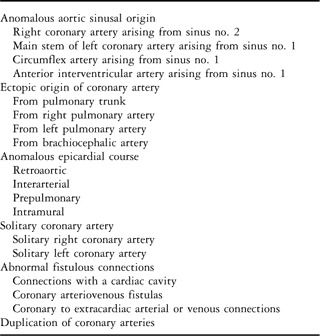
Coronary arteries can have multiple anomalous origins, and epicardial courses, in the otherwise normally structured heart. Recently developed techniques, such as computed tomography, now show the origins and courses of the coronary arteries with exquisite accuracy. Use of these techniques, therefore, will increase the ability to diagnose the abnormal arrangements, hopefully casting more light on the known relationship between some patterns and sudden cardiac death, particularly in competitive athletes.Reference Corrado, Basso and Thiene1–Reference Montagnana, Lippi, Franchini, Banfi and Guidi3 The anomalous patterns are relatively frequent, with an investigation using coronary arteriography as the diagnostic modality showing a prevalence of abnormal arrangement in around 1% of a large cohort of more than 100,000 patients.Reference Yamanaka and Hobbs4 Not all these variations, of course, are clinically significant. Even some of the arrangements implicated as causing sudden death can remain asymptomatic throughout the lifetime of the individual. Management and treatment of the abnormal arrangements, nonetheless, will be enhanced by awareness of their frequency, emphasising the importance of accurate description. At present, there is no consensus as how best to classify the multiple variations. In the past, the lesions were considered to be of major or minor significance.Reference Ogden5 Many of the allegedly major variations fail to produce symptoms during life, so such a system is less than ideal. It is much better simply to account for the anomalous origins of the arteries, their anomalous epicardial course, and to take note of other abnormal situations, such as myocardial bridging, abnormal communications, solitary coronary arteries, and duplicated arteries (Table 1).Reference Loukas, Groat, Khangura, Owens and Anderson6
Anatomic variations in origin and course of the coronary arteries.

Anomalous aortic origin of the coronary arteries
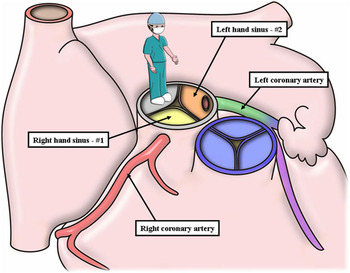
When the heart is otherwise normally structured, abnormal coronary arteries most frequently continue to have their origin from the aorta. It could be argued that the high take-off relative to the sinutubular junction, if excessive, should be considered an anomaly. The difficulty comes in determining the height of take-off judged to represent the abnormality. As origins minimally above the sinutubular junction are common,Reference Muriago, Sheppard, Ho and Anderson7 the arteries probably need to arise at least 1 centimetre above the junction in adults to be considered anomalous, with appropriate consideration given for growth when assessing the origins in infants and children. Abnormal sinusal origin is of far more significance, particularly if associated with a so-called intramural course.Reference Gittenberger-de Groot, Sauer and Quaegebeur8 Therefore, to describe the abnormal sinusal origin, it is first necessary to distinguish between the sinuses themselves without resorting to the normal arrangement. This is best achieved by distinguishing the coronary aortic sinuses as viewed by the observer standing, figuratively speaking, in the non-adjacent aortic sinus (Fig 1). Almost without exception, even if they have an anomalous origin, the coronary arteries will continue to arise from one or the other of the two sinuses adjacent to the pulmonary trunk. These can be distinguished as being to the right hand or left hand of the observer, with convention dictating that the right-handed sinus is considered to be no. 1, and the left-handed sinus no. 2.Reference Gittenberger-de Groot, Sauer, Oppenheimer-Dekker and Quaegebeur9 This convention has proved its value for surgeons transposing the coronary arteries as part of the so-called arterial switch procedure.Reference Lacour-Gayet and Anderson10 It is of equal relevance for those seeking a suitable means of describing the abnormal aortic sinusal origin in the otherwise normally structured heart. The convention also allows description of the anomalous sinusal origin of a major coronary artery from the non-adjacent sinus, albeit that this sinus would then no longer be a non-coronary aortic sinus. It would remain non-adjacent when considered relative to the pulmonary trunk.
The cartoon shows how, when the observer is standing, figuratively speaking, in the non-adjacent aortic sinus and is looking towards the pulmonary trunk, one of the remaining aortic sinuses is to the left hand, whereas the other is to the right hand. The right-handed sinus from this perspective is conventionally recognised as sinus no. 1, whereas the other sinus is no. 2.

Coronary arteries can also have an anomalous course relative to the arterial roots, irrespective of whether their sinusal origin is normal or abnormal. Such anomalous courses can be retroaortic, interarterial, or prepulmonary. Although the interarterial arrangement is often described as being “transseptal”, this is rarely the case, as in the otherwise normally structured heart there is no muscular septum between the aortic and pulmonary roots. The typical course for an artery running between the arterial roots is for the vessel to pass through the plane of the fibroadipose tissue existing between the aortic root and the free-standing subpulmonary infundibulum. During a part of this course, the coronary may traverse within the wall of the aorta, commonly described as representing an intramural arrangement. Of far greater significance when assessing an interarterial course is to establish the relationship with the attachment of the peripheral ends of the zones of apposition of the leaflets of the aortic valve to the sinutubular junction; this anatomical area is usually described as the commissure. If the coronary artery taking a so-called intramural courseReference Gittenberger-de Groot, Sauer and Quaegebeur8 also crosses the attachment of the leaflets at the sinutubular junction, then there is a greatly increased potential for narrowing the arterial orifice, with a concomitant increased chance of sudden cardiac death.
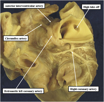
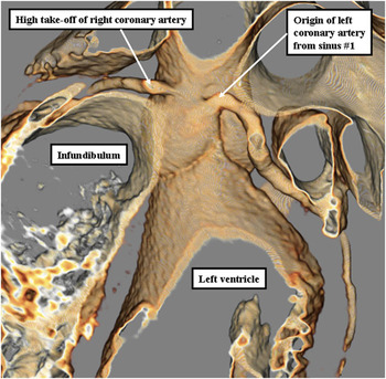
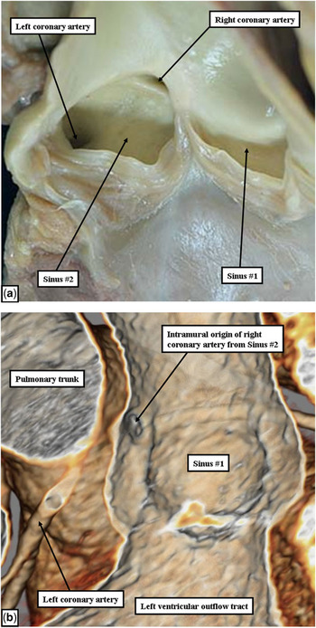
Although any possible anomalous course must be anticipated, certain combinations have been described as being frequent. The circumflex artery, for example, when arising anomalously from sinus no. 1, can take a retroaortic course, emerging into the left atrioventricular groove between the left atrial appendage and the pulmonary trunk (Fig 2). Arrangements such as this, with little likelihood of producing compression, are unlikely to be of clinical significance, as are those with prepulmonary courses. It is those arrangements associated with an interarterial course that are of greatest clinical significance, these being the ones most frequently associated with the intramural arrangement, and hence with the greatest potential for sudden cardiac death.Reference Corrado, Basso and Thiene1, Reference Corrado, Basso, Pavei, Michieli, Schiavon and Thiene2 There are then multiple anomalous patterns associated with such an interarterial course. In the most frequent patterns, the abnormal artery takes its origin from the opposite sinus when compared with the normal pattern. Thus, they are found when the main stem of the left coronary artery has its origin from sinus no. 1 (Fig 3), or the right coronary artery from sinus no. 2 (Fig 4). Although multiple hypotheses have been advanced to explain the potential for sudden death known to be associated with such patterns, none as yet is proven.Reference Loukas, Groat, Khangura, Owens and Anderson6 Should the artery taking an interarterial course also be a solitary vessel, supplying the entire cardiac arterial circulation, as when the main stem of the left coronary artery arises from sinus no. 1, passes between the arterial roots, and then supplies the entire coronary arterial circulation, then the risk of sudden death increases. Solitary arteries can also take a retroaortic course, this pattern being implicated with premature atherosclerotic disease.Reference Samarendra, Kumari, Hafeez, Vasavada and Sacchi11, Reference Angelini12
In this heart, the main stem of the left coronary artery arises from the aortic sinus giving rise to the right coronary artery, but then passes through the transverse sinus, behind the aortic root, before branching into the circumflex and anterior interventricular arteries. Note also the high take-off of the right coronary artery.

The computed tomogram shows origin of the left coronary artery from the aortic sinus giving rise to the right coronary artery. In this example, however, the left coronary artery passes between the aortic root and the subpulmonary infundibulum before it branches into the anterior interventricular and circumflex arteries.

(a) The aortic root opened and viewed from behind. The right coronary artery arises from the aortic sinus giving rise to the left coronary artery, and then crosses the zone of apposition between the aortic valvar leaflets before emerging within the right atrioventricular groove. This is the so-called intramural arrangement. The computed tomogram (b) shows the arrangement in a different patient, illustrating well the relationship to the sinutubular junction.

Ectopic origin of the coronary arteries
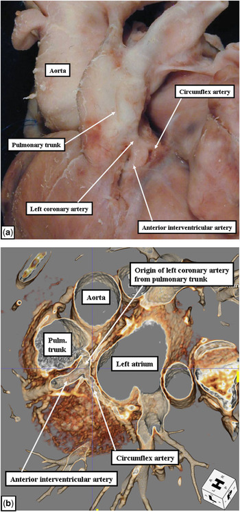
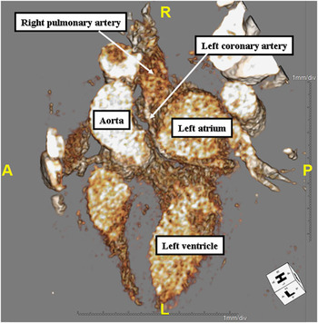
Although coronary arteries can rarely arise from a brachiocephalic artery, or one of its own branches,Reference Robiseck, Sanger, Daugherty and Gallucci13 the most significant ectopic origin is from the pulmonary trunk, or from the right or left pulmonary artery. Either the left or right coronary artery can arise from the pulmonary trunk. The anomalous origin of the left coronary artery is known as the Bland–White–Garland syndrome. This is one of the more common coronary arterial abnormalities encountered in children. Referred to as anomalous left coronary artery from the pulmonary artery, and abbreviated to “ALPACA”, it happens to occur from 1 in 250 to 1 in 400 of all congenitally malformed hearts, with an overall incidence of approximately 1 in 300,000 children.Reference Greenberg, Fish and Spindola-Franco14 The anomaly is more than twice as common in boys than it is in girls. Diagnosis and early surgical treatment are vital, as the anomalous coronary artery, and the myocardium it supplies, is fed with pulmonary rather than systemic blood. The anomalous artery, almost without exception, arises from the sinus of the pulmonary trunk adjacent to sinus no. 2 of the aortic root (Fig 5), although it may also arise from the right (Fig 6) or the left pulmonary artery.
The anatomical image (a), and a computed tomogram (b) from a different patient, shows origin of the left coronary artery from the pulmonary trunk, the so-called Bland–White–Garland syndrome.

In this patient, the anomalous left coronary artery arises from the right pulmonary artery, the anatomy shown with exquisite accuracy using computed tomography.

The clinical evolution of the patient with an anomalous origin of the left coronary artery from the pulmonary trunk depends on the extent of the development of a collateral circulation. Typically, the right coronary artery enlarges at its aortic origin, whereas the anomalous left artery tends to be small, and relatively thin walled. Should the collateral circulation be well developed and distributed, the left ventricle may remain well perfused, the heart retaining its essentially normal form and function. In situations in which the collateral circulation is poorly developed, the left ventricle becomes ischaemic, dilated, infarcted and fibrosed. The picture can then resemble dilated cardiomyopathy, as fibrosis can involve the papillary muscles and produce mitral valvar incompetence.
The right coronary artery can also have an anomalous origin from the pulmonary trunk, albeit far less frequently than the left. In two-thirds of the cases, the anomalous origin is found in association with other forms of congenital cardiac disease, such as aortopulmonary window, tetralogy of Fallot, or double outlet right ventricle. As with the origin of the left coronary artery from the pulmonary trunk, the anomaly may produce myocardial ischaemia, but often those affected are asymptomatic.Reference Williams, Gersony and Hellenbrand15
Duplication of coronary arteries
Any of the three major coronary arteries can be duplicated, most frequently the anterior interventricular artery. Such duplication, considered benign, is said to occur in up to 1% of the general population.Reference Spindola-Franco, Grose and Solomon16
Solitary coronary artery
The solitary origin of the three major coronary arteries can in itself be a substrate for sudden cardiac death as has already been discussed in relation to an interarterial course. Young patients with a solitary coronary artery are also known to be at risk of severe ischaemic cardiac disease, including infarction. When the left coronary artery is the solitary vessel originating from the aorta, it usually divides into its anterior interventricular and circumflex branches. The circumflex branch then typically continues through the right atrioventricular groove, supplying the myocardium usually fed by the right coronary artery.Reference Koizumi, Kawai, Honma and Kodama17 If the right coronary artery is the solitary vessel, it can take two patterns. The right artery itself can continue beyond the crux, running through the left atrioventricular groove and terminating as the anterior interventricular artery. Alternatively, the right artery can give rise to the main stem of the left coronary artery, which can then course in a retroaortic, interarterial, or prepulmonary manner before branching into the anterior interventricular and circumflex arteries. Should there be an interarterial and transmural pattern, then this carries the increased risk of sudden cardiac death, with the retroaortic pattern also allegedly potentiating to premature atherosclerotic disease.
Acknowledgement
We are indebted to Gemma Price, Institute of Child Health, University College, London, who produced the original cartoon for Figure 1.