Introduction
In vitro-produced (IVP) embryos have received a great deal of attention in recent decades, especially in bovines, for which good results have been obtained. However, some difficulties remain (Barceló-Fimbres & Seidel Jr., Reference Barceló-Fimbres and Seidel2007a). Although countless pregnancies have been obtained, IVP embryos have lower quality and viability when compared with in vivo-produced embryos because the in vitro conditions fail to reproduce the physiological conditions found in the female reproductive tract (Lonergan & Fair, Reference Lonergan and Fair2008).
Almost all media developed to support bovine embryo growth in vitro contain serum and/or BSA as a complementary protein and a source of growth factors (Mucci et al., Reference Mucci, Aller, Kaiser, Hozbor, Cabodevila and Alberio2006), and several authors have reported the beneficial effects of these substances (Carolan et al., Reference Carolan, Lonergan, Van-Langendonckt and Mermillod1995). However, other authors have demonstrated that serum has a biphasic effect on embryo development, inhibiting cleavage and development of early embryos and inducing compaction and blastocoele formation in older embryos (Pinyopummintr & Bavister, Reference Pinyopummintr and Bavister1991).
Several studies have demonstrated that IVP embryos are less resistant to cryopreservation procedures (Holm & Callesen, Reference Holm and Callesen1998), which is linked to morphological and physiological characteristics (Abe et al., Reference Abe, Otoi, Tachikawa, Yamashita, Satoh and Hoshi1999a,Reference Abe, Yamashita, Itoh, Satoh and Hoshib; Enright et al., Reference Enright, Lonergan, Dinnyes, Fair, Ward, Yang and Boland2000). Many of the differences in quality can be related to culture conditions, especially to the presence of serum, which has been reported to be responsible for the increase in the accumulation of cytoplasmic lipid observed in IVP embryos (Abe et al., Reference Abe, Yamashita, Satoh and Hoshi2002; Rizos et al., Reference Rizos, Guitiérrez-Adán, Pérez-Garnelo, De La Fuente, Boland and Lonergan2003; Moore et al., Reference Moore, Rodríguez-Sallaberry, Kramer, Johnson, Wroclawska, Goicoa and Niasari-Naslaji2007). However, the mechanism for the serum-induced lipid accumulation in embryos is not fully understood (Abe et al., Reference Abe, Yamashita, Satoh and Hoshi2002).
The capacity of an embryo to survive cryopreservation is a matter of concern for the transportation, commercialization and storage of bovine embryos. To improve cryotolerance, embryo freezing has been replaced with more rapid cooling techniques, such as vitrification (Sommerfeld & Niemann, Reference Sommerfeld and Niemann1999). During vitrification, a higher concentration of the cryoprotectant is used, inducing the solidification of the solution without ice formation and resulting in high cryopreservation success rates for oocytes and embryos from different species (Shaw et al., Reference Shaw, Kuleshova, MacFarlane and Trounson1997; Ptak et al., Reference Ptak, Dattena and Loi1999; Campos-Chillòn et al., Reference Campos-Chillòn, Walker, De La Torre-Sanchez and Seidel2006).
The manipulation of culture medium and the use of drugs to modify blastomere metabolism have also been used to produce embryos with greater resistance to cryopreservation. Among these options, the use of forskolin has been reported to induce an increase in pig embryo lipolysis (Men et al., Reference Men, Agca, Riley and Critser2006). Forskolin (7β-acetoxy-8,13-expoxy-1α,6β,9α-trihydroxy-Labd-14-en-11-one, C22H34O7), which is derived from the diterpene family (Seamon & Daly, Reference Seamon and Daly1981; Seamon et al., Reference Seamon, Padgett and Daly1981; Ho & Shi, Reference Ho, Shi and Qi-Huang1982; Bilodeau-Goeseels, Reference Bilodeau-Goeseels2006), is an adenylate cyclase activator that regulates cyclic adenosine monophosphate (cAMP) levels in cells (Seamon et al., Reference Seamon, Padgett and Daly1981; Ho & Shi, Reference Ho, Shi and Qi-Huang1982; Ek et al., Reference Ek, Arner, Bergqvist, Carlström and Wahrenberg1997; Arrese et al., Reference Arrese, Matthew, Gazard and Wells1999).
Forskolin (10 μM) significantly reduces the amount of cytoplasmic lipids in pig embryos, indicating that the addition of this drug to the culture medium can result in the production of IVP embryos that are more suitable for cryopreservation (Men et al., Reference Men, Agca, Riley and Critser2006). Therefore, the present experiments studied the effects of forskolin in culture medium with or without FCS to improve the cryotolerance of IVP zebu (Bos taurus indicus) bovine embryos after vitrification. As a positive control, in vivo-produced zebu embryos were used.
Materials and Methods
All chemicals utilized were purchased from Sigma (Sigma-Aldrich Corp., St. Louis, Missouri, USA), except when specified.
In vitro production of bovine embryos
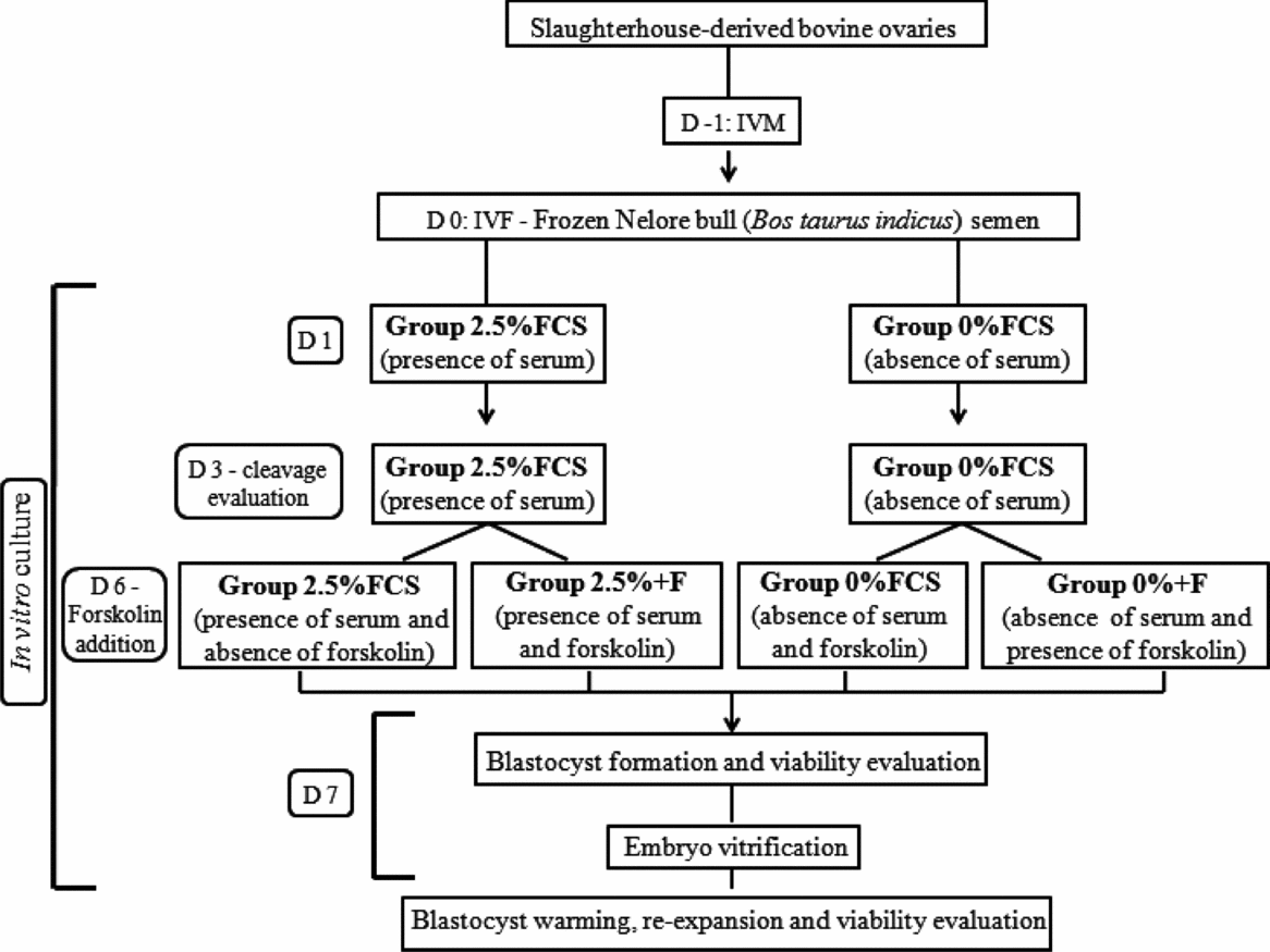
The experimental design for IVP is shown in Fig. 1. Ten replicates were used with a total of 2400 oocytes. Slaughterhouse-derived bovine ovaries were collected in 0.9% saline solution fortified with antibiotics at 35–37°C and carried within 2 h to the laboratory of School of Veterinary Medicine and Animal Science, UNESP, Botucatu-SP, Brazil. All follicles measuring 2–8 mm were aspirated with an 18-gauge needle attached to a 5-ml syringe. Oocytes with at least three intact layers of cumulus cells and homogenous cytoplasm were selected for in vitro maturation (IVM): oocytes were matured in medium consisting of Tissue Culture Medium 199 (TCM; Gibco, Grand Island, New York, USA) supplemented with 10% (v/v) FCS, 0.011 g/ml sodium pyruvate, 1 μg/μl FSH (Pluset®, Hertape Calier Saúde Animal, Juatuba, Brazil), 5 μg/μl LH (luteinising hormone) (Lutropin®, Bioniche Co., Belleville, Ontário, Canada), 100 IU/ml penicillin (Gibco, Grand Island, New York, USA) and 100 μg/ml streptomycin (Gibco, Grand Island, New York, USA).
Experimental design of in vitro-produced embryos. D: day; F: forskolin; FCS: fetal calf serum; IVF: in vitro fertilization; IVM: in vitro maturation.

An average of 30 oocytes were placed in 90-μl droplets of maturation medium overlaid with mineral oil and incubated in a humidified atmosphere with 5% CO2 at 38.5°C in an air incubator for 24 hours.
In vitro fertilization (IVF) occurred on day zero. Frozen Nellore bull (Bos taurus indicus) semen from a single batch was used throughout the study. Sperm cells were selected using a 45–90% Percoll method (Parrish et al., Reference Parrish, Krogenaes and Susko-Parrish1995), and the final concentration was adjusted to 2 × 10⁶ sperm cells/mL. IVM oocytes were washed twice and fertilized in Human Tubal Fluid medium (HTF; Irvine Scientific, Santa Ana, California, USA) supplemented with 0.5 mg/ml caffeine, 0.003 g/ml heparin, 0.0003 g/ml penicillamine, 0.00011 g/ml hypotaurine, 0.00018 g/ml epinephrine, 100 UI/ml penicillin and 100 μg/ml streptomycin. The oocytes were then incubated with the semen suspension under mineral oil in a humidified atmosphere with 5% CO2 at 38.5°C in an air incubator for 20 h.
Twenty hours post-IVF (day one), presumptive zygotes were denuded by repeated pipetting and transferred to culture plates in 90-μl drops of culture medium. The culture medium used was SOFaa (Holm et al., Reference Holm, Booth, Schmidt, Greve and Callesen1999) with 1% (v/v) non-essential amino acids (Gibco, Grand Island, New York, USA) and 1% (v/v) essential amino acids, supplemented with 5 mg/ml BSA, 0.011 g/mL sodium pyruvate, 100 UI/ml penicillin and 100 μg/ml streptomycin in the absence or presence of 2.5% (v/v) FCS, resulting in two groups (0% FCS and 2.5% FCS). The presumptive zygotes were cultured in the presence or absence of serum until day 7 and maintained in a humidified atmosphere of 5% CO2, 5% O2 and 90% N2 at 38.5°C.
Embryo cleavage was recorded on day 3 (72 h post-IVF) of growth. On day 6 (144 h post-IVF), the two groups were homogenously re-divided into four groups for the addition of forskolin (F; 10 μM; Sigma). The groups were allocated in relation to the number of embryos in each developmental stage, i.e., an equal distribution of embryos at each developmental stage for each group. Therefore, the four groups of blastocysts used in this study were: 2.5% FCS: n = 605 (with 2.5% FCS and without F); 0% FCS: n = 595 (without 0% and F); 2.5% + F: n = 605 (with 2.5% FCS and 10 μM F); 0% + F: n = 595 (without FCS and with 10 μM F). The embryos were cultured in forskolin medium from day 6 to day 7.
On the seventh day of blastocyst formation (168 h post-IVF), the quality grade of the blastocysts (grade 1 – excellent, 2 – regular, 3 – poor and 4 – degenerate structure) and scores related to the developmental stage of the embryos (4 – morula, 5 – early blastocyst, 6 – blastocyst, 7 – expanded blastocyst and 8 – hatched blastocyst) were verified, according to the classification of the International Embryo Transfer Society (1998). On the same day, the viability rates were also estimated.
In vivo production of bovine embryos
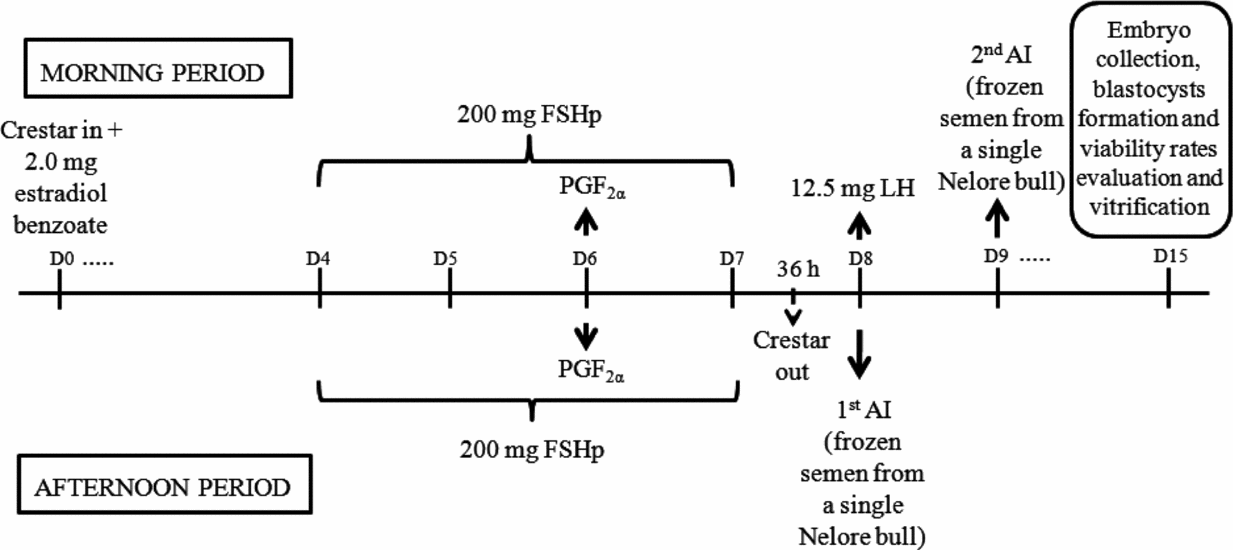
As a control, in vivo-produced embryos were collected from nine Nellore (Bos taurus indicus) cows with good body condition scores. On day zero, all cows were synchronized using an auricular implant containing 3 mg norgestomet (Crestar®, Intervet, Schering-Plough Animal Health Co., Cruzeiro, SP, Brazil) and 2.0 mg estradiol benzoate (Estrogin®, Farmavet, São Paulo, Brazil). Superovulatory treatment was performed using 200 mg FSHp (Follitropin®, Bioniche, Canada) per animal in decreasing doses administrated twice a day for 4 days. On day six, the animals received two treatments of PGF2α (Ciosin®, Intervet/Schering-Plough Animal Health, The Netherlands), once in the morning and once in the afternoon. Thirty-six hours after PGF2α injection, the auricular implant was removed. After 12 h, 12.5 mg LH (Lutropin®, Bioniche, Canada) was administered. The cows were artificially inseminated with frozen semen from a single Nellore bull (Bos taurus indicus) 12 and 24 h after the induction of ovulation.
Forty-five blastocysts were collected 7 days after the first artificial insemination using a 22G Foley probe and retrieved in a 75-μm plastic filter (Sudano et al., Reference Sudano, Landim-Alvarenga, Sartori and Machado2011a). The schemes for the induction of superovulation and embryo collection are shown in Fig. 2.
Induction of superovulation and collection of in vivo-produced embryos. AI: artificial insemination; D: day; LH: luteinizing hormone; FSHp: follicle-stimulating hormone; PGF2α: prostaglandin F2α.
Vitrification and warming of in vitro- and in vivo-produced embryos
Three hundred and ninety-four IVP embryos were vitrified in 10 replicates (2.5% FCS: n = 102; 0% FCS: n = 96; 2.5% + F: n = 101; 0% + F: n = 95), and 18 in vivo-produced embryos were also vitrified as the control. Embryo vitrification was performed according to the technique described by Campos-Chillòn et al. (Reference Campos-Chillòn, Walker, De La Torre-Sanchez and Seidel2006). Early, young and expanded blastocysts of grades 1 and 2 were used in the vitrification of IVP and in vivo-produced embryos, and the morula was discarded.
Briefly, the embryos were washed three times in holding solution (HS; Vigro®, Bioniche Co.) supplemented with 0.1% (w/v) polyvinyl alcohol (PVA) and then transferred to 500 μl of vitrification solution 1 [VS1: 5 M ethylene glycol (EG) in HS] for 3 min. The embryos were then transferred into 10 μl of vitrification solution 2 [VS2: 7 M EG in HS, 0.5 M galactose and 18% (w/v) Ficoll 70]. Straws were filled with 1 cm of galactose solution (GS; 1 M in HS), followed by 0.5 cm air, 7 cm GS and another 0.5 cm air. The embryos (1 or 2) were then loaded with 10 μl of VS2, followed by another 0.5 cm of air and sufficient GS to fill the straw. The time necessary for loading and closing a straw and transferring it into liquid nitrogen vapour was no longer than 45 s. The straws were held in liquid nitrogen vapor for 1 min. The embryos were stored for 1 to 6 months in liquid nitrogen (–196°C).
During warming, the straws were kept in air for 10 s and then transferred to a 35–37°C water bath for 15 s. The straws were slightly agitated to mix the columns of vitrification medium. The embryos were washed and cultured in SOFaa with 10% (v/v) FCS, 2.7 mM myo-inositol, 0.011 sodium pyruvate, 5 mg/ml BSA, 100 UI/ml penicillin and 100 μg/ml streptomycin for 12 h in an incubator with an atmosphere of 5% CO2, 5% O2 and 90% N2 to evaluate re-expansion (Sudano et al., Reference Sudano, Paschoal, Rascado, Magalhães, Crocomo, Lima-Neto and Landim-Alvarenga2011b) and cell viability.
Evaluation of cell viability
Two hundred and seventy-nine expanded IVP embryos were randomly selected for the evaluation of cell viability (139 fresh embryos = 2.5% FCS: n = 30; 0% FCS: n = 35; 2.5% + F: n = 40; 0% + F: n = 34 and 140 vitrified embryos = 2.5% FCS: n = 35; 0% FCS: n = 36; 2.5% + F: n = 34; 0% + F: n = 35). As a control, 28 in vivo-produced embryos (blastocysts and expanded blastocysts) were also used (15 fresh embryos and 13 vitrified embryos). The total number of cells, including intact and damaged cells, was counted, and the rate of damaged cells (the number of damaged cells, dyed red by propidium iodate, divided by the number of viable cells, dyed blue by Hoechst 33342) was assessed from each in vivo- and in vitro-produced embryo.
Intact embryos were incubated in 100-μl drops of a solution containing 125 μg/ml propidium iodate in PBS (phosphate-buffered saline) for 1 min, washed three times and transferred to a slide containing a 10-μl drop of Hoechst 33342 (1 mg/ml) diluted in glycerol. The drop containing the embryos was then covered with a cover slip and examined with an epifluorescence microscope equipped with a filter with 340–380 nm excitation and 430 nm emission wavelengths.
Statistical analysis
Data were analyzed with ANOVA (analysis of variance) using the general linear model (GLM) procedure with the SAS statistical software package (SAS Inst. Inc., Cary, NC, USA). The sources of variation in the model included FCS concentration (0% FCS and 2.5% FCS), forskolin addition (presence and absence) and first-order interactions; all factors were considered to be fixed effects. The arcsine transformation was applied to percentage data. If the ANOVA was significant, the means were separated by Tukey's test. The data are reported as untransformed least-squares means.
Results
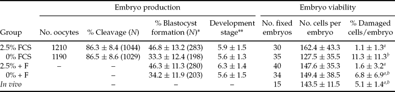
Embryos derived from IVF were randomly cultured in SOFaa with or without FCS. The omission of serum did not reduce the cleavage and blastocyst rate (P > 0.05; Table 1). On day 6 of growth, half of the embryos produced in the presence or absence of FCS were treated with the lipolytic agent forskolin, and the omission or addition of forskolin had no deleterious effects on blastocyst formation (P > 0.05; Table 1). The presence of FCS and/or forskolin also did not affect embryo development (P > 0.05; Table 1).
Mean ± standard deviation of cleavage rate (day three), blastocyst formation rate, development stage, total number of cells per embryo and percentage of damaged cells in in vitro-produced (evaluated on day 7) and in vivo-produced bovine blastocysts
a,bDifferent letters within columns represent significant differences (P < 0.05).
Blastocyst production rates were estimated considering the total number of oocytes in each maturation drop (IVF: day zero).
*After the estimation of cleavage rate on day 3, the two groups of presumptive zygotes were divided into four groups for forskolin addition on day 6 (2.5% FCS: n = 605; 0% FCS: n = 595; 2.5% + F: n = 605; 0% + F: n = 595).
**Developmental stage of day 7 embryos. Scores: 4 – morula, 5 – early blastocyst, 6 – blastocyst, 7 – expanded blastocyst and 8 – hatched blastocyst.
Number of in vivo-produced embryos: 45.
F: forskolin; FCS: fetal calf serum.
With respect to cellular viability, a similar number of cells was observed following all treatments of in vitro- and in vivo-produced embryos (P > 0.05; Table 1).
Although no difference in the percentage of damaged cells was observed between in vivo- and in vitro-produced embryos (P > 0.05; Table 1), a significantly greater percentage of damaged cells was observed in IVP embryos cultured in the absence of FCS (0% FCS) compared with those cultured with FCS (2.5% FCS; P < 0.001) and with FCS plus forskolin (2.5% + F; P < 0.01; Table 1).
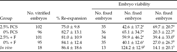
After vitrification, the re-expansion rate was similar between in vitro- and in vivo-produced embryos (P = 0.0989; Table 2). During the time of re-expansion evaluation, it was observed that in vivo-produced embryos had better morphology and a less degenerated shape than IVP embryos, independent of the treatment. The embryos were only analyzed by a stereomicroscope (under ×80 = magnification), and the results were confirmed using viability tests.).
Mean ± standard deviation of embryo re-expansion, total number of cells per embryo and percentage of damaged cells per embryo after vitrification of in vitro-produced (performed on day 7) and in vivo-produced bovine blastocyst
a,bDifferent letters within columns represent significant differences (P<0.05).
Blastocyst production rates were estimated considering the total number of oocytes in each maturation drop (IVF: day zero).
F: forskolin; FCS: fetal calf serum.
After warming, all experimental groups, 2.5% FCS (P < 0.001), 0% FCS (P < 0.05), 2.5% F (P < 0.05) and 0% + F (P < 0.01), had a significantly lower cell number compared with the in vivo-produced embryos (Table 2). Regarding the rate of damaged cells, the 2.5% FCS group had a greater percentage of damaged cells after vitrification compared with other groups (P < 0.05; Table 2), and the addition of forskolin in the presence of FCS reversed this deleterious effect.
Discussion
In the literature, many papers report the association of FCS with BSA (Thompson et al., Reference Thompson, Allen, McGowan, Bell, Lambert and Tervit1998; Rizos et al., Reference Rizos, Guitiérrez-Adán, Pérez-Garnelo, De La Fuente, Boland and Lonergan2003; Paschoal et al., Reference Paschoal, Sudano, Magalhães, Crocomo and Landim-Alvarenga2010a,Reference Paschoal, Sudano, Rascado, Magalhães, Crocomo, Lima-Neto and Landim-Alvarengab,Reference Paschoal, Sudano, Rascado, Magalhães, Crocomo, Lima-Neto, Martins and Landim-Alvarengac; Sudano et al., Reference Sudano, Paschoal, Rascado, Magalhães, Crocomo, Lima-Neto and Landim-Alvarenga2011b,Reference Sudano, Paschoal, Rascado, Lima-Neto and Landim-Alvarengac) but on the other hand, there are others papers that include either BSA or FCS (Pinyopummintr & Bavister, Reference Pinyopummintr and Bavister1991; Mucci et al., Reference Mucci, Aller, Kaiser, Hozbor, Cabodevila and Alberio2006) in embryo culture. In the present experiment, we used both conditions to culture IVP bovine embryos. When FCS was omitted, no deleterious effects on cleavage and blastocyst formation rates were observed (Table 1). Similar results were reported by Holm et al. (Reference Holm, Booth, Schmidt, Greve and Callesen1999), Rizos et al. (Reference Rizos, Guitiérrez-Adán, Pérez-Garnelo, De La Fuente, Boland and Lonergan2003) and Sudano et al. (Reference Sudano, Paschoal, Rascado, Lima-Neto and Landim-Alvarenga2011c), but contradictory results were observed by George et al. (Reference George, Daniaux, Genicot, Verhaeghe, Lambert and Donnay2008), who observed a higher rate of blastocyst formation in the absence of FCS.
Most published works focusing on cleavage and blastocyst assessment used taurine bulls or a crossbreed (taurine × zebu). This factor is one of the reasons that led to our experiment to highlight the cleavage and blastocyst rates compared with the results in the literature. In addition, these results reinforce the differences in breeds observed between zebu and crossbreed, as they behaved differently in relation to the addition of FCS to the culture medium (Paschoal et al., Reference Paschoal, Sudano, Rascado, Magalhães, Crocomo, Lima-Neto, Guastali, Maziero, Martins and Landim-Alvarenga2011).
The quality of IVP embryos is directly related to the culture conditions, and the addition of FCS to the culture medium has been investigated in several laboratories, sometimes showing a positive effect on embryo development and sometimes showing unfavorable results (Byrne et al., Reference Byrne, Southgate, Brison and Leese1999; Mucci et al., Reference Mucci, Aller, Kaiser, Hozbor, Cabodevila and Alberio2006).
The addition of FCS has been identified as the main factor responsible for morphological (Dorland et al., Reference Dorland, Gardner and Trounson1994; Abe et al., Reference Abe, Yamashita, Itoh, Satoh and Hoshi1999b) and physiological differences (Rizos et al., Reference Rizos, Guitiérrez-Adán, Pérez-Garnelo, De La Fuente, Boland and Lonergan2003) when compared with either embryos produced in the absence of FCS or in vivo-produced embryos. Some of these differences include the increase in the number and size of lipid droplets (Abe et al., Reference Abe, Otoi, Tachikawa, Yamashita, Satoh and Hoshi1999a,Reference Abe, Yamashita, Itoh, Satoh and Hoshib, Reference Abe, Yamashita, Satoh and Hoshi2002, Reference Abe, Shiku, Aoyagi and Hoshi2004; Rizos et al., Reference Rizos, Guitiérrez-Adán, Pérez-Garnelo, De La Fuente, Boland and Lonergan2003) and differences in embryo quality (Ferguson & Leese, Reference Ferguson and Leese1999; Sata et al., Reference Sata, Tsuji, Abe, Yamashita and Hoshi1999). From these data, Leivas et al. (Reference Leivas, Brum, Fialho, Saliba, Alvim, Bernardi, Rubin and Silva2011) proposed to use a low concentration of serum (2%) during various culture stages to obtain high-quality embryos, and this concentration is similar to the one we used in the present study.
Sudano et al. (Reference Sudano, Paschoal, Rascado, Macedo, Uliani and Landim-Alvarenga2008) reported in a preliminary study that embryos produced in the presence of 2.5% FCS had improved blastocyst development when compared with those grown in 10% FCS. When the quality was assessed as the number of cells per embryo, the addition of 10% FCS resulted in lower viability. In another study, it was observed that when the serum concentration increased from 0% to 10%, the cytoplasmic lipid accumulation also increased (Sudano et al., Reference Sudano, Paschoal, Rascado, Magalhães, Crocomo, Lima-Neto and Landim-Alvarenga2011b).
Because FCS increases the amount of lipid (Abe et al., Reference Abe, Otoi, Tachikawa, Yamashita, Satoh and Hoshi1999a,Reference Abe, Yamashita, Itoh, Satoh and Hoshib, Reference Abe, Yamashita, Satoh and Hoshi2002, Reference Abe, Shiku, Aoyagi and Hoshi2004; Rizos et al., Reference Rizos, Guitiérrez-Adán, Pérez-Garnelo, De La Fuente, Boland and Lonergan2003), the present work studied the combination of a medium with a low concentration (2.5%) of FCS with the use of the lipolytic agent forskolin.
Forskolin is an adenylate cyclase activator that can induce lipolysis in intact cells and is related to cAMP levels, which are important in the physiological functions of some systems (Seamon & Daly, Reference Seamon and Daly1981; Seamon et al., Reference Seamon, Padgett and Daly1981; Ho & Shi, Reference Ho, Shi and Qi-Huang1982; Ek et al., Reference Ek, Arner, Bergqvist, Carlström and Wahrenberg1997; Arrese et al., Reference Arrese, Matthew, Gazard and Wells1999; Men et al., Reference Men, Agca, Riley and Critser2006). Men et al. (Reference Men, Agca, Riley and Critser2006) observed that the treatment of pig embryos with 10 μM forskolin significantly reduces the amount of cytoplasmic lipids with no deleterious effect on embryo development. To our knowledge, the present work is the first to use forskolin in bovine IVP embryo culture, showing that the addition of 10 μM forskolin on day 6 of growth had no influence on bovine embryo production (Table 1).
The possible mechanism through which forskolin improves the cryosurvival of embryos is most likely due the partial reduction of the intracellular lipid content by stimulating intracellular lipolysis (Seamon et al., Reference Seamon, Padgett and Daly1981; Ho & Shi, Reference Ho, Shi and Qi-Huang1982). This effect is potentiated by all lipolytic hormones, such as epinephrine, adrenocorticotropic hormone and glucagon, and can be reversible (Ho & Shi, Reference Ho, Shi and Qi-Huang1982).
In biochemical terms, the binding of protein G with its specific receptor causes protein G hydrolysis, thereby releasing the α subunit. This subunit will stimulate the adenylate cyclase, an integral plasma membrane protein that catalyzes the conversion of ATP to cAMP. The cAMP is the most important system in the regulation of lipolysis (Lafontan & Berlan, Reference Lafontan and Berlan1993). When cAMP is formed, it stimulates a protein kinase (PKA) to catalyze the phosphorylation of target enzymes or transcription factors. In the case of lipids, PKA phosphorylates triacylglycerol lipase, which causes the lysis of triglycerides, thereby releasing fatty acids and forming glycerol (Nelson & Cox, Reference Nelson and Cox2002; Faherty et al., Reference Faherty, Fitzgerald, Keohan and Quinlan2007; Campbell & Farrel, Reference Campbell and Farrel2008).
In fat cells, there is a relationship between increased cAMP levels and the lipolysis of triglycerides (Honnor et al., Reference Honnor, Dhillon and Londos1985), and catecholamines (epinephrine and norepinephrine) are the regulators with five membrane receptors (β1, β2, β3, α1 and α2). When catecholamines bind to their receptors, the cAMP controller receptor increases, raising the intracellular cAMP levels (from ATP) and promoting PKA activation, which phosphorylates serine residues (Ser-563 in rats and Ser-551 in humans) in hormone-sensitive lipase (HSL). Then, HSL is transferred to the lipid droplet and stimulates the lipolysis of triglycerides (Honnor et al., Reference Honnor, Dhillon and Londos1985; Lafontan & Berlan, Reference Lafontan and Berlan1993).
In recent experiments, Barceló-Fimbres et al. (Reference Barceló-Fimbres, Anchondo-Garay, López-Franco, García-Quiñonez, Antillón-Ruiz, Rodríguez-Almeida and Seidel2010) produced IVP embryos with forskolin followed by cryopreservation and did not find satisfactory results in the pregnancy rate. One explanation for this low effect of forskolin on the pregnancy rate may be that the authors used a higher serum concentration than in the present experiment (up 2.5%), and thus, the lipolytic agent was not sufficient to induce lipolysis at the concentration used (10 μM). In our experiment, we did not transfer the embryos, but a decrease in the damaged cells rate was observed when forskolin was added.
Other drugs are used to reduce the serum lipid concentration, such as phenazine ethosulfate (PES) (De La Torre-Sanchez et al., Reference De La Torre-Sanchez, Kimberly and George2006; Barceló-Fimbres & Seidel Jr., Reference Barceló-Fimbres and Seidel2007a,Reference Barceló-Fimbres and Seidelb; Sudano et al., Reference Sudano, Paschoal, Rascado, Magalhães, Crocomo, Lima-Neto and Landim-Alvarenga2011b), pyrroline-5-carboxylate (De La Torre-Sanchez et al., Reference De La Torre-Sanchez, Kimberly and George2006) and 2,4-dinitrophenol (Rieger et al., Reference Rieger, McGrowan, Cox, Pugh and Thompson2002; Barceló-Fimbres & Seidel, Reference Barceló-Fimbres and Seidel2007a,b), which are embryonic metabolic regulators that can improve cryopreservation. Moreover, techniques such as embryo centrifugation and micromanipulation (Nagashima et al., Reference Nagashima, Kashiwazaki, Ashman, Grupen, Seamark and Nottle1994, Reference Nagashima, Kuwayamat, Grupen, Ashman and Nottle1996) or the use of lipolytic agents, such as dibutyryl cAMP (Ek et al., Reference Ek, Arner, Bergqvist, Carlström and Wahrenberg1997), epinephrine, norepinephrine, isoproterenol and theophylline (Seamon & Daly, Reference Seamon and Daly1981; Seamon et al., Reference Seamon, Padgett and Daly1981; Ho & Shi, Reference Ho, Shi and Qi-Huang1982; Ek et al., Reference Ek, Arner, Bergqvist, Carlström and Wahrenberg1997; Arrese et al., Reference Arrese, Matthew, Gazard and Wells1999; Men et al., Reference Men, Agca, Riley and Critser2006), can also remove the lipid drops in embryos by stimulating intracellular lipolysis.
The developmental stages of the embryos were evaluated through morphological analysis on day 7 (Table 1). Because the development of embryos in all treatment groups was very similar, it was possible to conclude that the omission of FCS and/or the addition of forskolin have no deleterious effects on embryo growth. These results resemble those obtained by Sudano et al. (Reference Sudano, Paschoal, Rascado, Lima-Neto and Landim-Alvarenga2011c) who observed that the omission of FCS did not influence embryo development. However, the low FCS concentration used in our experiment may be responsible for the similarity. In contrast, these results differ from those obtained by Gómez & Díez (Reference Gómez and Díez2000), who found that the addition of FCS promoted the earlier appearance of blastocysts and resulted in more embryos reaching the blastocyst stage on day 7, and that from Rizos et al. (Reference Rizos, Guitiérrez-Adán, Pérez-Garnelo, De La Fuente, Boland and Lonergan2003), who observed that the presence of FCS had a stimulatory effect on the speed of embryo development.
There have been reports showing that serum supplementation increased the cell number and, consequently, the quality of embryos (Van Wagtendonk-De Leeuw et al., Reference Van Wagtendonk-De Leeuw, Den Daas and Rall1997; Gardner et al., Reference Gardner, Pool and Lane2000), whereas others found a negative effect (Sudano et al., Reference Sudano, Paschoal, Rascado, Macedo, Uliani and Landim-Alvarenga2008) or no effect (Gómez & Díez, Reference Gómez and Díez2000; Paschoal et al., Reference Paschoal, Sudano, Silva and Landim-Alvarenga2009; Sudano et al., Reference Sudano, Paschoal, Rascado, Lima-Neto and Landim-Alvarenga2011c).
Pinyopummintr & Bavister (Reference Pinyopummintr and Bavister1991) and Mucci et al. (Reference Mucci, Aller, Kaiser, Hozbor, Cabodevila and Alberio2006) observed a greater number of cells in embryos produced in the presence of FCS. In the present study, no differences were observed among IVP groups in the total number of cells per embryo (Table 1). Gómez & Díez (Reference Gómez and Díez2000) also found this result, but in their experiment, the FCS was added 42 h after fertilization.
It is well recognized that bovine embryos derived in vivo, typically following superovulation, artificial insemination and non-surgical recovery, have a higher quality than those derived from in vitro maturation, fertilization and culture (Lonergan et al., Reference Lonergan, Fair, Corcoran and Evans2006), but no differences are observed in the present study between the number of cells in IVP embryos and in vivo-produced embryos. This result agrees with a study that argues that all media utilized were suitable for bovine embryo production (Pomar et al., Reference Pomar, Teerds, Kidson, Colenbrander, Tharasanit, Aguilar and Roelen2005). Similar results have been obtained by Enright et al. (Reference Enright, Lonergan, Dinnyes, Fair, Ward, Yang and Boland2000) who observed that IVP embryos in the presence of FCS have an average of 96.2 cells/embryo, whereas in vivo-produced embryos have 101.9 cells/embryo. In the present study, the number of cells in the embryos was greater than these reported values. This difference may be linked to the fact that only expanded blastocysts were analyzed in the present work.
The omission of FCS (0% FCS group) resulted in an increase in the percentage of damaged cells compared with embryos produced in the presence of FCS (2.5% FCS and 2.5% + F groups) (Table 1), agreeing with the literature showing that FCS is not essential for normal embryo development (Carolan et al., Reference Carolan, Lonergan, Van-Langendonckt and Mermillod1995; Abe et al., Reference Abe, Yamashita, Satoh and Hoshi2002). Although the absence of FCS in IVP culture decreases embryo quality, the embryos generated from this culture did not differ from in vivo-produced embryos.
Ice crystal formation is a problem in the cryopreservation of in vivo- and in vitro-produced embryos, but according to the literature, IVP embryos are more sensitive to cryopreservation than in vivo-produced embryos (Abe et al., Reference Abe, Yamashita, Satoh and Hoshi2002; Rizos et al., Reference Rizos, Ward, Duffy, Boland and Lonergan2002, Reference Rizos, Guitiérrez-Adán, Pérez-Garnelo, De La Fuente, Boland and Lonergan2003; Moore et al., Reference Moore, Rodríguez-Sallaberry, Kramer, Johnson, Wroclawska, Goicoa and Niasari-Naslaji2007).
The most commonly used procedure for the cryopreservation of in vivo-produced bovine embryos involves the equilibration of embryos in cryoprotectant for 5–10 minutes, seeding at approximately –6°C, cooling at approximately 0.5°C/min to approximately –32°C, followed by plunging into liquid nitrogen (Campos-Chillòn et al., Reference Campos-Chillòn, Walker, De La Torre-Sanchez and Seidel2006). Therefore, the morphological features of the cells of in vivo-produced embryos are better preserved under conventional freezing than under the vitrification process (Visintin et al., Reference Visintin, Martins, Bevilacqua, Mello, Nicácio and Assumpção2002).
In this way, the freezing process may vary in the resulting embryonic quality aspects after thawing, resulting in lower pregnancy rates (Zanenga, Reference Zanenga1993). IVP embryos are more sensitive to cryopreservation alterations, especially to slow freezing, than in vivo-produced embryos (Yu et al., Reference Yu, Deng, Liu, Li, Li, Zhang and Zan2010).
Because of these concerns, vitrification has been used as an alternative for the cryopreservation of IVP embryos, resulting in successful pregnancies after embryo transfer (Dinnyés et al., Reference Dinnyés, Carolan, Lonergan, Massip and Mermillod1996; Sommerfeld & Niemann, Reference Sommerfeld and Niemann1999; Campos-Chillòn et al., Reference Campos-Chillòn, Walker, De La Torre-Sanchez and Seidel2006). Despite this success, this technique is not very common for procedures with in vivo-produced embryos because more practical and reliable methodology and large field trials for evaluation are necessary (Campos-Chillòn et al., Reference Campos-Chillòn, Walker, De La Torre-Sanchez and Seidel2006). In the present study, we chose to vitrify in vivo-produced embryos to use as a control of IVP embryos.
Survival can be defined as re-expansion and continued development with normal morphology in culture following vitrification and warming (Johnson et al., Reference Johnson, Wroclawska, Bonilla and Moore2007). When the re-expansion rate was assessed, there was no difference between IVP groups in the presence or absence of FCS. However, Rizos et al. (Reference Rizos, Guitiérrez-Adán, Pérez-Garnelo, De La Fuente, Boland and Lonergan2003) and Mucci et al. (Reference Mucci, Aller, Kaiser, Hozbor, Cabodevila and Alberio2006) observed a higher re-expansion rate of embryos cultured in the absence of serum. The lack of a positive effect of serum supplementation may be a consequence of the differences in experimental conditions, such as the absence of a co-culture system during all developmental periods and the type and concentration of serum used (Mucci et al., Reference Mucci, Aller, Kaiser, Hozbor, Cabodevila and Alberio2006). Therefore, in another experiment, the presence of FCS led to a higher re-expansion rate of embryos (Johnson et al., Reference Johnson, Wroclawska, Bonilla and Moore2007).
Similarly, we did not find a difference in the re-expansion rate of in vivo- and in vitro-produced embryos. In the literature, in vivo-produced embryos had a higher rate of re-expansion than in vitro-produced embryos (Rizos et al., Reference Rizos, Ward, Boland and Lonergana2001, Reference Rizos, Guitiérrez-Adán, Pérez-Garnelo, De La Fuente, Boland and Lonergan2003). With regard to the hatching rate, it was observed that omitting serum from the medium of IVP embryos has a favorable effect on blastocyst formation, achieving the same rate as in vivo-derived embryos (Rizos et al., Reference Rizos, Guitiérrez-Adán, Pérez-Garnelo, De La Fuente, Boland and Lonergan2003). The ability of these blastocysts to withstand cryopreservation is used as a quality indicator (Rizos et al., Reference Rizos, Guitiérrez-Adán, Pérez-Garnelo, De La Fuente, Boland and Lonergan2003).
Traldi et al. (Reference Traldi, Leboeuf, Cognié, Poulin and Mermillod1999) showed that IVP goat embryos could survive cryopreservation and produce live offspring after embryo transfer. It appears that in vivo-produced and IVP small ruminant embryos are similar in their survival rates and developmental competence after cryopreservation and subsequent embryo transfer.
Recent data show that, within 24 h, IVP bovine embryos have a higher rate of re-expansion than in vivo-produced embryos, and after 48 and 72 h of survival, there is no difference between these embryos (Kuzmany et al., Reference Kuzmany, Havlicek, Wrenzycki, Wilkening, Brem and Besenfelder2011), proving that depending on the embryo culture and methodology applied, IVP embryos may have a similar or even a higher viability and quality than in vivo-produced embryos. The changes in the developmental competence and resistance to freezing damage of the embryos may be due to different media compositions or embryo production systems and could be used as a sensitive indicator of blastocyst quality (Rizos et al., Reference Rizos, Ward, Boland and Lonergana2001, Reference Rizos, Guitiérrez-Adán, Pérez-Garnelo, De La Fuente, Boland and Lonergan2003). Even when developmental rates are similar, blastocysts developing under different culture conditions can differ in their capability to survive freezing (Enright et al., Reference Enright, Lonergan, Dinnyes, Fair, Ward, Yang and Boland2000).
Although the re-expansion rates were similar between in vitro- and in vivo-produced embryos, the number of cells of in vivo-produced embryos after warming was higher compared with IVP embryos, indicating a greater sensitivity of IVP embryos to the vitrification process independently of the treatment group (Table 2).
In all studied groups, the total number of cells was lower after vitrification compared with fresh embryos from the same group, demonstrating that some of the blastomeres were destroyed during vitrification. Nevertheless, no differences were observed in the total number of cells among IVP embryo groups. Neither the omission of FCS nor the addition of forskolin reduced this effect.
Because FCS is not recommended for embryos that will be vitrified, it is important to emphasize the similarity between the 2.5% + F and 0% FCS groups, which means that the addition of forskolin to the embryos developed in serum medium led to similar cellular viability and cryotolerance as the embryos cultured in the absence of serum (Table 2). The same results were observed by Mucci et al. (Reference Mucci, Aller, Kaiser, Hozbor, Cabodevila and Alberio2006).
During vitrification, bovine cellular membranes undergo several types of damage (Wolfe & Bryant, Reference Wolfe and Bryant1999; Acker & McGann, Reference Acker and McGann2001), and the extent of cell damage could influence embryo survival. The membrane stability of IVP bovine embryos might be an important factor in determining their susceptibility to cryopreservation. In this study, the IVP embryos in the presence of FCS had a higher rate of cell damage than the embryos cultured in the absence of FCS (and in the presence of BSA), which agrees with the results of Mucci et al. (Reference Mucci, Aller, Kaiser, Hozbor, Cabodevila and Alberio2006).
It has been reported that the inclusion of unsaturated fatty acids (linoleic acid bound to BSA; linoleic acid albumin) in the culture medium improved blastocyst survival after cryopreservation (Imai et al., Reference Imai, Kobayashi, Goto, Dochi and Shimoshira1997; Hochi et al., Reference Hochi, Kimura and Hanada1999). The positive effects of linoleic acid albumin supplementation may be explained by the increased membrane fluidity (unsaturation level) due to the direct incorporation of these fatty acids into the plasma membrane (Imai et al., Reference Imai, Kobayashi, Goto, Dochi and Shimoshira1997).
The integrity of the plasma membrane may be restored in cells after cryopreservation as a consequence of the supply of membranes from intracellular vesicles, as the inhibition of vesicular transport strongly suppressed the restoration of the resistance to osmotic swelling after bovine embryo vitrification (Edashige et al., Reference Edashige, Asano, An and Kasai1999). The presence of large amounts of lipid droplets in the embryos developed in serum-supplemented medium probably damages the cellular mechanisms responsible for repairing plasma membranes after cryopreservation. The incorporation of saturated fatty acids, promoted by FCS, into the plasma membrane could probably induce changes in membrane composition, making the membrane more rigid and unable to withstand cryopreservation (Mucci et al., Reference Mucci, Aller, Kaiser, Hozbor, Cabodevila and Alberio2006).
The differences found between the in vitro- and in vivo-produced embryos confirm the high sensitivity of IVP embryos to vitrification, which is consistent with Sudano et al. (Reference Sudano, Paschoal, Rascado, Magalhães, Crocomo, Lima-Neto and Landim-Alvarenga2011b) who found that regardless of serum concentration, the in vivo-produced embryos had a greater number of cells per embryo after vitrification. Although the use of forskolin resulted in better-quality embryos after vitrification, the drug was not good enough to equalize in vivo- and in vitro-produced embryos in relation to the number of cells per embryo (Table 2).
Regarding the rate of damaged cells per embryo, there was greater injury in IVP embryos cultured in presence of FCS. Similar data are found in the literature (Mucci et al., Reference Mucci, Aller, Kaiser, Hozbor, Cabodevila and Alberio2006), confirming the negative effect of serum supplementation on vitrification. However, there is a favorable effect of 10 μM forskolin to serum-containing medium, significantly decreasing the damaged cell rate (group 2.5% + F), similar to the IVP embryos in the absence of FCS and to in vivo-produced embryos (Table 2).
Conclusion
According to the results of this study, serum omission produced embryos with lower quality and with a higher rate of damaged cells per embryo; however, after vitrification, the addition of FCS was extremely harmful to the embryos. This effect was reversible when forskolin was added to the serum-containing medium, which improved embryo survival without affecting embryo development and quality after vitrification.
Acknowledgements
Grant supported by Fundação de Amparo à Pesquisa do Estado de São Paulo FAPESP (São Paulo Res. Foundation).