Case description
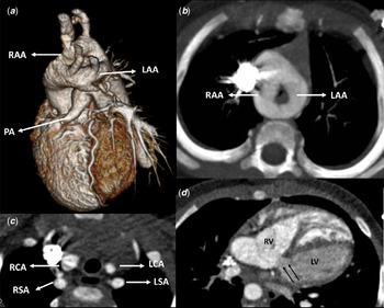
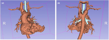
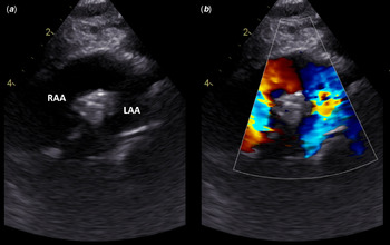
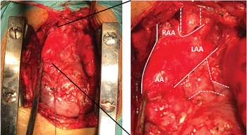
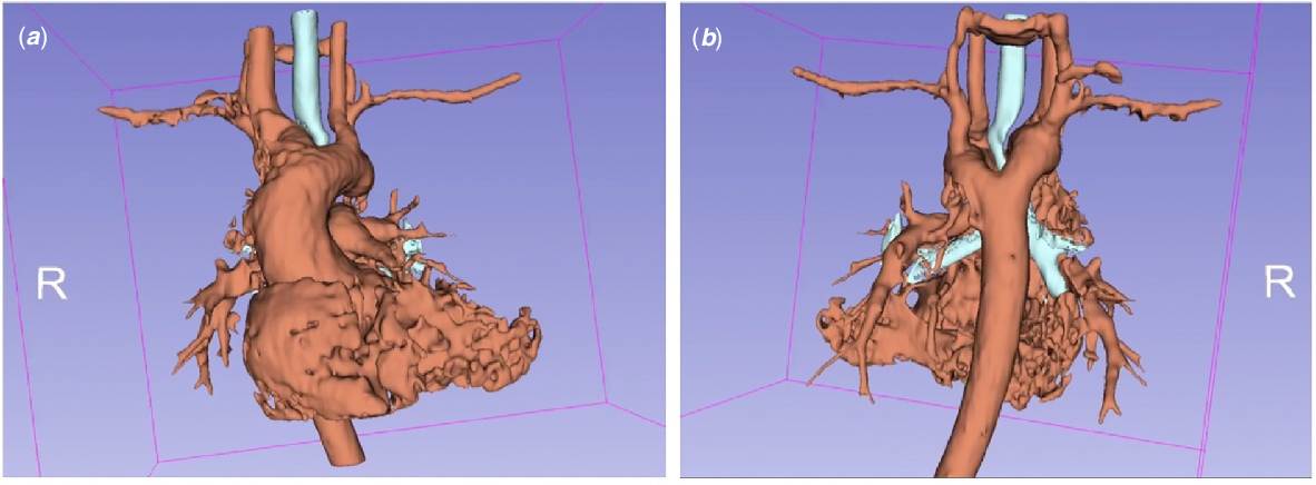
A male newborn with antenatal diagnosis of pulmonary atresia and ventricular septal defect presented with cyanosis upon birth. Antenatal diagnosis was challenging due to increased maternal body habitus and genetic testing was normal. Post-natal echocardiogram showed membranous pulmonary atresia with small but confluent pulmonary arteries, large ventricular septal defect, left aortic arch and large ductus arteriosus. At 4 days of life, he underwent a right-sided modified Blalock-Taussig shunt implantation and ductus ligation through right thoracotomy. After surgery, the patient developed refractory biphasic stridor, with no other symptoms. Otolaryngology examination showed only laryngeal oedema and tracheomalacia. Due to worsening cyanosis and syncope on exertion, a CT angiography was performed which showed a double aortic arch, forming a balanced vascular ring and causing tracheal compression (Figs 1 and 2). A detailed echocardiogram was also consistent with this diagnosis (Fig 3). Intracardiac anatomy proved to be adequate for surgical repair. Resection of the right aortic arch after the origin of the neck vessels, shunt division, ventricular septal defect closure and reestablishment of right ventricle to pulmonary artery continuity (using a conduit) were performed (Fig 4). The surgery was uneventful and currently, at 2.5 years old he remains asymptomatic.
CT angiography. ( a ) Tridimensional reconstruction (left-sided view) depicting the vascular ring, with similar size right (RAA) and left-sided aortic arch (LAA), the pulmonary atresia (PA) and the origin of the neck vessels; ( b ) Axial plane, maximum intensity projection (MIP) image depicting the right and left aortic arches encircling the trachea; ( c ) Axial plane, depicting the origin of the neck vessels and their relation with the trachea – RCA, right carotid artery; RSA, right subclavian artery; LCA, left carotid artery; LSA, left subclavian artery; ( d ) Axial oblique reformation, showing a large ventricular septal defect (black arrows) – RV, right ventricle, LV, left ventricle.

CT angiography tridimensional reconstruction of the double aortic arch (red) and its relation with the trachea and the bronchi (blue). ( a ) Anterior view; ( b ) posterior view. Tracheal compression by the double arch is evident. Please see also the video in the online supplementary material.
Suprasternal short axis echocardiogram view in 2D ( a ) and colour Doppler ( b ). LAA: left aortic arch; RAA: right aortic arch.

Intraoperative appearance demonstrating the double aortic arch. White line delimitates double aortic arch. Dashed white line delimitates the trachea and the main bronchi (*). AA: ascending aorta; LAA: left aortic arch; RAA: right aortic arch.

Double aortic arch is a rare occurrence, usually not associated with other forms of CHD. Reference Sahni and Franklin1–Reference Asada, Itoi and Hamaoka3 There are few reports describing its association with pulmonary atresia and ventricular septal defect. Reference Santos, Croti, Marchi and Sobrinho2,Reference Asada, Itoi and Hamaoka3 This diagnosis might be challenging and should be excluded in the setting of persistent unexplained respiratory symptoms.
Supplementary material
To view supplementary material for this article, please visit https://doi.org/10.1017/S1047951121004856
Acknowledgements
We thank Carla Saraiva, Radiology Department, José Pedro Neves, Cardiothoracic Surgery, and Duarte Martins, Paediatric Cardiology, for their contribution.
Financial support
This research received no specific grant from any funding agency, commercial or not-for-profit sectors.
Conflicts of interest
None.