

Introduction
A recent review on the effects of lipid and protein oxidation on broiler growth, oxidative status and meat quality dealt with many different potential sources for oxidative stress suggested that organic selenium (Se) yeast might be one dietary supplement that could reduce the incidence of oxidative stress (Estévez, Reference Estévez2015). After the first reports that organic Se yeast (Sel-Plex™) supplementation to broiler diets resulted in less drip loss from refrigerated breast meat (Edens et al., Reference Edens, Lyons and Jacques1996; Edens, Reference Edens, Lyons and Jacques1996; Edens et al., Reference Edens, Parkhurst and Sefton2000b), a progressive interest for the use of Sel-Plex™ as a reliable dietary source of Se to improve meat quality due to its antioxidant activity in cells and tissues has resulted in the development of a substantial body of research. Examples of the chronology of this developing data base for poultry meat quality can be reviewed in many different scientific papers (Downs et al., Reference Downs, Hess and Bilgili2000; Naylor et al., Reference Naylor, Choct and Jacques2000; Hess et al., Reference Hess, Downs, Bilgili, Lyons and Jacques2003; Choct et al., Reference Choct, Naylor and Reinke2004; Upton et al., Reference Upton, Edens and Ferket2008; Perić et al., Reference Perić, Milošević, Žikić, Kanački, Džinić, Nollet and Spring2009; Puvača and Stanaćev, Reference Puvača and Stanaćev2011). The use of organic Se yeast as a nutritional means to improve meat quality has extended beyond poultry to pork (Mahan et al., Reference Mahan, Cline and Richert1999), veal (Skřivanová et al., Reference Skřivanová, Marounek, De Smet and Raes2007), beef (Juniper et al., Reference Juniper, Phipps, Ramos-Morales and Bertin2008; Cozzi et al., Reference Cozzi, Prevedello, Stefani, Piron, Contiero, Lante, Gottardo and Chevaux2011), lamb (Vignola et al., Reference Vignola, Lambertini, Mazzone, Giammarco, Tassinari, Martelli and Bertin2009), goats (Sethy et al., Reference Sethy, Garg, Mishra, Biswal, Behera, Sahoo, Satapathy, Meher and Nayak2014), and salmon and other fish (de Lyons, 1998) as examples of that interest. Organic Se bound in meat protein has been considered an important issue in maintenance of beef and pork meat quality and stability due to its support of the enzymatic activities of certain glutathione peroxidases (Daun et al., Reference Daun, Johansson, Önning and Åkesson2001; Edens and Sefton, Reference Edens and Sefton2016), and it appears that organically bound Se in poultry meat is similarly important (Daun et al., Reference Daun, Lundh, Őnning and Åkesson2004).
Surai (Reference Surai2002) pointed out that in order to improve meat quality, producers had to combat pre- and post-slaughter oxidative stress effects on meat quality and stability. At the time of his review, producers had already begun to explore the use of dietary supplements to provide higher dietary levels of vitamin E or ascorbic acid, which are known to have powerful antioxidant properties and/or pre-slaughter withdrawal of dietary transition trace minerals that have pro-oxidant properties.
It has been a long-held belief that non-haem iron and copper have pro-oxidant influence in fresh and cooked meat, and many producers have routinely removed iron and copper from finisher diets as an attempt to improve oxidative stability to poultry meat. Contrary to the belief that transition metals have a negative influence on meat quality, Yang et al. (Reference Yang, Sun, Li, Wu and Yao2011) reported that dietary iron, copper, zinc and manganese generally improved meat quality by increasing lightness and yellowness values and water-holding capacity of breast or leg muscle. However, the transition metals apparently increased the shear force (toughness) for breast meat and had variable non-significant effects on thigh muscle shear force, which cast a shadow on the transition metal effects on tenderness of the meat. Yang et al. (Reference Yang, Sun, Li, Wu and Yao2011) made no mention about the influence of dietary transition metals on the Se status and oxidative status of the chicken muscles. Nevertheless, Shadhidi and Hong (Reference Shadhidi and Hong1991) reported earlier that Fe+2, Fe+3, Cu+1, and Cu+2 all had pro-oxidant activity in cooked pork stored at 4 °C for 21 days and that Fe+2 and Cu+1 had more oxidative potential than Fe+3 and Cu+2. Ruiz et al. (Reference Ruiz, Perez-Vendrell and Esteve-Garcia2000) reported that removal of iron and copper from finisher diets was related to less oxidative activity in cooked broiler leg meat. The improvement in antioxidant status due to removal of iron and copper is probably due to lower content of both iron and copper in the meat. It is of interest that the remaining iron and copper in muscle and meat retain pro-oxidant potential. Bekhit et al. (Reference Bekhit, Hopkins, Fahri and Ponnampalam2013) reported that these transition metals are bound to protein rendering these minerals to limited reactive ability.
After an animal has been slaughtered and the muscle matures to meat during refrigeration and retail display, biochemical alterations, such as pH shifts, protein and fat denaturation, and oxygen availability, which contribute to oxidative activity, can free those metals from the protein allowing them to decrease meat quality as they generate free radicals. It is at this point that interceptive reactions from the activity of Se-dependent enzymes, glutathione, and other antioxidant systems can reduce those newly generated free radicals.
In this review, it is important to address the issue of Se forms that have an influence on meat quality. Additionally, it was important to attempt to explain how selenoproteins such as certain glutathione peroxidases play an integral role in maintenance of meat quality, and why organic Se has a greater positive influence on meat quality than inorganic Se in selenite or selenate forms.
Selenium: an essential trace element
The use of organic Se in yeast cell wall protein (Sel-Plex™) to improve meat quality seems, at first, to be outside the primary role of Se as the integral entity, selenocysteine or in active sites of selenoproteins/enzymes (Holben and Smith, Reference Holben and Smith1999). Edens and Gowdy (Reference Edens, Gowdy, Lyons and Jacques2004) observed that, among the known 25–28 selenoproteins and enzymes in animals, many have antioxidant potential, but many others have functions yet to be determined (Kryukov et al., Reference Kryukov, Castellano, Novoselov, Lohanov, Zehtab, Gulgó and Gladyshev2003; Kieliszek and Błażejak, Reference Kieliszek and Błażejak2013; Brigelius-Flohé and Maiorino, Reference Brigelius-Flohé and Maiorino2013).
The role of Se as an essential trace element in animal nutrition has been reviewed extensively by various authors (Edens, Reference Edens, Lyons and Jacques1996; Edens and Sefton, Reference Edens and Sefton2016; Surai, Reference Surai2002) and is known to be required for maintenance of health, growth, prevention of disease in both young and old individuals and myriad biochemical-physiological functions (Scott et al., Reference Scott, Nesheim and Young1982). The form of Se is important in its uptake, storage and functionality within the animal. Organic Se (selenomethionine) is abundant in plants and meat (Burk, 1976; Olson and Palmer, Reference Olson and Palmer1976; Levander 1986; Cai et al., 1995) and is the natural form that most animals ingest. It is now known that selenomethionine is actively incorporated into proteins, randomly substituting for methionine (Pan et al., Reference Pan and Tarver1964; Hansson and Jacobson, Reference Hansson and Jacobsson1966; Markham et al., Reference Markham, Hafner, Tabor and Tabor1980; Ip and Hayes, Reference Ip and Hayes1989; Schrauzer, Reference Schrauzer1998, Reference Schrauzer2000). The selenoaminoacids are bound in protein, principally as selenomethionine and selenocysteine and constitute 50 to 80% of the total Se in plants, grains (Butler and Peterson, 1967) and in Sel-Plex™, the organic Se-enriched yeast cell wall protein (Kelly and Power, 1995).
Non-ruminant animals are unable to synthesise selenomethionine from either selenite or selenate forms of inorganic Se (Cummins and Martin, Reference Cummins and Martin1967; Olson and Palmer, Reference Olson and Palmer1976; Sunde, Reference Sunde1990). However, selenomethionine and inorganic selenate and selenite selenium can be converted to selenocysteine, which is found in body tissues of all animals (Esaki et al., Reference Esaki, Nakamura, Tanaka, Suzuki, Morino and Soda1981; Beilstein and Whanger, Reference Beilstein and Whanger1986). In muscle tissue, the abundance of selenocysteine is not unexpected since selenomethionine, selenite and selenate Se are readily converted to selenocysteine, which can be specifically inserted into selenoproteins such as selenoproteins SelW, SelM, SelV, SelT, SelH, Sep15, SelN, the GSH-px, the thioredoxin reductases many of which function as antioxidants (Ip and Hayes, Reference Ip and Hayes1989; Schrauzer, Reference Schrauzer1998; Rederstorff et al., Reference Rederstorff, Krol and Lescure2006; Lescure et al., Reference Lescure, Rederstorff, Krol, Guicheney and Allamand2009), and methionine sulfoxide reductase (Sepx1), which functions in reduction of oxidised methionine residues in proteins (Lee et al., Reference Lee, Dikiy, Kim and Gladyshev2009). Dietary selenomethionine is readily incorporated non-specifically, especially into muscle cell myofibrillar protein and other cellular proteins, becoming a structural component of the substituted myofibrillar proteins serving as a rich source of stored Se, which can be redistributed to other Se containing proteins as the original muscle cell myofibrilar protein degrades (Hansson and Jacobson, Reference Hansson and Jacobsson1966; (Olson and Palmer, Reference Olson and Palmer1976); Ip and Hayes, Reference Ip and Hayes1989; Schrauzer, Reference Schrauzer1998, Reference Schrauzer2000). Beilstein and Whanger (Reference Beilstein and Whanger1986) injected young rats with either [75Se] selenite or [75Se] selenomethionine and examined the compartmentalisation of each tracer in erythrocyte and whole liver protein acid hydrolysates. [75Se] selenocysteine was the principle form of selenium in [75Se] selenite-injected animals at one and 20 days post-injection. In the liver of [75Se] selenomethionine-injected rats, both [75Se] selenomethionine and [75Se] selenocysteine were found at one day post-injection and, at 20 days post-injection, most of the [75Se] selenomethionine had been converted to [75Se] selenocysteine. Glutathione peroxidase contained 75Se as selenocysteine regardless of the selenium compound injected. Haemoglobin of [75Se] selenomethionine-injected animals contained principally [75Se] selenomethionine at both one and 20 days post-injection, indicating the deposition of this selenoaminoacid into protein. In acid hydrolysates of whole liver 75Se was recovered principally as [75Se] selenocysteine from animals injected with [75Se] selenite or [75Se] selenomethionine. No differences were found in deposition of 75Se in liver, kidney, testes, erythrocytes or plasma in rats injected with labelled selenite or selenomethionine, but a significantly greater retention was found in muscle of selenomethionine-injected rats as compared to those given selenite. Additionally, it has been determined via 77Se (a non-radioactive isotope form) that selenium metabolites such as methylselenol and Se-methylselenocystiene (Ip, Reference Ip1998) can be found in tissues and function in molecular actions related to redox changes leading to inhibition of cellular proliferation (Fleming et al., Reference Fleming, Ghose and Harrison2001).
Schrauzer (Reference Schrauzer2000) referred to work in which the replacement of methionine by selenomethionine usually did not alter protein structure but might influence the activity of enzymes if selenomethionine replaced methionine in the vicinity of an enzyme's active site. The CH3-Se group of selenomethionine is more hydrophobic than the CH3-S-moiety of methionine, and in these selenomethionine-substituted enzymes, substrate access was affected via alterations of the kinetic parameters by factors of 40 to 400% (Boles et al., Reference Boles, Cisneros, Weir, Odom, Villafranca and Dunlap1991; Bernard et al., Reference Bernard, Wells, Cleasby, Borlat, Payton and Proudfoot1995). Nevertheless, if a large number of methionine residues are replaced with selenomethionine in certain enzymes by more than 50%, the enzyme could become inactive (Boles et al., Reference Boles, Cisneros, Weir, Odom, Villafranca and Dunlap1991).
The retention and metabolism of organic and inorganic Se is discussed in Edens and Sefton (2016a, b) and should be accessed for in-depth discussion. However, activity of organic Se, functioning as an antioxidant and organic Se ability to accumulate in tissues and cells, particularly membranes, is key to its potential in terms of promoting meat quality and preventing drip loss via improved robustness of cell membranes. The following section discusses the importance of Se in sustaining meat quality.
Factors influencing consumer preferences in the purchase of meat
Wood et al. (Reference Wood, Enser, Fisher, Nute, Richardson and Sheard1999) described meat quality as the ‘attractiveness’ of meat to the consumer. Despite other consumer considerations regarding purchases, physical/visual appearance plays the first major role in either acceptance or rejection of the meat on a packing tray. This is often based on a subjective assessment of the colour in both white and red meats, marbling, and the amount of liquid in the packing tray. When the issue of meat quality is discussed, this includes multiple factors that influence the perceived tenderness/toughness, juiciness/moisture content, firmness/moisture, protein content and functionality, appearance/colour and apparent hydration, and economic value of meat (Northcutt et al., Reference Northcutt, Foegeding and Edens1994).
One of the primary factors that cause consumer rejection of meat and meat products at the food market is the apparent loss of meat-held water via weepage or drip loss (Huff-Lonergan and Lonergan, Reference Huff-Lonergan and Lonergan2005). Consumers observe meat colour, which changes as water is lost from the meat, as well as relative firmness and moisture, as relating to perceived tenderness of the meat.
Moisture within meat is held in association with protein residing within myofibrils of sarcomeres, between the myofibrils, and between myofibrils and the cell membrane, among muscle cells and muscle bundles. It has been reported that, when muscle matures, the retained moisture can change in response to biochemical processes within the muscle and in response to handling of the product (Honikel, Reference Honikel, te Pas, Everts and Haagsman2004; Honikel and Kim, Reference Honikel and Kim1986). Oxidative stress within the muscle and subsequently in the matured meat can damage cell membranes, proteins within the myofibrils, and bundles of muscle fibres, which will then decrease moisture holding capacity of meat. The antioxidant effect of both GSHpx-1 and GSHpx-4 in muscles and meat is well documented (Daun et al., Reference Daun, Johansson, Önning and Åkesson2001 and Reference Daun, Lundh, Őnning and Åkesson2004). A recent study (Chen et al., 2011) has shown that increased GSHpx-4 mRNA expression and elevated enzyme activity with improved antioxidant status in muscle are correlated with increased water holding capacity (WHC) and reduced drip loss in pork meat.
In food animals, there are extensive antioxidative processes that function to control production and detoxification of reactive oxygen (ROS) and nitrogen (RNS) species (Surai, Reference Surai2002). Oxidative and nitrosative stressors have a significant impact on the stability of tissue lipids and polyunsaturated fatty acids in both living animals and in post-slaughter meat quality, and the negative influence on meat quality is the potential for increased moisture loss from the meat. Oxidative processes, which begin in the preslaughter animal, continue in post-slaughter muscle as it converts to meat in both refrigerated (DeVore et al., Reference DeVore, Colnago, Jensen and Green1983; Ryu et al., Reference Ryu, Rhee, Lee and Kim2005 and Reference Ryu, Rhee, Lee, Lee and Kim2006; Chen et al. 2011) and frozen states (Combs and Regenstein, Reference Combs and Regenstein1980; Abdel-Kader, Reference Abdel-Kader1996).
Selenium involvement in reduction of drip loss
Edens (Reference Edens, Lyons and Jacques1996) made the original observation that supplementation of organic Se in the commercial product Sel-Plex™ decreased drip loss from broiler breast meat. Numerous additional studies have examined the influence of Sel-Plex™ and inorganic selenite Se on drip loss from poultry, swine, bovine and ovine meats (Table 1).
Influence of supplemented dietary selenium as sodium selenite (NaSe) compared with selenium yeast (Sel-Plex™ or other organic selenium products), or selenomethionine (SeMet) on relative drip loss rates, an indicator of meat water holding capacity, from refrigerated meats.

ND- Not determined.
aNatural selenium in basal diet.
1Brewer's Yeast selenium.
2Alkosel.
Although the tabulation of the effects of sodium selenite, Sel-Plex™ and selenomethionine is not exhaustive, it does demonstrate the effectiveness of selenomethionine and Sel-Plex™ in the reduction of drip loss in refrigerated, maturing meats. Since selenomethionine is the primary selenoaminoacid in selenised yeast in Sel-Plex™ and other similar products, there is a likely possibility that meats from poultry fed such products have improved antioxidant status. With this improvement, WHC of maturing meat should be enhanced because there is less cell membrane and muscle fibre damage from free radical attack.
Perhaps it is not the presence of selenomethionine that improves meat quality as assessed by drip loss, but is rather the presence of the pro-oxidant sodium selenite that is the cause of increased drip loss (Upton et al., Reference Upton, Edens and Ferket2008). This concept was supported by research published by Perić et al. (Reference Perić, Milošević, Žikić, Kanački, Džinić, Nollet and Spring2009) who found that breast meat from broilers fed Sel-Plex™ at 0.3 ppm had less drip loss than breast meat from broilers fed sodium selenite. As combinations of sodium selenite with Sel-Plex™ were compared, breast meat from broilers fed a lower selenite and higher Sel-Plex™ diet had lower drip loss than breast meat from broilers fed a higher selenite and lower Sel-Plex™ diet. An indicator of cellular stability is the measure of blood alanine aminotransferase and aspartate aminotransferase from the liver, and in the work by Perić et al. (Reference Perić, Milošević, Žikić, Kanački, Džinić, Nollet and Spring2009) those enzyme activities were less in those broilers fed Sel-Plex™ compared to sodium selenite fed broilers.
As shown in Table 1, there can be variable responses when Sel-Plex™-fed veal calves were compared to those fed no Se (Marounek et al., Reference Marounek, Skřivanová and Skřivanová2006; Skřivanová et al., Reference Skřivanová, Marounek, De Smet and Raes2007). The reason for these differences is not clearly understood since procedures for the two reports were exactly the same.
In beef cattle, Cozzi et al. (Reference Cozzi, Prevedello, Stefani, Piron, Contiero, Lante, Gottardo and Chevaux2011) found improved body Se and antioxidant status, which were related to improved meat quality characteristics in bulls given Se yeast while held in pens in a barn, although basal diets contained very little Se (0.04 to 0.06 ppm). Juniper et al. (Reference Juniper, Phipps, Ramos-Morales and Bertin2008) reported improved Se and antioxidant status in beef cattle fed Se yeast, but did not find any difference between inorganic and organic Se feeding on meat oxidative stability. A major difference in the basal diet provided by Juniper was that the Se concentration was greater (0.16 ppm) than that in Cozzi's diet. The basal diet Se level in both studies was derived from plant-based feed ingredients, which primarily contain Se as selenomethionine and some selenocysteine (Burk, 1976; Olson and Palmer, Reference Olson and Palmer1976). The lower levels of Se in Cozzi's basal diet were borderline deficient and, as such, could have minimal influence on induction of glutathione peroxidases (GSHpx-1 and GSHpx-4). In Juniper's study, the basal Se content of 0.16 ppm was at a concentration where GSHpx-4 would have been approaching maximised activity in other animals (Lei et al., Reference Lei, Evenson, Thompson and Sunde1995 and Reference Lei, Dann, Ross, Cheng, Combs and Roneker1998; Sunde and Hadley, Reference Sunde and Hadley2010; Zoidis et al., Reference Zoidis, Pappas, Georgiou, Komatis and Feggeros2010). Assuming that beef cattle respond to graded levels of dietary Se with induction of both GSHpx-1 and GSHpx-4, the animals in Juniper's study would have been expressing nearly maximised antioxidant capacity due to GSHpx-4 in cell membranes and could continue to increase GSHpx-1 activity with increased sodium selenite and certainly with increases in selenomethionine from Sel-Plex™. Therefore, it was possible for Juniper to observe improved tissue antioxidant capacity, probably through increased GSHpx-1 activity, and not see any difference in meat oxidative stability because GSHpx-4 was maximised by basal diet Se concentration. In contrast, Cozzi's animals had a control group that probably did not reach maximised GSHpx-4 activity, and added selenite and the selenomethionine in the Se-yeast product were capable of increasing both GSHpx-1 and GSHpx-4. The difference between selenite and selenomethionine drip loss observation might have been associated with the dynamics of digestion and assimilation of the sodium selenite from the gastrointestinal tract in Juniper's experimental animals. It has been reported that bioavailability of Se in ruminants is very low because inorganic selenite Se is easily reduced to elemental Se and selenides in the rumen environment (Wright and Bell, Reference Wright and Bell1966; Spears, Reference Spears2003). Harrison and Conrad (1984ab) found Se availability to range from 17% to 50% in non-lactating dairy cows given a variety of diets. Assuming an availability of 33% for sodium selenite in beef cattle, the amount absorbed by the cattle in Juniper's study would be roughly 0.1 ppm, which would not increase further the GSHpx-4 activity but would be sufficient for growth.
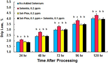
Upton et al. (Reference Upton, Edens and Ferket2008) examined drip loss from male broiler breast meat from birds fed a variety of Se supplemented diets (see Figure 1). The data suggested that selenite Se may be associated with an oxidative process that promotes post-mortem development of compromised cell membranes and facilitates increased moisture loss from processed breast meat. It appeared again that the presence of sodium selenite induced the highest drip loss rate (17%) in broiler breast meat (Figure 1). These data are in agreement with Mahan's (Reference Mahan, Cline and Richert1999) observations with swine (13.7% reduction with organic sources of Se) and those of Downs et al. (Reference Downs, Hess and Bilgili2000) and Hess et al. (Reference Hess, Downs, Bilgili, Lyons and Jacques2003) in broiler chickens (47% decrease in drip loss 24 hours post-mortem). Naylor et al. (Reference Naylor, Choct and Jacques2000) reported decreased drip loss rate (20–27% decrease in drip loss in Sel-Plex™-fed compared with sodium selenite-supplemented broilers). Upton's results are important for the poultry industry in many parts of the world because, in poultry processing facilities, the processed carcass is chilled in a hypotonic ice-water bath. The flesh of the carcass usually absorbs the water from this ice bath due to the fact that the cytoplasmic compartment of the muscle cell is hypertonic to the ice water bath. Therefore, the muscle cells will absorb water, swell and many instances rupture if the amount of water absorbed exceeds the capacity of the cells. Sodium selenite has been implicated in ROS production (Edens and Gowdy, 2005), and cells that contain larger amounts of ROS experience compromised cellular membrane integrity. Therefore, animals fed sodium selenite have a high probability for increased drip loss by reason of ROS production. The use of Sel-Plex™ as a source of supplemental dietary Se provides a more efficiently utilised form of organic Se and facilitates a greater antioxidant enzyme presence in glutathione peroxidase (Edens and Gowdy, 2005), which then acts to more readily reduce peroxides and other free radicals that compromise cell membranes.
Effect of selenium source on drip loss from male broiler breast meat (from Upton et al. Reference Upton, Edens and Ferket2008). Different letters above the histogram bars indicates significant differences among treatment means. P ≤ 0.05.

Water holding capacity, pH, and colour of meat as influenced by selenium
Drip loss from fresh meat is an easily measured quantification of the water holding capacity (WHC) of the meat, which is probably the most important of the meat quality characteristics (Huff-Lonergan, Reference Huff-Lonergan2010; Huff-Lonergan and Lonergan, Reference Huff-Lonergan and Lonergan2005). The WHC of fresh meat is its ability to retain inherent hydration and is an important property of fresh meat as it affects both the yield and the quality of the end product (Huff-Lonergan, Reference Huff-Lonergan2010; Pearce et al., Reference Pearce, Rosenwold, Andersen and Hopkins2011). WHC is influenced by both the ultimate pH of the muscle conversion to meat and the amount of space in the myocyte where water resides among the myofibrils (Allen et al., Reference Allen, Russell and Fletcher1997; Pearce et al., Reference Pearce, Rosenwold, Andersen and Hopkins2011). Decreased WHC has many negative impacts on marketers of fresh meat, causing loss of retailed meat mass, loss of protein in the drip/purge loss and poor appearance (colour) of the meat (Offer and Knight, Reference Offer, Knight and Lawrie1988). Factors that affect WHC include metabolic state of the food animal prior to slaughter, packaging and product handling, cuts of meats, rate of post-mortem muscle temperature decline, sarcomere length, ionic strength, osmotic pressures, development of rigor, and cold storage and freezing temperature, which all can alter water content in cellular and extracellular compartments (Offer and Knight, Reference Offer, Knight and Lawrie1988).
Organic Se supplementation to pigs is known to improve the juiciness and tenderness of meat and reduces fat content and water loss (Cole, Reference Cole, Lyons and Cole2000). Since organic Se in Sel-Plex™ can reduce drip loss (Table 1), it is plausible to conclude that WHC could also be affected by dietary Sel-Plex™.
Water in muscle tissue is held in several compartments, which includes spaces within and around myofibrils, within cellular membrane-bound structures, the cytoplasm and in the interstitial fluids. It is the interaction of these fluids with protein in the myofibrils that dictates WHC (Honikel, Reference Honikel, te Pas, Everts and Haagsman2004; Honikel and Kim, Reference Honikel and Kim1986), and even though water is held in various muscle cell compartments, the final content of skeletal muscle is roughly 75–88% of total muscle mass (Offer and Cousins, Reference Offer and Cousins1992). The intracellular water content of myocytes plays a crucial role in determining meat quality associated with toughness, juiciness, firmness, colour, and appearance, which then affects the consumer's perceived economic value of that meat (Ranken, Reference Ranken1976; Offer and Knight, Reference Offer, Knight and Lawrie1988).
It is well known that lower pH of fresh meat is associated with poor WHC (Barbut, Reference Barbut1997; Huff-Lonergan et al., Reference Huff-Lonergan, Baas, Malek, Dekkers, Prusa and Rothschild2002; Zhang and Barbut, Reference Zhang and Barbut2005; Chan et al., Reference Chan, Omama and Betti2011a,Reference Chan, Omama and Bettib). Ashgar et al. (Reference Ashgar, Gray, Booren, Gomaa, Abouzied, Miller and Buckley1991) reported that lower pH will more likely lead to shrinkage of myofibrils and increased myocyte permeability due to increased lipid peroxidation that occurs in conversion of muscle to meat (Macit et al. (Reference Macit, Aksakal, Emsen, Esenbuga and Aksu2003). However, ultimate pH of poultry breast meat has not been consistently found to be altered by dietary Se form or concentration, which suggests that benefits seen with organic Se might be due to other factors (Perić et al. (Reference Perić, Milošević, Žikić, Kanački, Džinić, Nollet and Spring2009). Similarly, Stef et al. (Reference Stef, Stef, Jianu, Druga, Daniel, Alda and Alda2011) found that pH of breast and leg meat was not altered by the feeding of neither sodium selenite nor Sel-Plex™, but drip loss from both breast and leg meat was decreased with Sel-Plex™. Wang et al. (Reference Wang, Pan, Peng, Zhao and Zhao2009) did not measure breast meat pH in response to the feeding of Se yeast but found decreased drip loss from the meat, which was associated with lower thiobarbituric acid reactive substances (TBARS) attributed to the feeding of Se yeast. The influence of the Se yeast had no effect on meat lightness (L* values) and yellowness (b* values) but redness (a* values) of the breast meat was increased by the Se yeast. Wang et al. (Reference Wang, Zhan, Zhang, Wu and Yuan2011) compared sodium selenite, D-selenomethionine, and L-selenomethionine on breast meat drip loss and oxidative stability. Selenomethionine did not alter breast meat ultimate pH but decreased drip loss compared to sodium selenite. The redness (a* values) of the breast meats were not different among Se treatments. In another study comparing Se source influence on meat quality, both sodium selenite and selenomethionine caused decreased pH and increased drip loss compared to the control group fed no supplemental Se (Dhumal et al., Reference Dhumal, Nikam, Khose and Ingle2013). In all treatments, TBARS increased with storage time, but selenomethionine treatments caused a slower rate of TBARS elevation.
In pigs, enhanced WHC of meat was associated with increased expression of the Sepw1 gene (encodes selenoprotein W, an antioxidant selenoprotein (Loflin et al., Reference Loflin, Lopez, Whanger and Kioussi2006)), Selenoprotein W plays a major role in the control of white muscle disease in lambs (Whanger, Reference Whanger2000) and is responsive to dietary Se yeast, which was reported to improve antioxidant status as well as reduce drip loss and TBARS in muscle and meat (Li et al., Reference Li, Zhou, Zhao, Lei, Xia, Gao and Wang2011). In the Li et al. (Reference Li, Zhou, Zhao, Lei, Xia, Gao and Wang2011) study, provision of 0.3 and 3.0 ppm dietary Se yeast resulted in higher pH of muscle and meat than that measured in samples from basal-fed pigs, but meat quality assessed by colour was not different between Se deficient and Se-fed pigs. Feeding excess Se did not improve meat quality characteristics except for lower drip loss.
In young Charolais bulls, feeding Se yeast maintained a more alkaline pH in meat aged for six days than meat from animals fed sodium selenite, and decreased drip loss in meat aged for 11 days (Cozzi et al., Reference Cozzi, Prevedello, Stefani, Piron, Contiero, Lante, Gottardo and Chevaux2011). Meat from Se-fed bulls was lighter in colour (higher L* value; suggesting greater water content) with no differences in redness (a* value) or yellowness (b* value). Shear force of meat from Se-fed bulls was less, indicating more tender meat with better WHC (Cozzi et al., Reference Cozzi, Prevedello, Stefani, Piron, Contiero, Lante, Gottardo and Chevaux2011).
In veal, Skřivanová et al. (Reference Skřivanová, Marounek, De Smet and Raes2007) saw no differences in pH or colour of meat despite feeding Sel-Plex™ or a combination of Sel-Plex™ and vitamin E, and drip loss from the veal was not altered in response to either treatment, even though GSHpx activity was increased by Se supplementation.
The WHC of goat meat was improved by 1.4% due to feeding Se yeast, even though pH was not different between treatments, and, additionally, shear force values were less in the meat from Se fed goats (Sethy et al., Reference Sethy, Garg, Mishra, Biswal, Behera, Sahoo, Satapathy, Meher and Nayak2014).
From published work, the influence of dietary Se, especially organic Se, on meat quality appears to be somewhat different between food animal species. In poultry, regulation of WHC (as related to rigor development through higher muscle and meat pH) does not appear to be as important in mammalian food animals, where ultimate pH is often an indicator of WHC. Thus, the relationship of dietary Se and development of ultimate pH in poultry meat is not as clear-cut as that in red meats.
It has been recognised that there are differences between fast twitch muscle fibres (e.g. poultry breast and wing muscles used for rapid movement and high production of small amounts of glycolytic energy) and slow twitch muscle fibres (e.g. poultry leg and thigh muscles used for prolonged/endurance movements, such as standing and walking, and slower production of large amounts of oxidative energy) (Xiong, Reference Xiong1994). However, breast, leg, and thigh muscles are heterogeneously composed of both glycolytic and oxidative muscle fibres, with mostly glycolytic fibres in breast and oxidative fibres in thigh and leg (Xiong, Reference Xiong1994). The different muscle fibre types have different sensitivities to pH changes during the conversion of muscle to meat, with fast twitch fibres being more acid-labile than slow twitch fibres (Bárány, Reference Bárány1967). Sams (Reference Sams1987) reported that male broiler oxidative muscle fibres developed rigor mortis more quickly than glycolytic muscle fibres, which was attributed to greater anaerobic capacity in the oxidative muscle fibres than that in glycolytic muscle fibres. Northcutt et al. (Reference Northcutt, Foegeding and Edens1994), working with pre-slaughter heat conditioned broiler chickens on a sodium selenite supplemented diet, noted that drip loss from oxidative muscles from the leg was less and had greater WHC than the same assessments in glycolytic muscle fibres from breast meat. The differences in drip loss and WHC appeared to be directly associated with final pH of the glycolytic and oxidative muscles, with ultimate pH in glycolytic muscles being significantly more acidic than in the oxidative muscles.
Allen et al. (1998) reported a strong relationship between breast meat colour and indicators of meat quality, with dark breast meat having higher pH, less drip loss, greater WHC, and lower shear force values than light breast meat. A report by Qiao et al. (Reference Qiao, Smith and Northcutt2001) confirmed these observations, showing that lighter than normal breast meat had lower pH, higher moisture, and lower WHC. The meat used by Allen et al. (1998) was from broilers from an unknown dietary Se background, but it was assumed that the birds had been fed sodium selenite. In another study with broilers from an unknown dietary Se background, breast meat colour reflected its WHC (Bowker and Zhang, 2013). After segregating breast fillets into light (L* = 62.5; a* = 0.3; b* = 13.6) and dark (L* = 45.5; a* = 1.2; b* = 9.4) classes, Bowker and Zhang (2013) noted that pH of dark breast fillets was significantly greater than that in light fillets, and that dark fillets had slightly higher WHC than light fillets. Furthermore, in the dark fillets drip loss was less than in light fillets. While direct comparisons between oxidative and glycolytic muscle fibres were not made in this investigation, the differences in WHC between light and dark breast fillets might be attributed to a larger presence of oxidative or intermediate muscle fibres in dark fillets than in light fillets.
Feeding high levels of dietary sodium selenite plus vitamin E to chickens did not alter L*, a*, and b* values of refrigerated breast and thigh meat (Ryu et al., Reference Ryu, Rhee, Lee and Kim2005), and sodium selenite and vitamin E feeding did not provide any protection against discolouring of breast and thigh meat due to accumulation of surface metmyoglobin in refrigerated breast and thigh meat. Furthermore, after 12 days refrigeration, cholesterol oxidation in thigh muscle was increased by sodium selenite even when fed in conjunction with 73.53 mg vitamin E/kg diet. In glycolytic breast muscle, the combination of sodium selenite with 73.53 mg vitamin E/kg diet did not provide any significant additional protection against cholesterol oxidation compared to supplementation with vitamin E alone. Thus, these data suggest that stability of oxidative and glycolytic muscles might actually be decreased by feeding sodium selenite. The implication of this is that meat quality parameters would probably decrease in refrigerated meats, and the meat quality parameters affected would include meat colour, WHC, and rancidity. No data were presented to compare the response to organic Se.
Medeiros et al. (Reference Medeiros, Oba, Shimokomaki, Pinheiro, de Silva, Soares, Pissinati and de Almeida2012) fed organic Se (0 to 0.6 mg/kg diet) from the cell walls of Candida pelliculosa to broiler chickens from hatch to 42 days of age. They determined that a linear increase in dietary organic Se resulted in a corresponding significant linear increase in breast meat pH, which was associated with a significant linear increase in WHC. Their results indicated that there was a significant improvement in tenderness of the breast meat as seen by a significant linear decrease in shear force of the breast meat. Breast meat colour analysis showed that there was a significant quadratic increase in L* values of the meat, but a* and b* were not influenced by organic Se in the diet.
Rajashree et al. (Reference Rajashree, Muthukumar and Karthikeyan2014) fed organic Se from Saccharomyces cerevisiae to broiler chickens (Rajashree and Muthukumar, Reference Rajashree and Muthukumar2013) and noted that selenomethionine at 0.5 ppm/kg diet significantly increased WHC of breast meat compared to sodium selenite and unsupplemented dietary groups. However, improved WHC was not associated with a more alkaline breast meat pH. Supplementation with organic Se was associated with significantly higher GSHpx activity and lower TBARS in the breast meat.
Feeding selenomethionine to broilers certainly has an effect on WHC of breast meat as exemplified by higher pH and decreased drip loss, which has been demonstrated repeatedly, but consensus on its effect on breast meat colour is not always consistent. In a report by Jiang et al. (Reference Jiang, Lin, Zhou, Luo, Jiang and Chen2009) a progressive increase in dietary selenomethionine caused darker colouration as shown by significantly decreased L* values with no changes in a* and b* values. Shear force values were not affected significantly by dietary selenomethionine, and antioxidant status of the breast meat was significantly improved. Similar results were reported by Boiago et al. (Reference Boiago, Borba, Leonel, Giampietro-Ganeco, Ferrari, Stefani and de Silva2014) who supplemented broiler diets with either sodium selenite or selenomethionine. In this investigation selenomethionine feeding caused the production of darker (lower L* values) breast meat than sodium selenite with no effects on a* and b* values for either treatment. Seven days after storage, breast meat from selenomethionine fed broilers had lower TBARS than breast meat from the sodium selenite fed birds. When Se was fed at 0.5 ppm/kg feed, shear force was less than in meat from the control group, and this was associated with higher breast meat pH. However, WHC was not influenced by pH or Se source.
Chen et al. (Reference Chen and Li2014) compared the effects of sodium selenite to Sel-Plex™ and another Se yeast product (Jiaotianle) on broiler breast meat quality. There were no differences between treatment groups, but meat from Sel-Plex™-fed birds had the least drip loss compared to sodium selenite and Jiaotianle, which had the highest drip loss. There was no influence of treatments on meat colour. However, GSHpx activity was increased by the Se yeast supplementation compared to sodium selenite, but TBARS values were increased in the breast meat from Se yeast-fed birds, which appeared to be an inconsistent response based on research from other meat scientists reported herein.
Meat quality of Grey geese in response to Sel-Plex™ supplementation was examined by Baowei et al. (Reference Baowei, Guoqing, Qiaoli and Bin2011). WHC was improved by increasing dietary levels of Sel-Plex™ and drip loss was significantly reduced, while shear force of the breast muscle was decreased and antioxidant status was elevated.
Juniper et al. (Reference Juniper, Phipps and Bertin2011) compared high and low dietary levels of Sel-Plex™ to sodium selenite and unsupplemented diets fed to commercial turkeys. The background level of natural Se in the unsupplemented diet was 0.11 mg Se/kg. Drip loss from the breast meat was not affected by the dietary Se treatments, even though breast GSHpx activity was elevated. It is assumed that GSHpx-4 activity might have been maximised in the cell membranes and that cytosolic GSHpx-1 could have been approaching maximal activity in all turkeys and that supplementation of either Sel-Plex™ or sodium selenite would only have minimal influence under these experimental conditions, which resulted in no differences in drip loss due to Se treatments.
Zhan et al. (Reference Zhan, Wang, Zhao, Li and Xu2007) fed pigs either a control diet with no supplemental Se (natural plant based organic Se at 0.045 ppm/kg basal feed), a diet supplemented with sodium selenite (0.3 mg/kg feed), or supplemented with selenomethionine (0.3 mg/kg feed). They found that muscle GSHpx activity was elevated by both sodium selenite and selenomethionine compared with the control and that there was a bi-phasic TBARS response in muscles, with selenomethionine maintaining significantly lower TBARS than control and sodium selenite. This was associated with an eight fold increase in muscle TBARS. The pH of the loin was not altered significantly by the dietary treatments, but WHC of the loin meat was significantly improved in the selenomethionine treatment group compared with control and sodium selenite treatments. The redness of the meat was increased by the selenomethionine treatment but not by sodium selenite treatment. These results strongly suggested that sodium selenite, due to its pro-oxidant properties, has the potential to decrease meat quality associated with increased oxidative instability.
Interactions with vitamin E
An investigation by Wang et al. (Reference Wang, Zhan, Zhang, Wu and Yuan2011) examined the effectiveness of sodium selenite, L-selenomethionine and D-selenomethionine on oxidative stability of breast meat and other tissues from broiler chickens that had been fed 10 mg vitamin E/kg of diet. Both selenomethionine isomers reduced drip loss more than sodium selenite, but neither a* values nor pH were affected by any of the Se treatments.
The role of GSHpx in maintenance of meat quality has been discussed above, from which one can conclude that GSHpx-1, GSHpx-4, GSH, and vitamin E are all necessary for maintenance of oxidative stability in muscle cells and in the maintenance of meat quality. However, it is the unique relationship between GSHpx-4 and vitamin E in membranes that plays a pivotal role in the maintenance of meat quality. Among the different GSHpx species, only GSHpx-4 resides in the cell membrane (Ursini et al., Reference Ursini, Maiorino and Gregolin1985). In plasma membranes, the GSHpx-4 interaction with GSH and vitamin E allows for nearly complete inhibition of lipid oxidation (Ursini and Bindoli, Reference Ursini. and Bindoli1987). Furthermore, GSHpx-4 activity, which prevents free radical generation from lipid hydroperoxides, can influence the vitamin E requirement necessary to inhibit lipid peroxidation (Ursini and Bindoli, Reference Ursini. and Bindoli1987). It is the interaction between GSH, GSHpx-4 and vitamin E in the cell membranes that supports maintenance of meat quality associated with lipid oxidation, odour, colour and even WHC related to cell membrane integrity and protein integrity in muscle fibres.
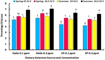
The influence of Sel-Plex™ on meat quality is positive, but relatively little has been done to address the interaction of dietary vitamin E and Sel-Plex™ on meat quality. In earlier work (Edens, Reference Edens, Lyons and Jacques1996; Upton et al., Reference Upton, Edens and Ferket2008) when vitamin E was added at 24.3 mg/kg diet and Se as either sodium selenite or Sel-Plex™ was included at 0.1 and 0.2 mg/kg broiler diet, it was determined that organic Se improved meat quality. From an unpublished component of early research on the influence of Sel-Plex™ on poultry meat quality (Edens et al., Reference Edens, Carter, Parkhurst and Sefton2000a), it was seen that vitamin E was crucial to the maintenance of WHC in breast meat (Figure 2). In that experiment conducted in mild spring versus hot summer climatic conditions, vitamin E was supplemented in broiler diets at either 24.3 mg/kg diet or 12.13 mg E/kg diet, and Se was provided as either sodium selenite (0.1 and 0.3 mg/kg diet) or Sel-Plex™ (0.1 and 0.3 mg/kg diet). The influence of season was significant, whereby drip loss in summer was greater than in spring due to preslaughter exposure to high environmental temperatures, which reflected the findings of Pingel et al. (Reference Pingel, von Lengerken and Knust1995), but there was no difference in drip loss within season due to dietary Se level. In this experiment, there was a significant Se source effect on breast meat drip loss, in which broilers given sodium selenite had significantly greater drip loss than breast meat from broilers fed Sel-Plex™. There was a significant season x Se source interaction, which indicated that, in both seasons, broilers fed sodium selenite had higher breast meat drip loss than that from broilers fed Sel-Plex™. It was of interest that broilers fed 0.1 mg sodium selenite/kg diet had lower drip loss in both spring and summer than broilers fed 0.3 mg sodium selenite/kg diet, but in broilers given Sel-Plex™, even though there was no difference in drip loss between dietary Sel-Plex™ levels, meat from broilers fed the 0.1 mg Sel-Plex™/kg diet tended to have slightly higher drip loss than those fed 0.3 mg/kg. When the data were segregated by level of dietary vitamin E supplementation, there was a significant season x vitamin E interaction, showing a decrease in breast meat drip loss due to the higher level of dietary vitamin E (24.3 mg E/kg diet) compared with the lower level (12.13 mg E/kg diet). The lowest drip loss percentage was found in breast meat from broilers fed the higher level of vitamin E (24.3 mg E/kg diet) during the spring season compared to Spring-12.13 mg E/kg diet and to Summer-24.3 mg E/kg diet and Summer-12.13 mg E/kg diet, which had the highest drip loss overall (Figure 2). A significant season x Se source interaction demonstrated lower drip loss in all Sel-Plex™ treatments compared to sodium selenite treatments, with the very lowest drip loss being associated with the Sel-Plex™ -Spring-24.3 mg E/kg diets.
Influence of season (spring vs. summer), dietary selenium source as either sodium selenite (NaSe) or Sel-Plex™ (SP), and supplemental vitamin E (24.3 or 12.13 mg E/kg diet) on 120 hours drip loss from broiler breast meat. Different lower case letters over the histogram bars indicates significant differences among the treatment means (P ≤ 0.05).

The mechanism whereby Se and vitamin E interact to reduce oxidative damage to muscle cells appears to be unequivocal, but there are still many questions that require answers. Apart from the Se x vitamin E interaction in the maintenance of meat quality, a large knowledge base has been amassed describing the vitamin E influence on meat quality in poultry but mostly without addressing Se involvement (Fellenberg and Speisky, Reference Fellenberg and Speisky2006).
Protein carbonylation, protein sulfhydryls, methionine sulphoxide reductase, and meat quality
Carbonylation of meat protein and loss of sulfhydryls have been receiving increased interest over the past 25 years as factors that might be involved in poor meat quality (Estévez, Reference Estévez2011; Lund et al., Reference Lund, Heinonen, Baron and Estévez2011). The reviews by Estévez (Reference Estévez2011) and Lund et al. (Reference Lund, Heinonen, Baron and Estévez2011) provide an insight on the problem of protein oxidation in meat.
Protein oxidation is precipitated by loss of antioxidant protection, which appears to be preceded by lipid peroxidation (Estévez, Reference Estévez2011; Lund et al., Reference Lund, Heinonen, Baron and Estévez2011). Lipid peroxidation occurs in response to oxidative stress, and a diversity of aldehydes, such as 4-hydroxyalkenals and malonaldehyde, are formed when lipid hydroperoxides are elevated in biological systems. These aldehydes are highly reactive and may be considered as toxic secondary messengers, which disseminate and augment initial free radical degenerative events. The aldehydes most intensively studied so far are 4-hydroxynonenal, 4-hydroxyhexenal, and malonaldehyde. The cellular reactions of 4-hydroxyalkenals and malonaldehyde with biomolecules, such as amino acids, proteins and nucleic acid bases, often lead to cytotoxicity, genotoxicity, chemotactic activity, inhibition of cell proliferation and gene expression, and ultimately, protein carbonylation (Esterbauer et al., Reference Esterbauer, Schaur and Zollner1991).
Protein carbonylation is a process describing the covalent adduction of lipid aldehydes, often containing six, nine or 12 carbons, to the side chains of protein lysine, histidine and cysteine residues (Esterbauer et al., Reference Esterbauer, Schaur and Zollner1991). Lipid aldehydes are produced from hydroperoxidation of polyunsaturated fatty acyl groups followed by non-enzymatic Hock cleavage. The resultant aldehydes can undergo Schiff-base formation with lysine residues, but more commonly are subject to Michael addition reactions that produce a lipid acyl group containing free carbonyls. Such carbonyl groups are capable of secondary Schiff-base formation with an adjacent amine or cyclisation, but in many cases the free aldehyde remains unmodified, thereby allowing for its detection using a variety of hydrazide-based reagents or, in some cases, using antibodies directed to nine-carbon acyl derivatives such as 4-hydroxy 2,3 trans nonenal (Curtis et al., Reference Curtis, Hahn, Long, Burrill, Arriaga and Bernlohr2012).
Delles et al. (Reference Delles, Xiong, True, Ao and Dawson2014) investigated the influence of dietary antioxidants and oil quality on the oxidative and enzymatic properties of chicken broiler breast meat stored in oxygen-enriched packaging (HiOx: 80% O2/20% CO2) in comparison with air-permeable polyvinylchloride (PVC) or skin packaging systems during retail display at 2 to 4 °C for up to 21 d. Broilers were fed either a diet with a low-oxidised (LO) or high-oxidised (HO) oil, supplemented with or without Se yeast (Sel-Plex™) and an organic mineral antioxidant pack for 42 d. In all packaging systems, lipid oxidation was inhibited by up to 32.5% with the antioxidant-supplemented diet when compared to control diets particularly in the HiOx and PVC systems. Protein sulfhydryls were significantly protected by 14.6 and 17.8% for LO and HO dietary groups, respectively, in PVC 7 day samples by antioxidant diets. However, muscle tissue protein carbonyl content increased during storage for all dietary treatments and all packaging conditions. The carbonyl level in HO samples, irrespective of packaging, was higher than those in LO samples. However, muscle samples from birds fed antioxidant-supplemented diets had lower carbonyl content compared with the basal group. The effect of packaging systems and storage time on protein carbonyl formation was overall similar to that of TBARS, suggesting a possible relationship between lipid oxidation and protein carbonyl formation. Glutathione peroxidase, catalase, and superoxide dismutase activities were significantly higher in samples from birds fed antioxidant-supplemented diets compared to the basal diet, regardless of oil quality. Also, serum carbonyls were lower in broilers fed a LO, antioxidant-supplemented diet. The results demonstrated that dietary antioxidants can minimise the oxidative instability of proteins and lipids, and protection may be linked to improved cellular antioxidant enzymatic activity.
It has been reported that in addition to lipid peroxidation, carbonylation of proteins can be induced when there is elevation of ROS and RNS radicals such as O2 ·− , H2O2, ·OH, NO·, and ONOO − . These can interact with protein side chain residues, such as lysine, arginine, proline, threonine, and glutamic acid, yielding carbonyl aldehyde and ketone adduct formations (Dalle-Donne et al., Reference Dalle-Donne, Rossi, Giustarini, Milzani and Columbo2003 and Reference Dalle-Donne, Aldini, Carini, Columbo, Rossi and Milzani2006; Dean et al., Reference Dean, Fu, Stocker and Davies1997; Moskovitz and Oien, Reference Moskovitz and Oien2010). Moskovitz and Oien (Reference Moskovitz and Oien2010) discussed the fact that ROS content in tissues is reflected in parallel by protein-carbonyl content. The carbonylation of protein amino acid side chains in muscle tissue results in impaired conformation of myofibrillar proteins, which contributes to denatured protein and loss of functionality after protein amino acid side chains are oxidised (Burcham and Kuhan, Reference Burcham and Kuhan1996; Amici et al., Reference Amici, Levine, Tsai and Stadman1989). Moskovitz and Oien (Reference Moskovitz and Oien2010) noted that an enzymatic reversal process of protein carbonylation has not yet been identified, but there is a unique enzymatic reversal of protein-methionine sulphoxide that is mediated by the selenoprotein methionine sulphoxide reductase (MsrB) and the non-Se MsrA.
Although protein-methionine sulphoxide can be reduced by methionine sulphoxide reductase, the role of methionine sulphoxide, as an indicator of loss of sulfhydryl groups in association with protein oxidation and its role in maintenance of meat quality remains unclear. Nevertheless, there is ample evidence to suggest that Se plays a role in the maintenance of methionine sulphoxide reductase B activity. In mice fed a Se adequate diet there was no significant increase in protein methionine sulphoxide formation, but those fed a Se deficient diet showed significant methionine sulphoxide formation in association with significant protein carbonyl derivative accumulations in tissues such as the liver, kidney, cerebrum, and celebellum (Moskovitz and Stadtman, Reference Moskovitz and Stadtman2003). Methionine sulphoxide formation is associated with protein carbonylation, which is apparently precipitated by increased lipid peroxidation resulting from general oxidative stress. This review and others in this series (Edens and Sefton, 2016a, b) discuss the protective effect of Sel-Plex™ and even inorganic Se against lipid peroxidation in meats.
Based on evidence presented herein, it might be safe to conclude that methionine sulphoxide reductase (MsrB) might not play a direct, but an indirect role, in maintenance of meat quality. However, from an evolutionary point of view, methionine sulphoxide reductase probably plays a more important role in the maintenance of protein conformation and protein functionality in living systems. Loss of protein conformation and functionality causes loss of enzyme activity and failure of cells, tissues, organs and physiological systems.
Proteins with a large number of methionine residues, which is the most hydrophobic of all the amino acids, tend to exist within the cell membrane lipid bilayer. Some of those methionine residues are exposed to the aqueous exterior of the cell membrane and are vulnerable to oxidation. Once oxidised, methionine sulphoxide residues can be reduced back to methionine by the enzyme methionine sulphoxide reductase. Thus, an oxidation-reduction cycle occurs in which exposed methionine residues are oxidised (e.g., by H2O2) to methionine sulphoxide residues, which are subsequently reduced (Levine et al., Reference Levine, Mosoni, Berlett and Stadtman1996). This cycle is important because methionine sulphoxide accumulation can alter the structural conformation and function of protein and promote carbonylation of hidden amino acid residues (Brot and Weissbach, Reference Brot and Weissbach1991; Oien and Moskovitz, Reference Oien and Moskovitz2008). With protein conformational change and loss of functionality, cellular, tissue and organ functions can be lost, which can be equated indirectly to loss of meat quality. Collectively, such oxidative processes lead indirectly to low meat quality attributes characterised by increased drip loss, decreased WHC, development of rancidity, loss of colour, and loss of desirable qualities associated with cooked meat.
Estévez (Reference Estévez2011) has reported that several processing factors, such as irradiation, cooking, dry-curing, fermentation and hydrostatic pressure, influence protein carbonylation and meat quality. These factors appear to be influenced by antioxidants in feed and in the meat, feeding regimes, and even the kind of packaging and storage conditions. Estevez (Reference Estévez2011) discussed the possible effects of protein carbonylation on nutritional value, texture traits, colour, aroma, flavour, WHC and biological functionality of meat proteins, and found that each characteristic of meat quality was affected significantly. It has been known for some time that meat proteins play a major role in meat quality characteristics (Lawrie, Reference Lawrire and Lawrie1998).
Although lipid peroxidation and free radicals are intimately involved in the induction of protein carbonylation and oxidation of thiol groups in methionine and cysteine, relatively little has been done to explore the possibility that Se-based antioxidants might preserve meat quality via protection of membrane lipids and prevention of excess carbonylation with progressive loss of thiol groups in muscle foods (Korzeniowska et al., Reference Korzeniowska, Króliczewska and Kopeć2015). Estévez (Reference Estévez2011) reviewed nutritional strategies, such as limiting oxidised fat in animal diets, as a means to minimise oxidative stress and loss of meat quality. He noted in many studies that the nature of dietary fat seemed to have a higher impact on lipid oxidation than on protein carbonylation.
From a practical point of view, protein oxidation in breeder egg albumen can be influenced by the addition of methionine and Sel-Plex™ in breeder diets (Wang et al., Reference Wang, Pan, Zhang, Peng, Zhao and Zhou2010). The addition of methionine elevated the carbonyl content of the egg albumen, but the combination of methionine (4.0 g/kg) with Sel-Plex™ (0.6 mg Se/kg) protected the integrity of albumen protein by minimising the albumen protein carbonyl content (Wang et al., Reference Wang, Pan, Zhang, Peng, Zhao and Zhou2010). Korzeniowska et al. (Reference Korzeniowska, Króliczewska and Kopeć2015) concluded that the improved albumen protein condition was due to a higher GSH content and GSH-px activity attributed to Sel-Plex™ (Pappas et al., Reference Pappas, Acamovic, Sparks, Surai and McDevitt2005) that decreased the rate of protein oxidation due to lower rates of lipid peroxidation.
Earlier, Wang et al. (Reference Wang, Pan, Peng, Zhao and Zhao2009) studied the influence of DL-methionine and Sel-Plex™ supplementation in breeder hen diets on meat quality of broiler progeny. The Se content of broiler breast meat increased with increasing Sel-Plex™ supplementation to the breeder diets. The carbonyl content of myofibrillar protein in broiler breast meat decreased with increasing DL-methionine supplementation to the breeder hens, and the levels in breast meat from the 0 mg Se/kg diet was significantly higher than the meat from birds fed Sel-Plex™ at 0.3 mg Se /kg feed. Selenium supplementation to the breeder hen diet at 0.30 and 0.60 mg/kg decreased broiler malondialdehyde content compared with that of 0 mg of Se/kg diet. Adding 4.0 and 5.4 g of DL-methionine/kg to feed decreased malondialdehyde content compared with samples from birds fed 3.2 g DL-methionine/kg diet. Supplementation of DL-methionine at 5.4 g/kg increased meat a* value colour compared to 3.2 and 4.0 g DL-methionine/kg diet. Supplementation of Sel-Plex™ at 0.6 mg/kg significantly increased a* value compared 0 and 0.3 mg Sel-Plex™/kg diet, and 0 mg Sel-Plex™/kg diet increased b* value compared with 0.30 and 0.60 mg Sel-Plex™ /kg diet. Sel-Plex™ supplemented at 0.30 and 0.60 mg Se/kg diet decreased drip loss compared with 0 mg Sel-Plex™/kg diet, and 4.0 and 5.4 g of DL-methionine/kg diet decreased drip loss compared with 3.2 g of DL-methionine/kg diet, respectively. Wang et al. (Reference Wang, Pan, Peng, Zhao and Zhao2009) concluded that methionine and Se yeast supplementation to the maternal diets could improve colour, WHC and oxidative stability of meat from male offspring. Thus, broiler muscle protein oxidation and carbonylation was inhibited by the addition of Sel-Plex™ to breeder diets. The protective effect again can be attributed to transference of Se from the dam to the progeny, which modified the antioxidant properties in the progeny. The interaction between Sel-Plex™ and DL-methionine possibly helped to sustain the integrity of thiol groups associated with muscle protein methionine and cysteine residues.
Aladrović et al. (Reference Aladrović, Ljubić, Tur and Pluzarić2013) fed sodium selenite (0.15 mg Se/kg diet) and Sel-Plex™ (0.3 mg/kg diet) to broiler chickens to investigate the influence of inorganic and organic Se on oxidative damage in different tissues before and after a 48 hour period of fasting. Since there was an unequal amount of Se provided by the feeding of inorganic vs. organic Se in this experiment, the lower oxidative influence from inorganic selenite might have biased potentially for some of the data collected by these scientists. Lipid peroxidation and carbonylation of proteins in liver, kidney, and small intestine resulted in a variety of responses, which appeared to be linked to dietary Se form and concentration and was not expected in light of other studies comparing the antioxidant effects of inorganic and organic Se fed to broiler chickens. Kidney lipid peroxides were elevated in chickens given Sel-Plex™ before fasting, but after fasting for 48 hours, there were no differences between selenite- and Sel-Plex™-fed kidney lipid peroxide levels. Protein carbonylation was elevated in kidney tissue by feeding Sel-Plex™ compared to sodium selenite before fasting, but after fasting protein carbonylation in selenite-fed broilers was elevated, although there was no change seen in Sel-Plex™-fed broilers. In liver and small intestine, neither tissue from either selenite- or Sel-Plex™-fed broilers differed in lipid peroxidation, and, after fasting, broilers fed Sel-Plex™ had increased liver lipid peroxidation while the small intestine showed a decrease in lipid peroxidation. There were no differences in liver and small intestine carbonylation before fasting, but. after fasting. liver carbonylation in broilers fed Sel-Plex™ was decreased compared to those fed selenite. Before fasting, carbonylation in the small intestine was greater in broilers fed Sel-Plex™ compared to those fed selenite, but, after, fasting carbonylation was slightly greater in selenite- and Sel-Plex™-fed broilers. The carbonylation results from this investigation by Aladrović et al. (Reference Aladrović, Ljubić, Tur and Pluzarić2013) did not appear to be correlated with lipid peroxidation in all tissues examined. Furthermore, it was apparent that protein carbonylation and lipid peroxidative varied significantly among the tissues examined.
Korzeniowska et al. (Reference Korzeniowska, Króliczewska and Kopeć2015) provided a definitive study on the influence of dietary Se modulation on carbonyl and sulfhydryl groups in chicken meat proteins. These authors discussed that the potential effect of protein oxidation on meat quality and health is still not fully understood, supporting a conclusion by Estévez (Reference Estévez2011), and pointed out that there were few studies of protein carbonylation studies with poultry meat. In their study, sodium selenite was compared to Se yeast as potential antioxidants to control carbonylation and loss of sulfhydryl groups in fresh, chilled and frozen breast and leg meat. The supplementation of organic Se from yeast increased breast and leg meat Se concentration compared to sodium selenite. In fresh meat, Se yeast was not found to produce significant effects on carbonylation in breast meat and actually increased carbonylation in leg meat compared to sodium selenite. Sulfhydryl groups were not altered by either Se treatment in fresh breast and leg meat. Under chilled storage, carbonylation was not modified in meat from control birds. However, it was reported that both sodium selenite (0.38 and 0.50 mg Se/kg diet) and Se yeast (0.38 mg Se/kg diet) had protective effects against carbonylation in chilled breast meat similar to the effects reported by Wang et al. (Reference Wang, Pan, Peng, Zhao and Zhao2009). Dietary sodium selenite and Se yeast supplementation were associated with increased carbonylation in chilled leg meat. Increased carbonylation was attributed to pro-oxidant effects of Se, at least for sodium selenite, but the pro-oxidant effect of Se yeast, which has not been found in many other investigations (Balogh et al., Reference Balogh, Weber, Erdelyi and Mezes2004; Petrovic et al., Reference Petrovič, Boldižárová, Faix, Mellen, Arpašová and Leng2006; Petrovic et al., Reference Petrovič, Marcinčák, Popelka, Nollet and Kováč2009), should be explored further. Sulfhydryl groups were not modified in either chilled breast or leg meat by dietary Se supplementation.
Korzeniowska et al. (Reference Korzeniowska, Króliczewska and Kopeć2015) noted that dietary Se supplementation protected protein reactive groups against carbonylation of frozen breast and leg meat proteins in comparison to control meats from broilers fed no supplemental Se. Neither sodium selenite nor Se yeast had any effect on breast and leg sulfhydryl groups during frozen storage. Although the data suggested that Se supplementation had some protective effects against carbonylation and loss of sulfhydryl groups in fresh, chilled and frozen poultry breast and leg meat, additional studies are needed to explore dietary Se influence on meat protein oxidation processes.
An earlier study by Petrovic et al. (Reference Petrovič, Marcinčák, Popelka, Nollet and Kováč2009) examined the influence of trace elements and inorganic (sodium selenite) vs. organic Se (Sel-Plex™) on lipid peroxidation in chilled and frozen breast and thigh meat. These scientists observed significant lipid peroxidation in both chilled and frozen breast and thigh meat in association with dietary inorganic Se, but Sel-Plex™ supplementation held lipid peroxidation at a significantly lower level. Their observations on lipid peroxidation in chilled and frozen breast and thigh meat were in agreement with other studies revealing that there was a pro-oxidative property associated with the use of inorganic Se in broiler diets (Balogh et al., Reference Balogh, Weber, Erdelyi and Mezes2004; Petrovic et al., Reference Petrovič, Boldižárová, Faix, Mellen, Arpašová and Leng2006).
Soyer et al. (Reference Soyer, Özalp, Dalmis and Bilgin2010) examined the effect of freezing temperature and duration on lipid and protein oxidation in chicken leg and breast meat. The meats were frozen at −7, −12, or −18 °C for periods up to six months duration. Both lipid and protein oxidation can be considered processes of autoxidation, and it was not surprising that both processes developed in the frozen meats. Lipid peroxidation products increased during the first two to three months of frozen storage at all temperatures, and in both leg and leg meat the malodialdehyde concentrations increased throughout the six months of frozen storage. On the other hand, phospholipid concentrations within the frozen breast and leg meat declined progressively over the frozen storage time. Protein oxidation (carbonyl adducts accumulation) in the meats increased progressively over the duration of frozen storage mirroring the peroxidation product accumulation in the meat. Additionally, protein sulfhydryl groups decreased over the duration of frozen storage. These observations suggested that the earlier development of lipid peroxidation during frozen storage might be a stimulus for the protein oxidative processes (Estévez, Reference Estévez2011; Lund et al., Reference Lund, Heinonen, Baron and Estévez2011). Thus, if lipid peroxidation, which is responsive to Se-based antioxidants, is a stimulus for protein oxidation, it is reasonable to think that Se-based antioxidant enzymes might play an effective role in inhibition of protein oxidation in meat development.
In view of the fact that carbonylation appears to develop in parallel with lipid peroxidation and that, in most studies, lipid peroxidation is inhibited by Se-based antioxidant activities, it follows that Se source-based antioxidants and other feed-grade antioxidants might be important in the control of protein oxidative processes and maintenance of protein sulfhydryls. Assessment of the involvement of protein oxidation and lipid peroxidation on meat quality characteristics is a difficult endeavour complicated by variability in myofibrillar solubilities and in differences in the types of muscle being examined. Yet, it is important that the relationships among protein oxidation, lipid peroxidation, and antioxidants be established.
Conclusions
The consumer driven market continues to demand that farming provides the highest quality poultry and red meats, and this will become the basis for competitive positions in national and international markets. Consumers will reject meats that have obvious levels of drip loss. Feeding strategies involving Se impact on post-mortem muscle protein functionality, specifically the moisture retention capacity of the meat as influenced by rate of pH change. It is clear from the information available that neither vitamin E nor organic Se, provided in Sel-Plex™, can totally ameliorate post-mortem meat problems associated with loss of WHC , colour changes, myofibril loss of functionality, altered pH, lipid oxidation and more when used alone in diets. It is the interaction between Sel-Plex™ (as an inducer of GSHpx-4), GSH, and vitamin E in cell membranes that provides the most efficient means to combat quality issues such as drip loss in poultry meat. Additionally, protein oxidation develops in parallel with membrane lipid peroxidation. The greater the damage done to lipids by reactive substances the greater the content of muscle carbonyls and loss of sulfhydryls in low quality meat, and these characteristics might be circumvented by adequate feeding of selenium-based antioxidants.
Acknowledgements
None.
Declaration of interest
Dr. A. E. Sefton is an employee of Alltech, Inc.