Lithium carbonate has been the standard pharmacotherapy for adults since the 1950s for a variety of psychiatric disorders. At present, it is the most commonly prescribed mood stabiliser to treat mania and depression associated with bipolar illness.Reference Goodwin and Athanasios 1 , Reference Schou 2 Cardiac effects of lithium on the heart are common. They can range from benign T-wave flattening and inversions on the surface electrocardiogram to hypotension and cardiovascular collapse. This drug is increasingly being used in the paediatric population as adjunct therapy for certain behavioural, anxiety, attention-deficit hyperactivity, and bipolar disorders.Reference Kafantaris 3 If indicated, and found to be effective, lithium is usually administered as a part of the child’s maintenance therapy. Given the narrow toxicity/therapeutic ratio of this agent in children, and the lack of pharmacotherapeutic guidelines for its use in paediatrics, lithium poisoning remains a constant threat in this age group.Reference Geller and Luby 4 Thus, there is a need for paediatric cardiologists following-up children on this drug to be knowledgeable about the risks as well as the benefits associated with this form of therapy.
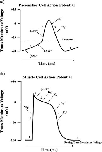
Healthcare providers should be aware of factors predisposing the patient to lithium toxicity. In this study, we present the case of a child on supplementary lithium for a bipolar condition, who presented with severe intoxication and a markedly elevated serum level of the drug. We present this case primarily to exemplify the profound clinical manifestations that can arise from lithium usage in young children, despite them being on therapeutic amounts of the drug. The cell membrane appears to be the main site of lithium’s action. Therefore, to better understand the cardiac pathophysiology associated with this agent, we reviewed the effects of lithium ion (Li+) on membrane electrogenic pumps and channels, determining the distribution and passage of sodium (Na+), potassium (K+), and calcium (Ca++) ions across the cardiac cell sarcolemma. The corresponding transmembrane Na+, K+, and Ca++ currents are involved in the genesis of the action potentials governing auto-rhythmicity (Fig 1a) and contractile activity (Fig 1b) of the heart. Insomuch as the electrocardiogram (ECG) reflects the summated effects of individual cardiac cell electrical activity, we related observed changes in the ECG from lithium to alterations in the underlying cardiac action potentials.
Cardiac cell action potentials are short-lived electrochemical events that are collectively organised to determine the auto-rhythmicity and contractile activity of the heart. An action potential is elicited in a cell when membrane Na+ channels transiently open and K+ channels close, which leads to depolarisation: the transmembrane voltage becomes more positive. A few milliseconds later, the Na+ channels close and Ca++ channels open to maintain the state of depolarisation. Finally, the Ca++ channels become inactivated and K+ channels re-open, resulting in re-polarisation: the transmembrane voltage becomes more negative. When a channel opens, there is increased electrical conductivity of specific ions through that channel, thereby creating the transmembrane currents giving rise to the action potentials. The ionic channels are embedded in the cell’s plasma membrane. Their opening and closing is contingent upon the transmembrane voltage attaining precisely defined threshold values. Conduction of ions can be rapid or slow, depending on the phase of the action potential. ( a ) The cardiac pacemaker cell action potential represents the transmembrane voltage, as a function of time, in a pacemaker cell. Phase 4, the so-called pacemaker potential, corresponds to depolarisation of the cell. It arises primarily from an inward current of Na+ through slow voltage-dependent (f-Na+) channels, in conjunction with a residual outward leak of K+, as the K+ channels associated with phase 3 continue to close. These so-called funny (f-Na+) channels open when the cell membrane voltage is most negative, and then subsequently conduct less current as the voltage progressively rises. This interplay between membrane voltage and decreasing Na+ influx allows the threshold voltage, required for auto-rhythmic firing, to be reached within a required time period, without external electrical stimulation. As the membrane potential approaches the threshold value, an inward current of Ca++ through transient fast voltage-dependent (T-Ca++) channels helps drive the voltage to the threshold value, typically~−30 mV. The (f-Na+) and (T-Ca++) channels then close. Phase 0 of depolarisation begins with an inward current of Ca++ through slow long-lasting voltage-dependent (L-Ca++) channels. This inward current of Ca++ initiates the action potential. Phase 3 corresponds to re-polarisation of the cell. It evolves from a combination of an outward current of K+ through slow delayed rectifier (KS +) and fast delayed rectifier (KR +) channels. The (L-Ca++) channels then close. The membrane voltage is restored to a baseline value, typically~−70 mV, and the spontaneous depolarisation process repeats. ( b ) The cardiac muscle-cell action potential represents the transmembrane voltage, as a function of time, in muscle cells. Phase 4, the resting membrane potential, corresponds to the membrane voltage in the unstimulated state of the cell. Phase 0 of depolarisation begins with an inward current of Na+, down its steep concentration gradient, through fast short acting voltage-dependent (NaF +) channels. The speed of conduction of electrical activity through the myocardium reflects the maximum rate of rise in membrane voltage (V) during phase 0, with respect to time, (dV/dt)max. Phase 1 involves inactivation of most of the (NaF +) channels, but with continuation of a weak outward current of K+. Phase 2 represents the “plateau phase” of the action potential. It is maintained largely by an inward current of Ca++ through slow long-lasting voltage-dependent (L-Ca++) channels and a combination of an outward current of K+ through slow delayed rectifier (KS +) and fast delayed rectifier (KR +) channels. The corresponding influx of Ca++ triggers electromechanical contraction of the myocyte. Phase 3 corresponds to re-polarisation of the cell. It evolves from a combination of an outward current of K+ through fast delayed rectifier (KR +) and an inward current of K+ through additional rectifier (KI +) channels. The (L-Ca++) channels close. The (KS +) channels close somewhat later. The (KR +) channels close as the membrane potential is restored to baseline value, ~−100mV. In phase 4, the current IKI helps in setting the resting membrane voltage close to the equilibrium voltage for K+.

Illustrative case
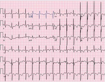
A five-year-old boy, with pervasive attention-deficit hyperactivity disorder and a bipolar condition, presented in an obtunded state from a week-long illness involving poor fluid intake, fever, vomiting, and diarrhoea. He had been on lithium carbonate (300 mg am and 450 mg pm) for three months. In retrospect, by history, he had signs of lithium toxicity after two months of treatment – namely, muffled speech, poor muscle tone, and a shuffled gait. These symptoms worsened following a change in his supplementary medication from methylphenidate (Ritalin) to amantadine (Symmetrel). On presentation, the child was agitated, disoriented, and combative. Other notable signs were dysarthria, hand tremors, and truncal ataxia. He was afebrile, with a blood pressure of 116/69 mmHg. Cardiac rhythms were normal and there were no murmurs. Laboratory data revealed a white blood cell count of 18.46 K and serum electrolyte levels of 130 mmol/L sodium, 3.2 mmol/L potassium, 93 mmol/L chloride, 29 mmol/L carbon dioxide, 1.7 mg/dl magnesium, and 9.2 mg/dl calcium. Blood urea nitrogen was 27 mg/dl, and the creatinine level was significantly elevated at 1.8 mg/dl. Thyroid function tests showed a high TSH level at 8.4 mIU/L but a normal free T4 level at 1.35 ng/dl. Urinalysis and urine toxicology screen were non-contributory. Figure 2 shows the patient’s ECG on admission. It revealed a heart rate of ~100 beats per minute, with minimal variability. The P-waves were diminutive but arose from the sinus node. The atrial rate was less than the ventricular rate, suggesting sinus node depression with a junctional escape rhythm. The QRS duration was 128 ms. There were ST segment depressions of 1–2 mm with T wave inversions in the precordial leads. The heart rate-corrected QT (QTc) interval was 590 ms. It is hard to be certain what ECG changes were due to Li+ alone and what might have been attributable to the electrolyte abnormalities present on admission, although the two are likely related. The serum lithium level was markedly elevated at 5.4 mEq/L; the therapeutic range is 0.8–1.2 mEq/L.
The 12-lead electrocardiogram (ECG) recorded at the time of presentation to the emergency room, after three months of lithium carbonate therapy (See text for analysis).

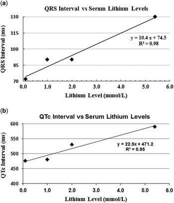
The patient was treated by discontinuing home medications and by initiating continuous veno–venous haemodialysis with ultrafiltration – a recognised mode of treatment for severe lithium intoxication. Serum lithium levels and 12-lead ECGs were obtained daily. The lithium levels steadily declined with dialysis. Haemodialysis was discontinued after 48 hours. There was recovery of sinus rhythm with P-waves of normal amplitude and width, but with first-degree block. Figure 3 shows the QRS durations and QTc intervals versus serum lithium levels. Both were initially significantly prolonged, but decreased directly in proportion to the falling lithium levels. Using linear regression analysis, the QRS duration was found to decrease more rapidly than the QTc interval. We observed no significant rebound serum lithium levels, which is occasionally encountered after acute haemodialysis sessions. The remaining treatment consisted of aggressive hydration and electrolyte replacement to avoid volume depletion, preserve serum electrolyte balance, and enhance residual lithium clearance.
QRS duration and corrected QT (QTc) interval (ordinate) versus serum lithium levels (abscissa).

Discussion
Lithium has been the standard pharmacotherapy for bipolar disorder in adults since the 1950s. It has a wide range of clinical applicability. The drug has been proven to be efficacious in 70–80% of patients with mania, and it usually does not compromise intellectual function. Thus, lithium has become an indispensable part of current pharmaceutical psychiatric therapy. It is well absorbed in the gastrointestinal tract; however, it diffuses slowly from the extracellular to the intracellular space, taking at least one week of maintenance therapy to equilibrate across the cell membrane.Reference Okusa and Crystal 5 Consequently, many young children started on normally prescribed amounts of the drug experience side-effects during the first few weeks of the treatment. Lithium is eliminated almost exclusively by the kidneys. Lithium is potentially nephrotoxic; it can produce electrolyte abnormalities, diabetes insipidus unresponsive to vasopressin, and renal failure.Reference Baldesarini and Tarazi 6 , Reference Ward, Musa and Bailer 7 Any medication that alters the glomerular filtration rate may worsen lithium toxicity.
At present, lithium is increasingly being used in children to reduce deviant behaviour, improve attentiveness, control hyperactivity, and stabilise mood, depending on their psychiatric illness.Reference Findling, Kafantaris and Pavuluri 8 Clinical signs of lithium intoxication include gastrointestinal symptoms – nausea, vomiting, and diarrhoea – and CNS effects – disorientation, confusion, slurred speech, tremors, muscle weakness, and altered gait – all of which were present in our patient.Reference Singer and Rotenberg 9 Children have a narrow toxicity/therapeutic ratio of the drug. Therefore, the Food and Drug Administration has not approved the labelling of lithium for use in patients under 12 years of age.Reference Hagino, Weller and Weller 10
Lithium toxicity in children can arise as a consequence of dose adjustments, concomitantly used psychoactive agents, thiazide diuretics, elevated blood lithium levels, non-steroidal anti-inflammatory drugs, and chronic illnesses such as cystic fibrosis, diabetes mellitus, and anorexia nervosa. Studies have shown that children are more vulnerable to lithium intoxication than their adult counterparts.Reference Hagino, Weller and Weller 10 Lithium, as a natural salt, distributes throughout body water. Insomuch as total body water is proportionately greater in children than in adults, this allows a more extensive distribution of toxic metabolites at relatively lower doses.Reference Campbell, Silva and Kafantaris 11 The danger of lithium intoxication is increased with sodium depletion, as renal retention of Li+ increases. In this setting, as compensation for low serum sodium, the kidneys selectively re-absorb Li +, as the renal tubules do not adequately differentiate between Li+and Na+. Consequently, any condition resulting in a decrease in intra-vascular volume and/or a natriuresis will stimulate proximal renal tubule re-absorption of sodium (and thereby lithium), resulting in increased serum lithium levels. Lithium’s small ionic size, non-protein binding, absence of metabolites, and high water solubility render it quite suitable for removal by dialysis. Dialysis is indicated when serum lithium levels exceed 4 mEq/L, regardless of the patient’s appearance, or when serum levels are between 2 and 4 mEq/L and the patient has evidence for end-organ involvement.
Precautions regarding lithium usage in children
Before initiating lithium therapy, a thorough medical evaluation of the child or adolescent should be undertaken. Cardiac history is important, as it may reveal risk factors making an otherwise relatively safe drug become dangerous. The patient and parents should be questioned about the presence in the child of the following: palpitations, inappropriate slow or rapid heart rates, lightheadedness or dizziness, unexplained syncope or seizures, and pre-existing thyroid, kidney, or heart disease. A family history of cardiac rhythm disturbances, sudden death, and prolonged QT and/or Brugada syndrome is highly relevant. Laboratory studies should include urinalysis, complete blood count, and thyroid- and kidney-function tests: Lithium inhibits thyroid hormone synthesis, which can result in an increased serum TSH and a borderline low free T4 level, as seen in our patient. A baseline ECG and 24-hour Holter monitor are essential. During treatment, an important caveat is that the child can develop lithium toxicity during a febrile illness. The condition should be aggressively treated with Tylenol and fluids, and attention should be given to any loss of electrolytes, especially sodium and potassium, caused by vomiting, diarrhoea, or an eating disorder.
Lithium usage during pregnancy
Lithium is not recommended during pregnancy, especially in the first trimester. Lithium crosses the placenta, with blood concentrations equilibrating between the maternal and foetal circulations. Although lithium is not a major human teratogen, it is classified as category D by the FDA. Data from birth registries show an increase primarily in cardiac anomalies due to maternal use of the drug. Congenital Ebstein’s anomaly of the tricuspid valve is a well-known complication.Reference Weinstein and Goldfield 12 Lithium is excreted and it accumulates in breast milk. Lithium toxicity in breast-fed infants has been associated with hypotonia, bradycardia, atrial flutter, myocardial contractile dysfunction, hypothyroidism, and diabetes insipidus. It is possible that toxicity may, in part, be related to immature kidney function in neonates.
Lithium’s electrocardiac effects (background)
In this review, we focussed on the electrocardiac effects of lithium. In adults, extensive experience attests to lithium’s deleterious actions on the heart. These include the following: ECG evidence for sinus node dysfunction, atrial rhythm disturbances, atrioventricular block, fascicular block, T-wave abnormalities, QTc interval prolongations, ventricular ectopy, ventricular tachycardia, and an unmasking of a Brugada-like and prolonged QT syndrome.Reference Mitchell and Mackenzie 13 – Reference Schou, Amdisen and Trap-Jensen 15 Cardiac contractile dysfunction and cardiomyopathy have also been reported.Reference Schou, Amdisen and Trap-Jensen 15 In some cases, there may not be a clear-cut relationship between serum lithium levels and severity of cardiac symptoms. In children, cardiac effects of lithium have been less well defined, although an isolated case of sinus node dysfunction,Reference Moltedo, Porter, State and Snyder 16 ventricular tachycardia,Reference Francis, Hamzeh and Lantin-Hermoso 17 and cardiomyopathyReference Dietrich, Mortensen and Wheeler 18 has been reported.
Lithium’s effects on action potentials and the electrocardiogram
Bradycardia associated with sinus or junctional rhythm
Our patient was in junctional rhythm with a heart rate of ~100 beats per minute, which exhibited little variability, despite his highly agitated state. Low heart rates have been reported to be associated with lithium toxicity.Reference Mitchell and Mackenzie 13 , Reference Moltedo, Porter, State and Snyder 16 There appears to be an inverse correlation between heart rate and serum lithium levels.Reference Weintraub, Hes, Rotmensch, Soferman and Liron 19 Low intrinsic heart rates associated with lithium most likely stem from this ion’s known suppression of cardiac muscle automaticity. Carmeliet,Reference Carmeliet 20 using isolated hearts from various animal species, showed that substitution of Li+ for Na+ in the perfusion medium reduces the frequency of spontaneous beating, eventually leading to complete cardiac arrest in diastole.Reference Carmeliet 20 On a cellular level, Li+, because of its physiochemical similarity to Na+, gives rise to characteristic alterations in phase 4 of the pacemaker action potential, which can have a profound impact on the frequency of cardiac contractions. At the end of phase 3, re-polarisation, the transmembrane voltage becomes highly negative (~−70 mV). This signals the opening of slow voltage-dependent (f-Na+) channels: these channels have the property that they open or become activated when the cell membrane becomes hyperpolarised. Lithium ion attenuates the inward Na+ current in two ways. First, it renders the resting membrane voltage less negative, which causes some of the voltage-dependent (f-Na+) channels to remain closed (inactive). Second, Li+ directly competes with Na+ for passage through the (f-Na+) channels, as these cation channels do not adequately discriminate between Li+, Na+, or K+. The reduced inward Na+ current decreases the rate at which the membrane voltage rises – becomes less steep – during phase 4. Consequently, a longer period of time is required for the voltage to reach threshold, which slows the frequency of cell depolarisation.
The PR interval
Our patient converted to sinus rhythm with first-degree AV block (PR interval 184 ms), after ~48 hours of haemodialysis; however, the prolonged PR interval required ~6 days to fully resolve. It has been reported that 30–40% of adult patients receiving lithium develop PR interval lengthening, even on therapeutic doses of the drug. It appears that increasing doses of lithium give rise to longer PR intervals, which usually revert to normal values upon discontinuing the medication. First-degree AV block is considered a relatively benign conditionReference Jaffe 21 and usually requires no specific treatment; however, sustained conduction delay across the atrial myocardium and/or a prolonged inhibition of the AV node can lead to atrial rhythm disturbances and/or higher degrees of AV block.Reference Martin and Plascik 22
The QRS complex
Our patient initially had a QRS duration of ~128 ms. A QRS duration in excess of ~100 ms usually implies blockage of the fast voltage-dependent (NaF +) channels. The opening of these channels depends on the cell membrane voltage. Again, Li+ causes the resting membrane voltage to become less negative. As a consequence, some of the (NaF +) channels are closed (inactive) at the moment of cell excitation. Moreover, Li+ readily competes with Na+ for passage through the (NaF +) channels.Reference Carmeliet 20 In the presence of Li+, when (NaF +) channels are not fully active and/or insufficient Na+ enters the cell, action potentials can still be initiated; however, now the transmembrane ionic currents are likely to be carried by Li+ and/or Ca++. The resultant action potentials morphologically more closely resemble those of pacemaker cells, and thus can undergo spontaneous depolarisation, resulting in ventricular ectopy. In addition, as Li+ reduces the inward Na+ current, the slope of phase 0 of the action potential decreases, Figure (1b). Insomuch as phase 0 evolves over a longer period of time, the corresponding QRS complex widens. Although the action potential (V) created by Li+ can propagate, its amplitude and maximal rate of rise (dV/dt)max during phase 0 are markedly reduced.Reference Carmeliet 20 Conduction speed (Vc) of electrical activity through heart muscles is related to (dV/dt)max, as (Vc 2~(dV/dt)max). Consequently, there is a decrease in the speed by which these hybrid action potentials propagate, thereby contributing to lengthening of the QRS complex. These changes in QRS duration emphasise the importance of vigilant ECG monitoring of patients on lithium, as heterogeneous conduction of electrical impulses through ventricular muscles can give rise to more complex rhythm abnormalities.Reference Francis, Hamzeh and Lantin-Hermoso 17 In our patient, after being on lithium for two months, amantadine was included in the treatment protocol. As with lithium, this medication is known to affect cardiac action potential morphology.Reference Salata, Jalife, Megna and Alperovich 23 Amantadine slows down electrical conduction by decreasing (dV/dt)max, which can exacerbate the conduction delays caused by lithium, Figure (1b).
The ST segment
The ST segment is normally isoelectric and is determined primarily by an inward current of Ca++ through the slow long-lasting voltage-dependent (L-Ca++) channels, and partly by an outgoing current of K+ through slow delayed rectifier (KS +) channels, during phase 2 (plateau phase) of the action potential, Figure (1b). In non-ischaemic myocardial cells, ST segment depressions may be related to hypokalaemia, alterations in transmembrane Ca++ currents, or changes in the cell’s resting membrane voltage, each of which can arise as a consequence of Li+’s action.
The T-wave and QT interval
Our patient exhibited T-wave flattening and inversions in the precordial leads – the leads most sensitive to T-wave changes – along with QTc interval prolongation. Initial reports have indicated that a substantial number, ~30%, of adults treated with lithium exhibited T-wave morphology changes, similar to those seen with hypokalaemia, even when therapeutic serum levels of the drug and normal serum electrolytes were achieved.Reference Demers and Heninger 24 , Reference Mateer and Clark 25 This suggests that intracellular potassium [K+]i is low, as Li+ ’s entrance into a cardiac cell is known to provoke a marked efflux of K+.Reference Carmeliet 20 In most cases, T-wave alterations resolve after discontinuing the medication. Nevertheless, in a limited number of individuals, ventricular ectopy can ensue. This response suggests that patients with pre-existing ventricular irritability, ST-T segment abnormalities, and/or QT interval prolongations may be at risk for developing ventricular arrhythmias, most notably torsades de pointes, especially because Li+ alone can cause bradycardia.
Lithium’s unmasking of ventricular rhythm disturbances
The normal cardiac action potential requires a delicate balance between inflow (Na+ and Ca++) and outflow (K+) ionic currents. Li+ is a potent (NaF +) channel blocker, even at sub-therapeutic concentrations, and can disrupt this balance by reducing the inward (INa) current. This situation is similar to that in congenital Brugada syndrome, in which a mutation in the gene SCN5A encoding the (NaF +) channel decreases the inward (INa) current. Recently, a Brugada-like pattern on the ECGReference Yap, Behr and Camm 26 and a clinically expressed Brugada-like syndromeReference Parashar, Sheikh and Swank 27 have been reported in adults on lithium.Reference Yap, Behr and Camm 26 Lithium was found to create or unmask the classic J point and ST segment elevations and T-wave inversions in leads V1–V2, typical of the Type 1 Brugada pattern.Reference Crawford, Higdon, Casey, Good and Mungrue 28 In children, diagnosis of a lithium-induced Brugada-like syndrome may require assiduous analysis of ECGs. Probst et al,Reference Probst, Denjoy and Meregalli 29 for example, reported that in only 30% of the children (mean age 8 years), followed-up with known Brugada syndrome, was a Brugada-type pattern in V1–V2 realised at initial evaluation. In part, this may have been related to the QRS morphology pattern (RSr′ or rSr′) and T-wave inversions often seen in these leads in children and/or an intermittent expression of the Brugada ECG characteristics. Lithium can also significantly increase the QT interval. Part of this prolongation stems from Li+’s blockage of the fast delayed rectifier (KR +) channels, and their associated potassium currents (IKR ), which delays phase 3 re-polarisation of the action potential. In congenital long QT syndrome, it is a mutation of the gene HERG encoding for the (KR +) channel that reduces the (IKR ) current, which lengthens the QT interval. Lithium presents the additional risk of causing bradycardia, electrolyte imbalance, and hypothyroidism, all of which can further prolong the QT interval.Reference Wenzel-Seifert, Wittman and Haen 30
Lithium’s effects on electrogenic pumps and channels governing the distribution and passage of Na+, K+, and Ca++ across the cardiac cell membrane
Lithium ion’s cardiac toxicity stems from its effects on transport processes governing the distribution and passage of Na+, K+, and Ca++ across the cell membrane. Early experiments have shown that Li+ (ionic size~75% that of Na+) readily penetrates myocyte membrane barriers, likely through (NaF +) channels and, in doing so, accumulates in the interior of the cellReference Carmeliet 20 . Insomuch as Li+ does not equilibrate passively between the intracellular and extracellular spaces, it must be actively removed from the cells. There are two conceivable membrane protein complexes for transporting Li+ out of cells – namely, the Na+/K+-ATPase pump and the Na+/cation exchanger.
The Na+/K+-ATPase pump
This transporter would mediate Li+ removal by exchanging intracellular Li+ for extracellular K+; however, Li+’s affinity for binding to the intracellular Na+ sites on the pump is an order of magnitude less than that for Na+.Reference Gunther and Wright 31 , Reference Holstein-Rathlou 32 As Li+ is not handled precisely as Na+ or K+, substitution of Li+ on the Na+/K+-ATPase pump renders the co-transport process ineffective. As a consequence, not only Na+ but also K+ transport across the sarcolemma is impaired. A further complication is that restoration of normal ionic concentrations on either side of the cell membrane to pre-action potential levels requires a functioning Na+/K+-ATPase pump. As Li+ continues to build-up within a cell, a portion of intracellular K+ and Na+ diffuses into the surrounding extracellular medium to maintain charge balance. The lowered intracellular [K+]i and the altered transmembrane K+ currents are reflected in the ECG through T-wave morphology changes and QT interval prolongations. It is interesting that the Na+/K+-ATPase pump, which might have been construed as serving in the removal of Li+ from the cardiac cell, in fact contributes to its buildup and toxicity.
The Na+/cation exchanger
This transporter passively exchanges extracellular Na+ for an intracellular cation, such as Na+/Na+, Na+/H+, or Na+/Li+. Ionic transport by this pathway involves electro-neutral exchange, in either direction. Sodium/lithium counter-transport is driven by the transmembrane – inward Na+ and outward Li+ – electrochemical gradients. Extracellular Na+ running down its steep concentration gradient into the myocyte provides the energy required for uphill extrusion of intracellular Li+. Although Li+’s affinity for binding to this protein complex is similar to that of Na+, the maximal rate of Li+ transport across the cell membrane via Na+/Li+ exchange is more than two-fold lower than that for Na+/Na+ or Na+/H+.Reference Holstein-Rathlou 32 , Reference Ives, Yee and Warnock 33 Nevertheless, the Na+/Li+ counter-exchange process may be the most important pathway for Li+ to leave the cell. In this process, extracellular Na+ mediates the removal of intracellular Li+, which helps explain why sodium depletion in a patient can exacerbate lithium toxicity.
The standard membrane Na+/Ca++ exchanger is inert with respect to Li+; however, a novel Na+/Ca++ protein complex has been cloned from human cells, which has the structure to transport Li+ at a rate only slightly slower compared with Na+.Reference Palty, Ohana and Hershfinkel 34 The role of this transporter with respect to Li+ exchange is currently under active investigation, and may provide insight into treatment. Lithium can substrate-limit the activity of the native membrane Na+/Ca++ exchanger, in either direction.Reference El-Mallakh 35 As Li+ accumulates within the cell, it displaces a portion of intracellular Na+ to the extracellular space to maintain charge balance; however, this loss of Na+ may substrate-limit the Na+/Ca++ exchange process. Restricted functional capacity of the exchanger would impair maintaining normal differences in Na+ and Ca++ concentrations between the inside and outside of the plasma membrane, thereby affecting both the resting and active phase, depolarisation, of the cardiac action potential.
Lithium’s effect on cardiac contractile function
Lithium can impair cardiac contractile function because of its inhibitory effect on the Na+/Ca++ exchanger. In particular, immature heart myocytes have sparse and undeveloped sarcoplasmic reticula (T-tubular system), and thus rely primarily on transmembrane Ca++ influx to sustain cellular function. Moreover, the (T-Ca++) and (L-Ca++) channels, and their associated Ca++ currents, are deficient in neonatal cardiac myocytes.Reference Wetzel, Chen and Klitzner 36 Thus, the membrane Na+/Ca++ exchanger becomes of paramount importance in modulating coupling between myocardial excitation and contraction.Reference Chin, Christian, Caldwell and Thorburn 37 A lithium-related negative cardiac inotropic effect has been demonstrated in in vivo rabbitsReference Abdel-Zaher and Abdel-Rahman 38 and dogs.Reference Ricciutti, Lisi and Damato 39 It appears that Li+’s depression of contractile function may, in part, be related to its activation of adenosine triphosphate-sensitive potassium (K+ ATP) channelsReference Abdel-Zaher and Abdel-Rahman 38 . These channels are present in high number in membranes of myocytes and play an important role in cardiovascular regulation.Reference Nichols and Lederer 40 , Reference Terzic, Jahangir and Kurachi 41 Lithium opens (activates) (K+ ATP) channels, causing loss of intracellular K+ – a well-known feature of Li+’s action.Reference Carmeliet 20 This efflux of K+ can alter cell membrane voltage, so as to close voltage-dependent (L-Ca++) channels during phase 2 of the action potential. The corresponding reduction of Ca++ influx into the cell inhibits normal functioning of the contractile apparatus responsible for myocardial pressure development.
Conclusion
Lithium is gaining wide acceptance in paediatrics as supplementary treatment for certain behavioural, anxiety, and attention-deficit hyperactivity disorders, along with bipolar conditions. This report emphasises that children on lithium can become severely ill, even on therapeutic doses of the drug. Lithium commonly affects heart rhythm and electrical conduction. Thus, serial ECGs and/or Holter monitors may provide a means for early detection of lithium toxicity. The electrocardiac abnormalities associated with lithium have been reviewed in terms of Li+’s effects on electrogenic pumps and channels governing the distribution and passage of Na+, K+, and Ca++ across the cardiac cell membrane. The corresponding transmembrane currents ultimately give rise to the action potentials underlying auto-rhythmicity and contractile activity of the heart. Even in cases of severe lithium intoxication, most electrocardiac effects appear to be reversible, when the drug is expeditiously eliminated from the patient’s cardiovascular system. Despite growing evidence that chronic lithium therapy predisposes to cardiac, and other end-organ, dysfunction, no guidelines or evidence-based recommendations on monitoring for such effects have been established in children thus far. In view of recent published reports on lithium therapy being beneficial in selected children, its use may be indicated when more conventional psychotherapeutic modalities have failed. Nevertheless, the possibility of its adverse side-effects should be weighed against the consequences of the patient’s psychiatric illness going untreated. Healthcare providers and families of children being treated with lithium should be aware of the risk factors and the clinical manifestations associated with lithium toxicity.
Acknowledgements
None.
Financial Support
This research received no specific grant from any funding agency, commercial, or not-for-profit sectors.
Conflicts of Interest
The authors declare that they have no conflicts of interest.
Ethical Standards
This case study and review was approved by the institutional ethics committee and has been performed in accordance with the ethical standards.
Disclosures
None.