Introduction
TiO2 is known as the quintessential prototype photocatalyst. However, an obvious drawback of the different pure TiO2 phases is a wide band gap of 3–3.4 eV that limits the absorption of solar energy to only a few percent in the UV region. Since photocatalytic absorption is localized at the surface, surface modification appears as the direct route for both engineering the band gap and enhancing the photoactivity [Reference Lyu, Liu, Wang and Tian1, Reference Song, Li and Wang2]. The ideal surface modification would alter the surface of single crystal TiO2 such that it absorbs the entire spectrum of solar irradiation from the infrared to the UV. Intuitively, the most efficient light-harvesting photocatalyst appears black to naked eye. Black TiO2 first reported by Chen et al. [Reference Chen, Liu, Yu and Mao3] in 2011 is a nanomaterial that was created by surface modification and distinguished by its black color. This type of black TiO2 is produced by H2 reduction of white crystalline TiO2, exhibiting a narrow band gap and enhanced photoactivity attributed to surface disorder [Reference Chen, Liu, Yu and Mao3]. This material attracted great interest and remains the subject of intense research activity.
Recently, Chen et al. [Reference Chen, Liu and Huang4], Liu et al. [Reference Liu, Zhu, Wang, Yuan, Lin and Huang5], and Wang et al. [Reference Wang, Shen and Mao6] reviewed the synthesis, application, and the various photocatalytic mechanisms of black TiO2 based on experimental results and theoretical calculations. However, what constitutes black TiO2 remains intensely debated. First, great many forms of black TiO2 that have been reported do not conform to concise classification. The initially reported black TiO2 nanomaterials have been categorized as reduced, defective self-doped TiO2 [Reference Chen, Liu, Yu and Mao3]. This definition has been subsequently expanded to include black TiO2 obtained by elemental doping of TiO2 (H [Reference Wang, Yang, Lin, Yin, Chen, Wan, Xu, Huang, Lin, Xie and Jiang7, Reference Zheng, Huang, Lu, Wang, Qin, Zhang, Dai and Whangbo8, Reference Zeng, Song, Li, Zeng and Xie9], C [Reference Zeng, Song, Li, Zeng and Xie9], S [Reference Yang, Wang, Lin, Yin, Lu, Wan, Xu, Zheng, Lin, Huang, Xie and Jiang10], N [Reference Hoang, Berglund, Hahn, Bard and Mullins11], F [Reference Wang, Lu, Ni, Su and Xu12], N–F [Reference Wang, Lu, Ni, Su and Xu12, Reference Wang, Ni, Lu and Xu13], Co [Reference Yang, Kao, Liu, Sun, Yu, Guo, Liou and Hoffmann14], and X (H, N, S, I) [Reference Lin, Yang, Wang, Yin, Lü, Huang, Lin, Xie and Jiang15]), Al [Reference Song, Li, Lou, Ye and Zhu16], C–N–S tridoping [Reference Wan, Xing, Kuang, Li, Yin, Zhu and Zhou17] and oxidization processes [Reference Liu, Gao, Xu, Lou, Wang, Huang and Dai18, Reference Panomsuwan, Watthanaphanit, Ishizaki and Saito19, Reference Pei, Ding, Lin, Weng, Zheng, Hou and Liu20]. In addition, initially the core–shell structure was characteristic of what was considered “black TiO2,” but later work demonstrated completely amorphous TiO2 with black color [Reference Fan, Chen, Wang, Fu, Ren, Qian and Wang21]. Second, the electronic and crystal structure of the highly defective surface of black TiO2 is not well defined. For example, black TiO2 obtained by reduction processes has a crystalline core–amorphous shell structure. From a crystal structure perspective, the disordered shell in reduced titania can be titanium suboxides such as Magnéli phases and Ti2O3, or TiO2 with oxygen deficiency, which breaks the symmetry only in certain unit cells. The criterion for differentiating between oxygen-deficient TiO2 and titanium suboxides is given in terms of tolerance for oxygen nonstoichiometry “x” for TiO2−x, where x = 0.001, meaning that for x > 0.001 a different titanium oxide compound can form [Reference Szot, Rogala, Speier, Klusek, Besmehn and Waser22]. The elimination of oxygen vacancies is known to be associated with relaxation and formation of shear planes in reduced TiO2 [Reference Szot, Rogala, Speier, Klusek, Besmehn and Waser22]. Therefore, substantial oxygen vacancy accumulation in localized regions such as near the surface can result in the formation of new phases of titanium suboxides. However, whether this same rule of thumb can be applied to black titania formation by crystallization of amorphous structures requires experimental confirmation because the concept of “phase” in amorphous material is not defined.
The disordered regions contain a rich variety of defects, among which oxygen vacancies and Ti3+ are the two most dominant defect types in black TiO2. Zhang et al. [Reference Zhang and Park23] reviewed the influence of the disordered regions and defects on photocatalytic properties. Specifically, they discuss the role of oxygen vacancy and Ti3+ defect concentrations in the enhancement of photoactivity from various perspectives [Reference Tan, Zhao, Niu, Mao, Cao, Cheng, Feng and Sun24]. However, the role of oxygen vacancies and Ti3+ in stabilizing the morphology and the phases of black TiO2 remains poorly understood. These defects not just dictate the physical, chemical, and photocatalytic properties, but also have a strong influence on the mechanism of black TiO2 formation. For example, reduction in the particle size can significantly enhance the functionality dictated by the surface-to-volume ratio of the catalysts. Investigations of these properties are performed using a variety of imaging and spectroscopic techniques. We start the review with the introduction of the basic structure of TiO2 and the relevant characterization methods. We review and discuss how to understand and control the structure of black TiO2 from the perspective of defect contributions and departure from the fundamental properties of pristine particles of pure TiO2 phases.
The crystal structure-related electronic band structure of pure TiO2 phases
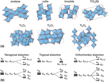
Rutile, anatase, and brookite are the three most common polymorphs of TiO2. Rutile and anatase have been used and studied most, partly because of their widespread availability. The electronic band structure of different phases is governed by the symmetry of the octahedral-shaped TiO6 fundamental building blocks. The crystal structures of rutile and anatase belong to the tetragonal space groups P42/mnm, a = b = 4.593 Å, c = 2.959 Å, and I41/amd, a = b = 3.785 Å, c = 9.514 Å, respectively [Reference Cromer and Herrington25]. There are a number of metastable forms of TiO2, including TiO2(B) [Reference Marchand, Brohan and Tournoux26], hollandite-like TiO2 [Reference Latroche, Brohan, Marchand and Tournoux27], and ramsdellite-like TiO2 [Reference Akimoto, Gotoh, Oosawa, Nonose, Kumagai and Aoki28], and the high-pressure forms: TiO2(II) [Reference Simons and Dachille29], baddeleyite-like TiO2 [Reference Sato, Endo, Sugiyama, Kikegawa, Shimomura and Kusaba30], cubic TiO2 (fluorite forms and pyrite form) [Reference Mattesini, de Almeida, Dubrovinsky, Dubrovinskaia, Johansson and Ahuja31], TiO2-OI [Reference Dubrovinskaia, Dubrovinsky, Ahuja, Prokopenko, Dmitriev, Weber, Osorio-Guillen and Johansson32], and cotunnite (PbCl2-like) TiO2-OII [Reference Dubrovinsky, Dubrovinskaia, Swamy, Muscat, Harrison, Ahuja, Holm and Johansson33]. The spatial representation of TiO2 polymorphs anatase and rutile, as well as suboxides such as Ti2O3, Ti4O7, and Ti5O9, is shown in Fig. 1(a). The fundamental difference in these oxides is related to the connectivity, volume, and shape of the TiO6 octahedra, where connectivity refers to the stacking pattern of neighboring octahedra, sharing tips, edges and, faces, and shape refers to the distortion of individual TiO6 octahedra. For perfect TiO6 octahedra such as in cubic TiO2, the site symmetry of both Ti and O is Oh. The tetragonal distortions in rutile and anatase lead to D2h and D2d symmetry of the central Ti ions. The TiO6 octahedra are subject to trigonal distortion in Ti2O3 and orthorhombic distortion in Ti4O7 and Ti5O9, leading to C3 and C1 site symmetry of Ti [Reference Stoyanov, Langenhorst and Steinle-Neumann34]. For example, the crystal field splitting (CFS) associated with the TiO6 octahedral coordination splits the 5-fold degenerate states of Ti-3d electrons into t2g (dxy, dxz, and dyz) and eg ( ${{\rm{d}}_{{x^2} - {y^2}}}$ and
 ${{\rm{d}}_{{z^2}}}$) bands. Figure 1(b) illustrates how different distortions further split the t2g and eg bands into sub-bands that are reflected in the electronic band structure of different phases. The various techniques used for the determination of the crystal and electronic structure of different titanium oxides are discussed in the next section.
(a) Comparison of the crystal structure of anatase, rutile, brookite, TiO2(B), Ti2O3, Ti4O7, and Ti5O9. These oxides consist of TiO6 octahedra with different connectivities, volumes, and shapes. (b) Tetragonal (anatase and rutile), trigonal (Ti2O3), and orthorhombic (Ti4O7 and Ti5O9) distortions of TiO6 cause the formation of different sub-bands in the electronic structure. Reprinted with permission from Ref. Reference Stoyanov, Langenhorst and Steinle-Neumann34.

Typical characterization methods used in black titania studies
Compared to bulk titanium dioxides whose structure is well understood, systematic studies devoted to understanding the structural changes of black TiO2 are more recent. The black TiO2 particles are a multicomponent system that typically contains a disordered surface layer, which makes characterization of the particle structure much more challenging. Although a variety of techniques have been used to characterize black TiO2, the interpretation of the results and the conclusions are biased by the limitations of the technique. For example, atomic resolution transmission electron microscopy (TEM) is the ideal tool for characterization of the intrinsic core–shell structure of black TiO2. However, for precise determination of the lattice parameter differences between black and pristine TiO2, X-ray diffraction (XRD) is more suitable than TEM imaging. Here we briefly introduce TEM techniques, X-ray photoelectron spectroscopy (XPS), and Raman spectroscopy, which are used most often for characterizing black TiO2, and comment on the advantages and shortcomings of each technique.
Characterization techniques
HRTEM
High-resolution transmission electron microscopy (HRTEM) imaging refers to the image governed by the phase contrast transfer function (CTF) damped by the envelope function. The scanning transmission electron microscopy (STEM) imaging in bright field (BF) mode is analogous to HRTEM imaging, which is also determined by the CTF. In comparison, Rutherford scattering makes the contrast in high-angle annular dark field (HAADF) mode of STEM atomic number (Z) dependent. Both HRTEM and STEM images can achieve atomic resolution as the information limit of HRTEM imaging, and the spatial resolution of STEM imaging after aberration correction nowadays routinely reaches sub-Ångström level. The TEM methods enable depth profiling of single particles to track the structural changes from the crystalline core to the shell, revealing the phase and the interface structure of individual black TiO2 particles. The high spatial resolution is a unique advantage of TEM imaging over other techniques.
An additional advantage of electron microscopy techniques is that associated spectroscopy techniques such as electron energy-loss spectroscopy (EELS) performed in STEM mode can be used to determine the electronic band structure with atomic scale spatial resolution. The excitation of electrons from the core level into the conduction band can be determined from the electron energy-loss near-edge structure (ELNES) in core-loss EELS. Therefore, the information from the Ti-3d states in the conduction band of titanium oxides can be obtained. For example, the CFS can be easily determined as the separation between t2g and eg peaks of the Ti-L2,3 edge. Not only that the splitting of eg bands due to different TiO6 octahedral distortions in anatase and rutile is clearly differentiated by the reversal of the “shoulder” features of Ti-L3 eg peaks, the unique features of the Ti3+ and Ti4+ L2,3 spectra can be differentiated using reference spectra to determine the ratio of Ti3+ to Ti4+ in black TiO2 [Reference Tian, Mahjouri-Samani, Eres, Sachan, Yoon, Chisholm, Wang, Puretzky, Rouleau, Geohegan and Duscher35]. The broad features after the energy range of the ELNES are called the extended electron energy-loss fine structure (EXELFS), where the bonding information of the local surroundings of the excited atom can be characterized. In principle, the local coordination in black TiO2 can be determined from the radial distribution function (RDF) derived from EXELFS, but this is yet to be demonstrated in practice. The same information about the electronic structure can be obtained from X-ray absorption spectroscopy (XAS) [Reference Zhou, Li, Wang, Qu, Yang, Xie, Zhang, Wang, Fu and Zhao36]. The ELNES and EXELFS are analogues of X-ray absorption near-edge structure (XANES) and extended X-ray absorption fine structure (EXAFS) in XAS. However, in contrast to electron microscopy techniques that are in principle capable of characterizing single particles, the information obtained by X-ray spectroscopy methods describes ensemble averages.
XPS
XPS is a spectroscopy technique capable of probing the 1–10 nm surface depth range depending on the incident photon energy [Reference Wagner, Briggs, Davis, Moulder and Muilenberg37]. The binding energy of an electron can be determined by measuring the kinetic energy of escaping photo-electrons and subtracting it from the incident X-ray photon energy and correcting it for the work function of the surface and the instrument. Here, local bonding information can be easily quantified so long as the chemical bonds of interest are distinguishable energetically, unlike EELS, which produces a joint density of states that is convoluted with multiple scattering. This advantage allows XPS to detect specific chemical bonds such as the hydroxyl group (Ti–OH) at about 532 eV for O 1s [Reference McCafferty and Wightman38, Reference Li, Han, Wang, Zheng, Wan and Ni39] on black TiO2 surface, which is difficult to obtain by EELS. An alternative method for obtaining chemical bonding information for molecular species is by Fourier transform infrared (FTIR) spectroscopy. Three distinct OH bands at 3718, 3669, and 3644 cm−1 have been observed for Ti–OH species [Reference Topsoe, Topsoe and Dumesic40]. XPS analysis is often combined with electron paramagnetic resonance (EPR) or electron spin resonance (ESR) [Reference Liu, Hou, Wang, Cui, Zhu and Wang41] to estimate the spatial distribution of defects in the core from that in the shell of black TiO2.
Raman spectroscopy
Raman spectroscopy provides information about vibrational, rotational, and other low-frequency modes in molecules based on inelastic scattering of monochromatic photons. The Raman spectrum is dictated by the symmetry of the materials, with each titanium di(sub)oxide exhibiting a unique fingerprint of transitions and relative intensities. Anatase has six allowed Raman transitions in the vibrational spectrum at low temperatures. The three Raman bands at 639, 197, and 144 cm−1 are assigned to the Eg modes, the 513 cm−1 band is assigned to the A1g mode, and two bands at 519 and 399 cm−1 correspond to the B1g modes [Reference Ohsaka, Izumi and Fujiki42]. At room temperature, 516 cm−1 band occurs because of the combination of A1g mode at 513 cm−1 and one of B1g modes at 519 cm−1. Rutile has four Raman active fundamentals: B2g mode at 826 cm−1, A1g mode at 612 cm−1, Eg mode at 447 cm−1, and B1g mode at 143 cm−1 [Reference Ohsaka, Izumi and Fujiki42]. The defect density, from oxygen deficiency [Reference Parker and Siegel43, Reference Li Bassi, Cattaneo, Russo and Bottani44] or structural disorder [Reference Wang and Saxena45], can also influence both the vibrational mode intensities and the peak position. Raman spectroscopy is a useful technique for quickly characterizing materials’ quality. However, it is an ensemble technique, which collects spectral information from all materials within the typical focus diameter size of 1–10 µm. This can be problematic for two reasons: it is not spatially resolved (nonspecific) and the spectral response is proportional to the optical path length (sensitivity). For example, the defective shell regions in black TiO2 prepared by the reduction method are typically limited to a thin 1–2 nm surface shell, resulting in a very weak signal that is obscured by the signal from larger nanostructures. Furthermore, a reference model for the molecular structure of each titanium di(sub)oxide is needed to fully understand the Raman spectra. Thus, Raman spectroscopy is more useful when investigating samples with high surface-to-volume ratio, such as nanostructures with small diameters <10 nm. However, the small size can present new challenges in the form of quantum confinement effects [Reference Zhu, Zhang, Chen and Yin46].
What is the size limit for ultra-small black TiO2 nanomaterials?
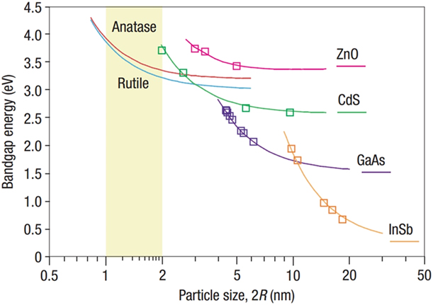
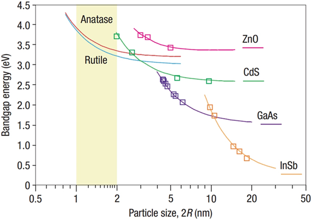
In general, the surface defects dominate functionality such as charge separation in photocatalysis, while the bulk defects act as charge recombination centers [Reference Li, Yan, Wang, Zhao, Zhang, Gong and Guan47]. Therefore, nanomaterials with core–shell structure, such as black TiO2 with defects concentrated in a thin surface shell, represent an ideal model system for studying photocatalytic processes. Furthermore, the high surface area of the nanoparticles amplifies photocatalytic activity [Reference Sang, Zhao and Burda48]. Synthesis of ultra-small TiO2 (1–4 nm) nanoparticles with high surface area is performed by hydrolysis or thermolysis [Reference Satoh, Nakashima, Kamikura and Yamamoto49, Reference Monticone, Tufeu, Kanaev, Scolan and Sanchez50, Reference Scolan and Sanchez51]. However, for particle diameters on the order of a few nanometers quantum size (Q-size), effects were reported to produce widening of the band gap [Reference Mills and Le Hunte52]. The size of the black TiO2 should be controlled to avoid Q-size effect-related interference on photocatalysis. Anpo et al. first observed the Q-size effect in 10-nm-diameter TiO2 particles by measuring the semiconductor photoactivity [Reference Anpo, Shima, Kodama and Kubokawa53]. Kormann et al. [Reference Kormann, Bahnemann and Hoffmann54] reported Q-size effects for TiO2 particles below 3 nm diameter, which exhibited increasing band gap with decreasing particle size. The critical dimension for the onset of Q-size effect in TiO2 is still debated, but generally it is not more than 2 nm [Reference Satoh, Nakashima, Kamikura and Yamamoto49, Reference Monticone, Tufeu, Kanaev, Scolan and Sanchez50]. The discrepancy of the observed critical size may arise from other factors such as surface states created by the defects. For example, Lin et al. [Reference Lin, Huang, Li, Ni, Shah and Tseng55] found that the band gap was reduced for particle size ranging from 29 to 17 nm, while nonproportionally increased band gap values were observed from 5.7 to 3.8 nm. They noted that surface states may also contribute to narrowing of the band gap [Reference Lin, Huang, Li, Ni, Shah and Tseng55]. Figure 2 shows that the critical dimension for Q-size effects in TiO2 is substantially smaller than that in other semiconductors [Reference Satoh, Nakashima, Kamikura and Yamamoto49]. Therefore, the photocatalytic activity of black TiO2 nanomaterials can be enhanced by reducing the particle’s size to approximately 2 nm without substantial interference from Q-size effects.
Comparison of the Q-size effect in rutile and anatase TiO2 with that in other semiconductors. The critical size corresponding to TiO2 is shaded by the colored band. Reprinted with permission from Ref. Reference Satoh, Nakashima, Kamikura and Yamamoto49.
Considering that most black TiO2 nanoparticles have some form of core–shell structure, the diameter of the black TiO2 nanoparticles with shells should be a few nanometers larger than the critical size for quantum size effects [Reference Chen, Liu and Huang4]. In the well-characterized two-step synthesis of black TiO2, where the first step is to produce amorphous precursors at room temperature, followed by the second step to convert the precursors into black TiO2 by annealing in a reducing environment, the particle size is controlled by the temperature. Particle growth and coarsening are difficult to avoid if the formation of black TiO2 involves processing above 400 °C. Thermal annealing combined with reduction in a H2 gas environment is often utilized to produce black TiO2 at low temperatures. Chen et al. reported that black TiO2 nanoparticles were produced in a 20 bar atmosphere of H2 gas at 200 °C [Reference Chen, Liu, Yu and Mao3]. There was almost no particle growth starting from pristine anatase of ∼8 nm and the resulting black anatase of ∼8 nm with a ∼1 nm amorphous shell after an annealing period of 120 h. In solution-based synthesis, adding a reducing agent can be used to decrease the formation temperature of black TiO2. For example, Ullattil and Periyat mixed titanium butoxide with a manganese acetate solution under microwave irradiation at 150 °C for 5 min to synthesize ∼5 nm size black TiO2 [Reference Ullattil and Periyat56]. In this case, the doped Mn2+ ions are key to formation of black anatase since it was found that only yellow anatase forms without Mn2+ ions [Reference Ullattil and Periyat56].
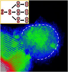
In contrast to the low-temperature case, at high temperatures, Chen’s group showed the growth of pure TiO2 nanocrystals after hydrogenation [Reference Xia, Zhang, Oyler and Chen57]. After hydrogenating pure TiO2 nanocrystals at 450 °C for 4 h, the crystal size increased from 19 to 45 nm for the anatase phase and 50 nm for the rutile phase. Chen’s group also summarized the size of TiO2 nanoparticles with different anatase/rutile ratio changed by annealing time in H2 and derived the specific crystalline interfacial area (SCIA) at different anatase/rutile ratios [Reference Xia, Zhang, Oyler and Chen58]. A strategy for suppression of the size of black TiO2 at high temperatures was discussed in our previous report [Reference Tian, Mahjouri-Samani, Wang, Puretzky, Geohegan, Tennyson, Cross, Rouleau, Zawodzinski, Duscher and Eres59]. The synthesis of ultra-small particles of 5 ± 2 nm diameter shown in Fig. 3 was achieved by annealing in Ar at 400 °C for two hours. In this work [Reference Tian, Mahjouri-Samani, Wang, Puretzky, Geohegan, Tennyson, Cross, Rouleau, Zawodzinski, Duscher and Eres59], we found three factors responsible for maintaining small particle size: (i) ultra-small 3 ± 1 nm starting amorphous nanoparticles; (ii) the formation of an amorphous Ti2O3 shell that prevents the agglomeration of neighboring particles; and (iii) using pulsed laser vaporization (PLV) to ensure high purity of the starting materials, thus eliminating the need for additional purification at high temperatures. In solution-assisted methods, there are residual impurities from the organic compounds that must be removed typically by using a high-temperature (∼500 °C) calcination step [Reference Lu, Yip, Wang, Huang and Zhou60, Reference Chen, Liu, Liu, Marcus, Wang, Oyler, Grass, Mao, Glans, Yu, Guo and Mao61].
Ultra-small ∼5 nm black anatase with a Ti2O3 shell obtained by annealing of ∼3 nm amorphous precursor particles in Ar. The Ti2O3 shell hinders lattice alignment and particle growth. The white dashed line is the interface between the core and the shell. Reprinted with permission from Ref. Reference Tian, Mahjouri-Samani, Wang, Puretzky, Geohegan, Tennyson, Cross, Rouleau, Zawodzinski, Duscher and Eres59.

The origin of lattice contraction and expansion
Deviations of lattice parameters from the known crystalline bulk phases have been reported widely for black TiO2. Both expansion and contraction of the lattice were observed, corresponding to changes in lattice constants a or c, or both. For example, Myung et al. [Reference Myung, Kikuchi, Yoon, Yashiro, Kim, Sun and Scrosati62] and Shin et al. [Reference Shin, Joo, Samuelis and Maier63] reported that black anatase has slightly larger lattice constants a and c than pristine anatase. However, Lu et al. [Reference Lu, Yip, Wang, Huang and Zhou60] reported lattice expansion only along the c direction. In contrast, lattice contraction (both a and c) was reported by Chen et al. [Reference Xia and Chen64] and Naldoni et al. [Reference Naldoni, Allieta, Santangelo, Marelli, Fabbri, Cappelli, Bianchi, Psaro and Dal Santo65]. However, few reports discuss the origin, and the interpretations were generally oversimplified, such as the lattice expansion resulting from the increased radius of Ti4+ versus Ti3+ [Reference Myung, Kikuchi, Yoon, Yashiro, Kim, Sun and Scrosati62]. To elucidate the mechanism, we consider the following three factors: (i) the surface structures, (ii) the Q-size effect of small nanoparticles, and (iii) the defect contributions.
First, the majority of black TiO2 is reported to have a core–shell structure. In this case, the lattice distortion is most likely related to the incoherent interface lattice match between the crystalline core and the amorphous shell. For example, in previous work, we observed a Ti2O3 amorphous shell on the rutile core [Reference Tian, Mahjouri-Samani, Eres, Sachan, Yoon, Chisholm, Wang, Puretzky, Rouleau, Geohegan and Duscher35], where the interface was found to be strained [Reference Tominaka66]. However, it is very difficult to determine whether the bulk TiO2 lattice is under compression or tension. One reason for this is heterogeneity of the interface-matching relationships because of the many facets of the nanoparticles. Even with TEM, it is unrealistic to assume that all the interfaces can be imaged because of the crystal orientation relative to the electron beam. Furthermore, the disordered structure of the shell cannot be always resolved, which then limits the validity of any model one could consider for explaining the interface matching. Second, for very small nanoparticles, the Q-size effect must be considered. For anatase, Swamy et al. [Reference Swamy, Menzies, Muddle, Kuznetsov, Dubrovinsky, Dai and Dmitriev67] systematically investigated the nonlinear size-dependent lattice parameter deviation on particle size by directly comparing their results with other reported data. He concluded that the Q-size effect was pronounced for particle sizes <10 nm. For rutile, Li et al. [Reference Li, Boerio-Goates, Woodfield and Li68] found that lattice expansion occurred for particle sizes 50 nm or smaller. The absence of Ti3+ and oxygen vacancies in these data suggests that the TiO2 used was completely stoichiometric. In the proposed “surface defect dipole model,” a great number of Ti–O dipoles formed at the surface with reduced particle size. The interactions of dipoles were attributed to negative pressure in ultra-small rutile nanoparticles [Reference Li, Boerio-Goates, Woodfield and Li68]. A more comprehensive review of the size effect on lattice deviations was published by Zhang and Banfield [Reference Zhang and Banfield69]. Third, the contribution of defects must be considered because of their abundance in black TiO2. The occupied sites and species of defects play an important role in lattice contraction and expansion. Not included in this discussion are “line defects” (dislocations) and “planar defects” (i.e., stacking faults and twin boundaries). Santara et al. systematically investigated undoped rutile nanomaterials with different morphologies by a series of structural and optical characterization techniques including SEM, HRTEM imaging, selected area electron diffraction (SAED), energy-dispersive X-ray spectroscopy (EDX), XRD, XPS, UV-vis-NIR absorption spectroscopy, photoluminescence (PL), and ESR [Reference Santara, Giri, Imakita and Fujii70]. They conclude that oxygen vacancies cause a relaxation of the Ti–Ti and Ti–O bonds and both oxygen vacancies and interstitial Ti play a role in lattice expansion. In contrast, the Columbic attraction between interstitial Ti ions and interstitial oxygen vacancies results in lattice contraction [Reference Santara, Giri, Imakita and Fujii70]. However, this model awaits to be verified by a corresponding simulation. Nevertheless, this research provides new insight into the origin of lattice deviation by considering the interaction between different defect types.
Phase control
Anatase to rutile transformation
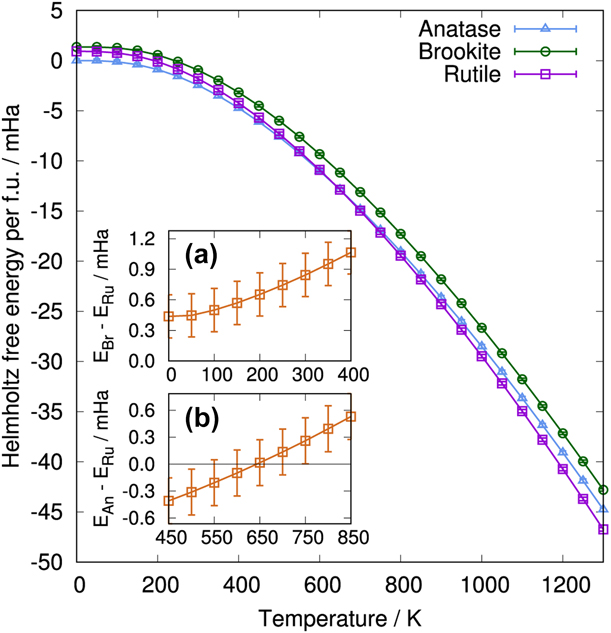
The most common phase of the black TiO2 core is rutile or anatase. Early researches showed rutile is thermodynamically more stable than anatase [Reference Smith, Stevens, Liu, Li, Navrotsky, Boerio-Goates and Woodfield71, Reference Mitsuhashi and Kleppa72, Reference Jamieson, Olinger, Dachille, Simons and Roy73] except at extremely low temperatures [Reference Muscat, Swamy and Harrison74]. For example, Smith [Reference Smith, Stevens, Liu, Li, Navrotsky, Boerio-Goates and Woodfield71] showed the free energy of anatase is uniformly negative for the complete temperature range at standard pressure, implying that anatase is a metastable form of TiO2. However, recently, Luo et al. [Reference Luo, Benali, Shulenburger, Krogel, Heinonen and Kent75] found that the Helmholtz free energy of anatase is smaller than that of rutile below 650 K ± 150 K, as shown in Fig. 4. Inset b in Fig. 4 shows the transition temperature occurs at 650 K and the uncertainty is estimated by the error bar. The anatase to rutile transition temperature (ART) is the key to controlling the phase during synthesis. However, the ART depends on many factors such as particle size [Reference Zhang and Banfield76], chemical environment [Reference Barnard and Curtiss77], and the presence of impurities, dopants, and defects. Since the properties of black TiO2 are generally dominated by the presence of oxygen vacancies and Ti3+, we only discuss their influence on the ART here.
Helmholtz free energy for rutile, anatase, and brookite at different temperatures. Insets (a) and (b) show the free energy differences between brookite and rutile from 0 K to 400 K and between anatase and rutile from 450 K to 850 K, respectively. Reprinted with permission from Ref. Reference Luo, Benali, Shulenburger, Krogel, Heinonen and Kent75.

Initial research [Reference Shannon and Pask78] showed that oxygen deficiency promotes the transformation of anatase to rutile, but the interstitial Ti3+ ions play a vital role in inhibiting the transition. The subsequent reports on reduced TiO2 provided further data that are consistent with these findings. For example, Vargas et al. [Reference Vargas, Arroyo, Haro and Rodríguez79] made a systematic investigation of the cation dopant effects on ART and attributed the promotion of the ART to increased oxygen deficiency. Riyas et al. [Reference Riyas, Krishnan and Mohan Das80] also reached a similar conclusion from investigations of the effect of transition metal oxides on ART.
From a structural perspective, there are two reasons why oxygen deficiency promotes the ART. First, oxygen vacancies facilitate the reconstruction of the Ti–O bonds because the anatase to rutile transformation is reconstructive [Reference Batzill, Morales and Diebold81]. In contrast to displacive transformations where Ti–O bonds are retained, the Ti–O bonds have to be broken and reformed, and this transformation involves volume contraction of a few percent [Reference Shannon and Pask78]. A high concentration of mobile oxygen vacancies is the key to facilitating the reconstruction of the interface in the ART [Reference Batzill, Morales and Diebold81]. Second, it promotes formation of titanium suboxides as distorted templates of rutile. During reduction, anatase can transform into suboxides, i.e., Magnéli phases [Reference Shannon and Pask78, Reference David, Trolliard and Maître82], which can be also considered as defective rutile [Reference Liborio and Harrison83, Reference Le Page and Strobel84]. Although the defects, oxygen vacancies and Ti3+, break the short-range order (SRO) of the rutile structure, the rutile-like slabs are maintained in these phases and can act as templates for rutile growth. In a recent report, we compared white TiO2 and black TiO2 produced from amorphous precursors annealed in oxygen-rich and oxygen-deficient Ar atmosphere, respectively [Reference Tian, Mahjouri-Samani, Wang, Puretzky, Geohegan, Tennyson, Cross, Rouleau, Zawodzinski, Duscher and Eres59, Reference Mahjouri-Samani, Tian, Puretzky, Chi, Wang, Duscher, Rouleau, Eres, Yoon, Lasseter, Xiao and Geohegan85]. These studies demonstrated that rutile formed at a relatively low temperature (500 °C) in Ar in combination with a Ti2O3 shell. However, the formation of rutile is highly suppressed when annealed in O2 gas, and instead TiO2(B) is readily formed. Even at high temperatures (900 °C), anatase is dominant with only a small portion of rutile.
The dopant effects on the anatase to rutile transformation were reviewed by Hanaor and Sorrell [Reference Hanaor and Sorrell86]. They concluded that the effects of dopants on ART depend on formation of oxygen vacancies and interstitial Ti3+. They presented a summary of the elements that inhibit or promote ART from the literature and their observations [Reference Hanaor and Sorrell86]. Although good agreement can be found for Zn reduction [Reference Zhao, Tan, Zhao, Lv, Zhou, Song and Sun87], the predictions are conflicting for Al reduction [Reference Yin, Lin, Yang, Wang, Zhu, Xu, Xie, Huang and Jiang88] and S doping [Reference Yang, Wang, Lin, Yin, Lu, Wan, Xu, Zheng, Lin, Huang, Xie and Jiang10]. It is possible that the dopant effects were suppressed by other factors such as annealing in vacuum, inert gas, or hydrogenation, where substantial amounts of oxygen vacancies and Ti3+ can form. Among various reduction processes, hydrogenation was the most often investigated synthesis technique for black TiO2 [Reference Chen, Liu and Huang4], where the H2-related promotion of ART was observed [Reference Naldoni, Allieta, Santangelo, Marelli, Fabbri, Cappelli, Bianchi, Psaro and Dal Santo65, Reference Wang, Wang, Ling, Tang, Yang, Fitzmorris, Wang, Zhang and Li89]. However, the balance between promotion and inhibition depends on a competition between oxygen vacancies and Ti interstitials. The formation of oxygen vacancies is usually accompanied by the presence of Ti3+. However, if Ti3+ is mostly at interstitial sites, no obvious ART can be observed. Therefore, producing a high concentration of reduced species, Ti3+, and oxygen vacancies by a strong reductant such as H2 is not always necessary for promoting the ART. As evidence for this, Shannon et al. [Reference Shannon and Pask78] showed that the formation of interstitial Ti3+ is suppressed in inert gas such as Ar. Although H2 is a stronger reducing agent than Ar, the anatase to rutile transformation occurs faster in Ar than in H2. This observation suggests that oxygen vacancies dominate promotion of ART, since interstitial Ti3+ formation is suppressed in Ar.
Interestingly, the formation of Ti3+ and oxygen vacancies by hydrogenation was challenged by Chen’s group [Reference Xia, Zhang, Oyler and Chen57], who synthesized a type of hydrogenated TiO2 containing no oxygen vacancies and Ti3+ characterized by electron spin resonance spectroscopy (ESR). They prepared pure crystalline TiO2 nanoparticles using titanium butoxide, ethanol, and deionized water as the precursor solution. The hydrogenated TiO2 nanoparticles were obtained by annealing in 5 bar H2 at 450 °C where anatase to rutile transformation occurred. Apparently, such low ART in this work is not associated with oxygen vacancies and Ti3+. Since TiO2 is known to reduce easily, the surface science of this material requires further exploration to elucidate the mechanism of stoichiometry preservation in such a strong reducing background, which could shed light on the protection of TiO2 from reduction.
Low-temperature synthesis of black rutile
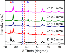
Although the anatase to rutile transformation in bulk typically occurs between 600 and 700 °C, solvothermal/hydrothermal methods have been shown to produce rutile near room temperature [Reference Hanaor and Sorrell86]. The actual temperature for formation of black rutile is higher because it involves a reduction step, consisting of postannealing at higher temperatures. For example, Zhang et al. [Reference Zhang, Zhang, Peng, Wang, Yu, Wang and Peng90] synthesized rutile particles using titanium butoxide reacting with HCl on fluorine-doped tin oxide glass slides at 170 °C for 6 h. To reduce rutile, the resulting material was annealed in a H2/Ar mixture of 1:4 ratio at 350 °C for 1 h. However, with the introduction of dopants, direct synthesis of black rutile at low temperatures becomes possible. This observation raises the question about the role of other impurities unintentionally present in the various synthesis methods. The dopants increase the oxygen vacancy concentration, which can both reduce the ART and produce the black color. For example, Zhao et al. [Reference Zhao, Tan, Zhao, Lv, Zhou, Song and Sun87] used a solvothermal method for preparing black rutile nanoparticles in the presence of Zn powder at 180 °C. They varied the amount of Zn powder to control the concentration of oxygen vacancies in TiO2 and the formation of black rutile. Without Zn powder, only white anatase was observed. The XRD spectra in Fig. 5 illustrate that above 1.5 mmol of Zn, only the peaks characteristic of pure rutile are observed. Below 1.5 mmol the fraction of anatase increases with decreasing concentration of Zn.
XRD spectra of black TiO2 with increasing amount of Zn in the solution from 0.5 to 2.5 mmol. The fraction of rutile increases and that of anatase decreases with increasing Zn concentration, and above 1.5 mmol of Zn, only rutile is observed. Reprinted with permission from Ref. Reference Zhao, Tan, Zhao, Lv, Zhou, Song and Sun87.

Other phases
Brookite and TiO2(B) are often present as by-products in the synthesis of anatase and rutile. Brookite is one of the three common polymorphs of TiO2 besides anatase and rutile, but it is less stable than anatase and rutile, as shown in Fig. 4 [Reference Luo, Benali, Shulenburger, Krogel, Heinonen and Kent75]. However, in contrast to anatase and rutile, potential applications are limited because of difficult synthesis [Reference Hanaor and Sorrell86, Reference Di Paola, Bellardita and Palmisano91]. Consequently, few reports exist on the synthesis of black brookite. A facile way to synthesize black brookite was demonstrated by direct reduction of precursors. For example, Zhu et al. [Reference Zhu, Lin, Lü, Zhao, Yang, Wang, Yin, Liu, Huang and Lin92] used TiOSO4·xH2O in water to react with NaOH and heated the precipitates at 220 °C for 48 h. The resulting brookite nanoparticles were reduced by Al powder. The Al and brookite were separately heated in a two-zone furnace in vacuum at 800 °C and 300–600 °C for 4 h. However, brookite is a metastable form of TiO2 that just like anatase transforms into rutile at high temperatures [Reference Li and Ishigaki93, Reference Rao, Yoganarasimhan and Faeth94, Reference Huberty and Xu95]. The introduction of self-doping defects was reported to stabilize brookite during high-temperature annealing. Xin et al. [Reference Xin, Xu, Wang and Wang96] mixed TiH2 into water and H2O2 and, after stirring, added the mixture to NaOH to control pH under hydrothermal treatment at 180 °C for 24 h using the reducing agent NaBH4. The as-synthesized brookite was found to be already reduced after washing, exhibiting a grey-blue color. By postannealing at 300, 500, and 700 °C, the powders acquired black color. The black brookite was found to be stable, and the phase transformation was not observed even with annealing to 700 °C.
The low-symmetry TiO2(B) structure is a less common phase of titania. This phase was discovered only recently (1980) [Reference Marchand, Brohan and Tournoux26], and it is not well known what factors influence its phase transformation temperature. TiO2(B) can be used to stabilize anatase [Reference Kumar and Rao97], and therefore black TiO2(B) often coexists with anatase. Li et al. [Reference Li, Shi, Tu, Qian, Li, Yang and Lu98] synthesized black bicrystalline anatase and TiO2(B) using TiO2·nH2O and K2CO3 as starting material. The mixture was heated at 860 °C for 4 h to produce K2TiO5. The hydrated titania was obtained by adding HCl. The black TiO2(B)/anatase mixture formed by heating the hydrated titania in pure H2 at 600 °C for 2 h. In comparison, annealing the hydrated titania in air yielded a mixture consisting of white TiO2(B)/anatase. Interestingly, no Ti3+ was found, but oxygen vacancies were reported at 0.58 wt% in both phases. The oxygen vacancies should enhance the ART, but instead inhibition of ART was observed, suggesting that the presence of TiO2(B) has a stabilizing effect on anatase. Note that increasing the temperature enhances the transformation of black TiO2(B) into black anatase instead of rutile [Reference Cai, Wang, Zhu, Wu, Zhang, Li, Jiang and Meng99].
The suboxide shell
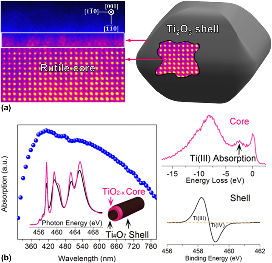
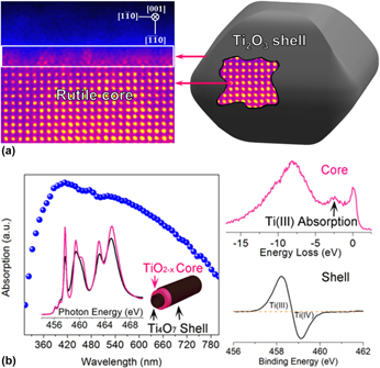
The thickness of the surface shell of black TiO2 is generally found to be around 1–2 nm. Such a thin surface layer makes characterization of the phase very challenging. For example, Zheng et al. [Reference Zheng, Hao, Zhao, Jia, Cao and Zhu100] made black TiO2 by bombarding commercial TiO2 nanoparticles by hot molecular species. The high-temperature and high-pressure hot molecular species were produced by explosive decomposition of TNP (trinitrophenol). Formation of a new metastable reconstructed surface crystalline phase that enhances photocatalysis was reported. However, the crystal structure of the surface layer could not be identified. The use of TEM techniques allows characterization of the shell providing both crystal structure and electronic band structure information on the atomic scale. Previously, we used a combination of TEM techniques including HRTEM, STEM, nanobeam electron diffraction (NBED), and EELS, to determine that the reconstructed shell of black TiO2 consists of Ti2O3 as illustrated in Fig. 6(a) [Reference Tian, Mahjouri-Samani, Eres, Sachan, Yoon, Chisholm, Wang, Puretzky, Rouleau, Geohegan and Duscher35]. The starting material consisted of stoichiometric amorphous TiO2 nanoparticles produced by PLV in O2. The black TiO2 nanoparticles were obtained by postannealing in Ar in a wide temperature range. Accumulation of a large concentration of oxygen vacancies in the surface shell was found to be the key factor driving a transition from TiO2 to reduced Ti2O3.
Illustration of black TiO2 consisting of a crystalline core and a suboxide shell. (a) Black rutile nanoparticles with a Ti2O3 shell. Reprinted with permission from Ref. Reference Tian, Mahjouri-Samani, Eres, Sachan, Yoon, Chisholm, Wang, Puretzky, Rouleau, Geohegan and Duscher35. (b) Black anatase nanotubes with a Ti4O7 shell. Reprinted with permission from Ref. Reference Li, Liu, Li, Wang, Shao, Wang, Sun, Pong, Guo and Sham101.

Similarly, Li et al. [Reference Li, Liu, Li, Wang, Shao, Wang, Sun, Pong, Guo and Sham101] reported the formation of amorphous Ti4O7 shells at the surface of black anatase. The phase characterization was carried out by a series of techniques, including XRD, TEM, X-ray photoemission spectroscopy (XPS), and synchrotron XAS. The material was synthesized by a two-step electrochemical anodization process. Amorphous nanotubes were first obtained by electrochemical anodization of a Ti foil. The nanotubes when annealed in NH3 at 450 °C transformed into crystalline anatase core and a Ti4O7 shell structure. When annealing was performed in air at 450 °C, the nanotubes transformed into pure anatase, as shown in Fig. 6(b) [Reference Li, Liu, Li, Wang, Shao, Wang, Sun, Pong, Guo and Sham101]. A few reports also discussed the formation of titanium suboxides such as Ti2O3 [Reference Zhou, Li, Wang, Qu, Yang, Xie, Zhang, Wang, Fu and Zhao36, Reference Lu, Zhao, Pan, Yao, Qiu, Luo and Liu102] and Magnéli phases [Reference Panomsuwan, Watthanaphanit, Ishizaki and Saito19, Reference Myung, Kikuchi, Yoon, Yashiro, Kim, Sun and Scrosati62, Reference Ishida, Doshin, Tsukamoto and Yonezawa103, Reference Fujiwara, Deligiannakis, Skoutelis and Pratsinis104] in black TiO2, but without supporting data from high-spatial-resolution techniques needed for precise characterization of the shell phase.
Summary and perspectives
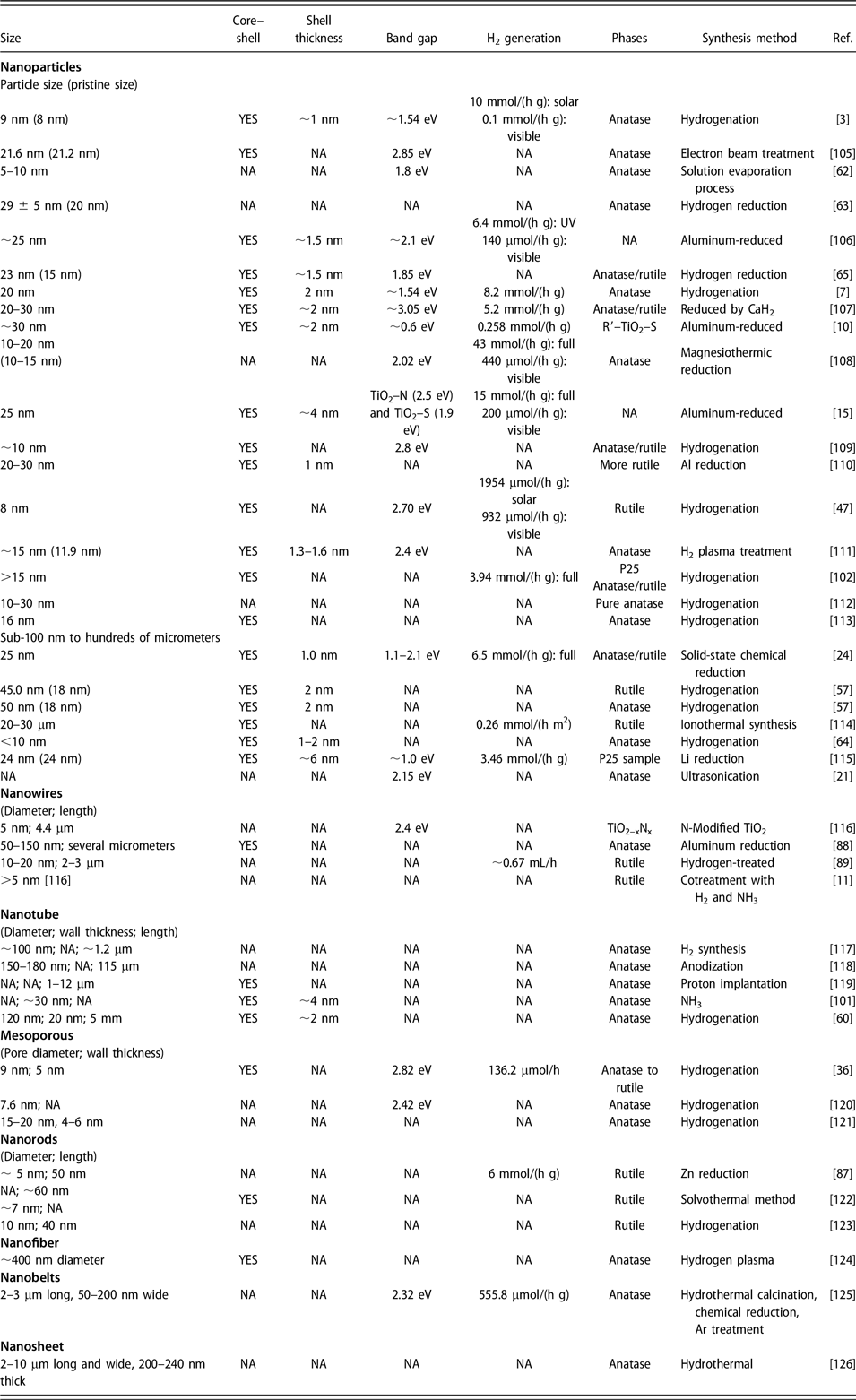
The structure of black TiO2 was found to depend strongly on the synthesis route, which explains why the corresponding formation mechanisms prove to be so difficult to establish. This review started with introducing the atomic structure of different TiO2 phases and a brief description of the strengths and the limitations of the most commonly used techniques used for characterization of black titania. We discussed particle dimensionality effects and size limits, lattice deviations, and the phase transformation of black TiO2 by comparing pristine and defective TiO2 focusing on Ti3+ and oxygen vacancies and summarized the corresponding approaches and various strategies for controlling synthesis. The representative examples of published research on black titania are compiled and presented in Table I.
Black titania synthesis-related properties.

NA = not available.
Chen et al. [Reference Chen, Liu and Huang4], Liu et al. [Reference Liu, Zhu, Wang, Yuan, Lin and Huang5], and Zhang et al. [Reference Zhang and Park23] published extensive reviews on the role of defects including oxygen vacancies, Ti3+, the Ti–H bond, and Ti–OH in photocatalytic property enhancement. However, few reports have investigated the atomic structure of the shell that is known to be the key to the performance of black titania. Perhaps, because of the lack of awareness that amorphous materials are not completely structureless, it is perceived to be unimportant. Specifically, Fan et al. [Reference Fan, Chen, Wang, Fu, Ren, Qian and Wang21] prepared black TiO2 by ultrasonic treatment of amorphous precursors. The amorphous features were retained after ultrasonication, and the “blackness” of the black TiO2 was controlled by the treatment duration. The changes of color were solely attributed to the presence of hydroxyls [Reference Fan, Chen, Wang, Fu, Ren, Qian and Wang21]. However, Liu et al. [Reference Liu, Zhu, Wang, Yuan, Lin and Huang5] suggested that this explanation may be too simplistic because the lack of clear support from the structure and simulations data presented. Indeed, Li et al. [Reference Li, Yan, Wang, Zhao, Zhang, Gong and Guan47] demonstrated that the hydroxyls only contribute ∼10% to the band gap narrowing of TiO2.
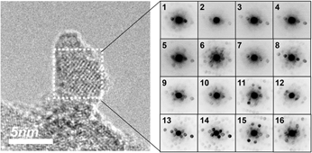
The definition of amorphous refers to the nature of disordered materials with no long-range order (LRO), while the SRO or medium-range order (MRO) might still be presented. The TiO6 octahedra represent the fundamental building blocks of TiO2 [Reference Zhang, Chen, Banfield and Waychunas127, Reference Prasai, Cai, Underwood, Lewis and Drabold128, Reference Van Hoang129]. The volume, shape, and connectivity of TiO6 octahedra dictate the properties of all the titanium oxide materials [Reference Rondinelli, May and Freeland130]. However, reconstruction of the amorphous structure from TiO6 octahedra presents an extremely difficult challenge because the nonequilibrium shapes and nonuniform distortions are very difficult to deduce by probing ensemble-averaged structural properties of disordered states using characterization techniques such as XRD, XAS, and EPR. In contrast, local probing by using NBED shows in a previously published work that the amorphous shell does not completely lose periodicity. To understand the meaning of the NBED diffraction patterns from amorphous TiO2 nanoparticles shown in Fig. 7, we consider the work by Hirata et al. [Reference Hirata, Guan, Fujita, Hirotsu, Inoue, Yavari, Sakurai and Chen131] who described similar diffraction patterns in the crystallization of Zr66.7Ni33.3 bulk metallic glass (BMG). The spots in the NBED diffraction pattern correspond to Zr–Ni polyhedra. The formation of NBED patterns originates from coherent scattering of a convergent electron beam. As the detected volume is changed from nanosize to atomic level by reducing the probe size from 7 to 0.36 nm, Hirata et al. showed that SRO of the BMG can be represented as joined polyhedra using a probe size of ∼0.36 nm in diameter. The various polyhedra with equilibrium shapes in the BMG were reconstructed by using Voronoi polyhedral analysis [Reference Borodin132, Reference Finney133].
Observation of MRO in NBED diffraction patterns (1 through 16) in TiO2 nanoparticles prepared by PLV shown in the HRTEM image in the left panel. Reprinted with permission from Ref. Reference Tian, Mahjouri-Samani, Wang, Puretzky, Geohegan, Tennyson, Cross, Rouleau, Zawodzinski, Duscher and Eres59.

It was shown that the well-resolved interface in HRTEM images between the core and the shell of black rutile has a symmetry commonly accepted as “added row Ti2O3” [Reference Onishi and Iwasawa134, Reference Onishi and Iwasawa135]. The shell is more accurately defined as “semi-crystalline,” meaning that the crystalline features are retained to recognizable degree. The EELS spectrum collected from the defective Ti2O3 shell is very similar to crystalline bulk, in agreement with the existence of Ti2O3 reflection points from NBED. Similarly, using high-spatial-resolution TEM techniques, Lu et al. [Reference Lu, Chen, Luo, Lu, Dai, Enriquez, Dowden, Xu, Kotula, Azad, Yarotski, Prasankumar, Taylor, Thompson and Jia136] characterized an oxygen-deficient amorphous shell synthesized on top of a crystal bilayer to simulate the structure of black TiO2 nanoparticles. It was found that the conductivity of the bilayer system was higher than that of a single amorphous layer, or a crystalline film, by a factor of two and five orders of magnitude, respectively. The increased conductivity was attributed to charge carrier accumulation at the interface because of band bending as the oxygen-deficient film contacts the pure TiO2 film [Reference Lu, Chen, Luo, Lu, Dai, Enriquez, Dowden, Xu, Kotula, Azad, Yarotski, Prasankumar, Taylor, Thompson and Jia136]. Careful examination of the STEM image in Fig. 1(e) in reference [Reference Lu, Chen, Luo, Lu, Dai, Enriquez, Dowden, Xu, Kotula, Azad, Yarotski, Prasankumar, Taylor, Thompson and Jia136] reveals that the periodicity of the crystalline side is retained on the amorphous side, which is very similar to previous results published by the authors of this review [Reference Tian, Mahjouri-Samani, Eres, Sachan, Yoon, Chisholm, Wang, Puretzky, Rouleau, Geohegan and Duscher35]. The nature of structure within amorphous or “semi-crystalline” materials presents many interesting opportunities and analysis challenges when short- and medium-range orders are taken into consideration. We hope that by raising awareness of the existence of order within the disordered shell, future studies will focus more attention on the atomic and electronic structure of the amorphous shell.
Acknowledgments
This work was supported by the U.S. Department of Energy (DOE), Office of Science, Basic Energy Sciences, Materials Sciences and Engineering Division.