Introduction
Traditionally, the genus Psoroma Ach. ex Michx. was the main genus of the Pannariaceae, containing species which typically had a green chlorococcoid alga as the primary photobiont. However, Jørgensen (Reference Jørgensen2001) showed that some of the large foliose austral species with a chlorococcoid photobiont previously included in Psoroma were better placed in Pannaria Delise ex Bory. He formally transferred three species to Pannaria and this process has been continued by other researchers (Elvebakk & Galloway Reference Elvebakk and Galloway2003; Passo & Calvelo Reference Passo and Calvelo2006). More recently, several segregate genera have been split off from Psoroma: the paleotropical species being transferred to Gibbosporina Elvebakk et al. (Elvebakk et al. Reference Elvebakk, Hong, Park, Robertsen and Jørgensen2016) and other Southern Hemisphere groups to Joergensenia Passo et al. (Passo et al. Reference Passo, Stenroos and Calvelo2008), as well as Psorophorus Elvebakk & S. G. Hong and Xanthopsoroma Elvebakk & S. G. Hong (Elvebakk et al. Reference Elvebakk, Robertsen, Park and Hong2010).
In this revised circumscription, the genus Psoroma is mostly confined to mid to high latitude regions, but also with representatives occurring at high altitudes in tropical countries such as Ecuador (Jørgensen & Palice Reference Jørgensen and Palice2010). The genus is most species-rich in the Southern Hemisphere; for example, the most recent lichen checklist for Chile (Galloway & Quilhot Reference Galloway and Quilhot1998) lists 35 taxa, although 13 of these have been removed to other genera and seven are synonyms (see above and Jørgensen (Reference Jørgensen2003)), leaving a total of 15. In contrast, only five species have been reported from the Northern Hemisphere (Henssen & Renner Reference Henssen and Renner1981; Jørgensen Reference Jørgensen2000 [2001], Reference Jørgensen2004; Timdal & Tønsberg Reference Timdal and Tønsberg2006; Elvebakk Reference Elvebakk2012; Elvebakk & Tønsberg Reference Elvebakk and Tønsberg2018) and two of these species are known only from single collections (P. cinnamomeum Malme from southern California and P. spinuliferum Elvebakk & Tønsberg from coastal Alaska).
Molecular investigation of the Pannariaceae by Ekman et al. (Reference Ekman, Wedin, Lindblom and Jørgensen2014) showed that another taxon, Psoroma tenue Henssen, was not congeneric with P. hypnorum (Vahl) Grey, the type species of Psoroma. However, they were unable to suggest the correct placement for this species because of the lack of resolution in that part of their phylogeny.
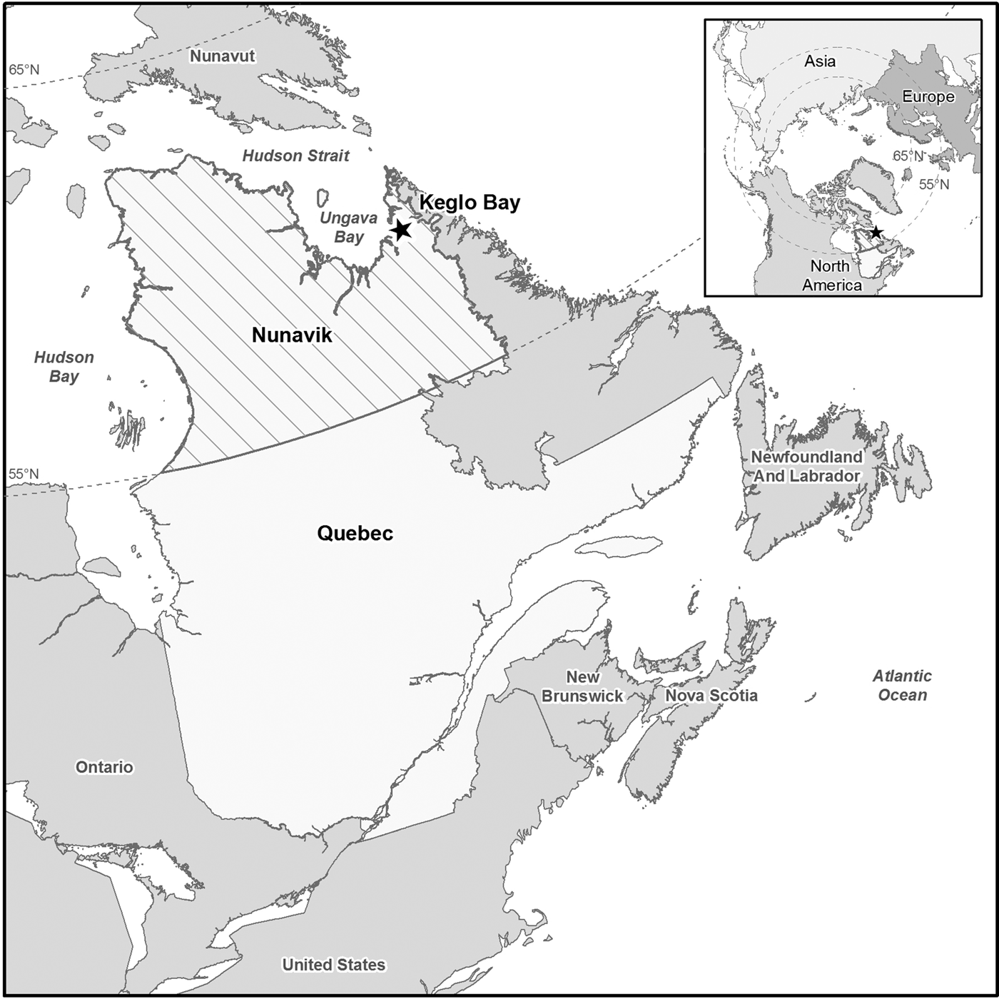
Here we describe a further species for the Northern Hemisphere that is known only from an area of late snow-lie in northern Québec, Canada (Fig. 1). We also discuss its systematic position within the Pannariaceae and provide a key to pannarioid lichens in arctic areas of North America.
Map showing the location of Keglo Bay in Québec, Canada from where the new species, Psoroma nivale, was collected. The hatched area is the region of Nunavik, a part of Québec above the 55 °N line.

Materials and Methods
Apothecial characteristics were examined by light microscopy on hand-cut sections mounted in water, 10% KOH (K), 50% HNO3 (N) or Lugol's reagent (0·15% aqueous IKI). Thallus sections were investigated in water and K. The ascus structure was studied in IKI, both without prior treatment and after pretreatment with K. Measurements of ascospores and paraphyses were made in water. Only ascospores liberated from asci were studied. Nomenclature of ascospore structures follows Nordin (Reference Nordin1997), and proper ascospores and perispores were measured separately. Sketches of a total of 20 ascospores and the IKI+ structures of c. 70 asci were drawn, and a selection made to represent the variation observed. Thin-layer chromatography followed the methods of Orange et al. (Reference Orange, James and White2001).
Additional collections examined: Austrella arachnoidea P. M. Jørg. French Southern and Antarctic Lands: Îles Kerguelen: Île Guillou, 49°28′57″S, 69°49′06″E, 14 m, feldfield, 2014, D. Ertz 18847 (MSC0242550).
Austrella isidioidea P. M. Jørg. & Fryday. Falkland Islands: West Falkland: Port Howard, Mt Maria, Lightning Rocks, 51·619028°S, 59·601849°W, 575 m, over Andreaea sp. in recess on exposed, E-facing rocks, 2015, Fryday 10918 (MSC0195379).
Fuscopannaria pacifica P. M. Jørg. USA: Alaska: Hoonah-Angoon District, Glacier Bay National Park, Bartlett Lake Road, 58·46350°N, 135·77520°W, mixed woodland, alder twigs, 2012, Fryday 9900 (MSC0256265).
Pannaria euphylla (Nyl.) Elvebakk & D. J. Galloway. New Zealand: South Island: at junction of Craigieburn River and Route 73, mature Nothofagus forest in ravine, 1971, H. A. Imshaug 48263 (MSC0140775).
Pannaria xanthomelana (Nyl.) Hue. New Zealand: Auckland Islands: Adams Island, slope of Magnetic Station, south-west of Camp Cove, Carnley Harbour, Metrosideros forest, 1973, H. A. Imshaug 57459 (MSC0242519).
Protopannaria hilaris (Zahlbr.) P. M. Jørg. Chile: Juan Fernandez Islands: Mas a Tierra, Portezuelo de Villagra, Villagra side of saddle, Drimys forest, 550 m, 1965, H. A. Imshaug 37627B (MSC0011743); ibid., 100–200 m E of the mountain pass between Quebrada Vaquería and Valle Ingles, 33°37′S, 78°51′W, 400 m, on Escallonia, 2006, Elvebakk 06:368 (TROM).
Protopannaria pezizoides (Weber) P. M. Jørg. & S. Ekman. USA: Wyoming: Big Horn Co., Bighorn National Forest, USFS 28 at North Fork of Crazy Woman Creek, 20·5 mi (33 km) SW of Buffalo, 44°09′33″N, 107°01′23″W, 8690 ft (2650 m), north-facing hillside with spruce [Picea] and few fir [Abies], 2007, C. M. Wetmore 96618 (Lichenes Exsiccati 215) (MSC0108446).—Norway: Troms: Karlsøy, Vanna, Djupvik, on limestone debris, 10 m, 25 vi 1994, Engelskjøn & Aasen (TROM).
Psoroma angustisectum Zahlbr. Chile: Juan Fernandez Islands: Mas Afuera, Los Inocentes, south summit, heath with Lophosoria, 1300 m, 1965, H. A. Imshaug 37416A (MSC0162040).
Psoroma buchananii (C. Knight) Nyl. Chile: Magallanes: Morro Chico, over bryophytes on a vertical S-facing cliff wall, 52°03′S, 71°24′W, 200 m, 1999, Elvebakk 99:1236 (TROM).
Psoroma hypnorum (Vahl) Gray. USA: Wyoming: Teton Co., Yellowstone National Park, Elephant Back Ridge, 1·5 mi (2·4 km) west of Fishing Bridge, 44°34′17″N, 110°24′27″W, 8600 ft, on ridge with lodgepole pine (Pinus contorta) and some whitebark pine (Pinus albicaulis), 1998, C. M. Wetmore 81768 (MSC0108449). Alaska: Hoonah-Angoon District, Glacier Bay National Park, Willoughby Island, 58·60809°N, 136·117358°W, terricolous, calcareous shoreline heath, 2011, Fryday 9715 (MSC0154284).
Psoroma polychidioides (Zahlbr.) P. M. Jørg. Falkland Islands: West Falkland: Hill Cove, NE base of French Peaks, UTM 21F TC 8188, 200 ft, stone run, 1968, H. A. Imshaug 41019 & R. C. Harris (MSC0136449).
Psoroma tenue var. boreale Henssen. Norway: Troms: Tromsøya, Telegrafbukta, on mosses between N-facing rocks, 2 m, just above the littoral zone, 2009, Elvebakk 09:039 & S. G. Hong (TROM).
Psoroma tenue var. tenue Henssen. Argentina: Tierra del Fuego: Ushuaia, Río Olivia, on rocks by the river, 1940, Santesson 971 (S).
‘Santessoniella’ arctophila (Th. Fr.) Henssen. USA: Alaska: Hoonah-Angoon District, Glacier Bay National Park, Muir Point, 58·83415°N, 136·05753°W, 4 m, terricolous/muscicolous, in flat area behind sea bank, Plantago zone, 2012, Fryday 10221, T. Spribille & M. Svensson (MSC).—Norway: Troms: Kåfjord/Gáivuona, 300 m N of the inlet Stuoraluokta of Lake Guolasjávri, on calcareous mineral soil on an exposed Dryas ridge, 69°22′N, 21°06′E, 840 m, 2011, Elvebakk 11:021 (TROM).
Xanthopsoroma contextum (Stirt.) Elvebakk & S. G. Hong. New Zealand: Auckland Islands: Auckland Island, tussock above Ranui Cove, mosaic of low scrub and tussock, 1972, H. A. Imshaug 56192 (MSC0136326).
The Species
Psoroma nivale Fryday, Elvebakk, F. L. Anderson & J. Gagnon sp. nov.
MycoBank No.: MB 831357
Superficially similar to P. hypnorum but differing in having a dark brown to black thallus without reddish hues, more finely-dissected squamules, less prominent internal IKI+ tube structures within the asci, the presence of strongly swollen paraphyses and narrow, elongate and ± bacilliform ascospores (l/w ratio: 2·5–3·0), without large, apical perispore extensions.
Type: Canada, Québec, Nunavik, Keglo Bay, 34·1 km east-northeast of Kangiqsualujjuaq, 15·2 km east-southeast of Lake Qarliik, 12·8 km south of Baudan River, 8·8 km south-southeast of Lake Angusik, 0·9 km west of an unnamed stream, 58·84867°N, 65·44511°W, 321 m, over Andreaea rupestris on low boulder below area of prolonged snow-lie, 1 August 2016, J. Y. Gagnon 31.1 (QFA: SF16-1315—holotype).
(Figs 2–4)
Thallus composed of small, horizontal to ascending, elongated, dissected and proliferating squamules, much branched with terminal lobes up to 1 mm long; end lobes c. 0·2 mm wide, mostly ±flat but becoming rounded, 0·1–0·2 mm thick; brownish black when dry becoming sordid yellow in shaded areas; lower surface sordid yellow. Cortex blackish brown pigmented, mostly composed of five or more layers of prosoplectenchyma; cells c. 10–15 × 3 µm, but cortex of upper surface c. 20 µm thick and composed of ± isodiametric cells (paraplectenchyma) to 10 µm across. Cephalodia present in basal parts of thalli, common but black and inconspicuous, pulvinate to irregularly coralloid, 0·2–1·0 mm diam. Primary photobiont Trebouxia, cells 9–15 µm with thick hyaline wall c. 2 µm wide, and finely papillose chloroplasts. Secondary photobiont Nostoc, individual cells blue-green, 5–8 × 3–7 µm, ellipsoid to irregularly angular, arranged in glomeruli without visible chain structures.
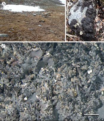
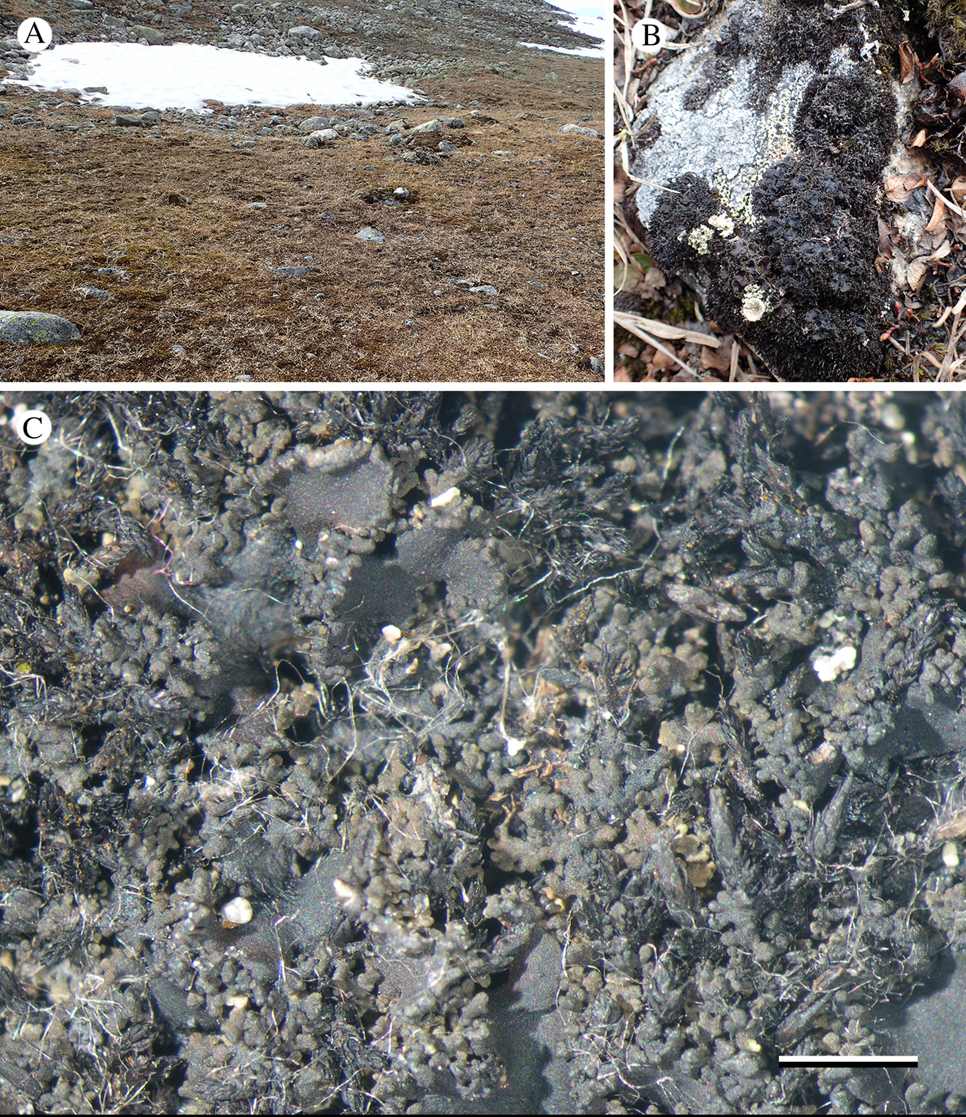
Psoroma nivale (holotype). A, habitat: Psoroma nivale occurred on a low boulder just below the centre of the snow patch; B, habit; C, thallus with apothecia. Scale = 2 cm. In colour online.
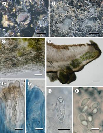
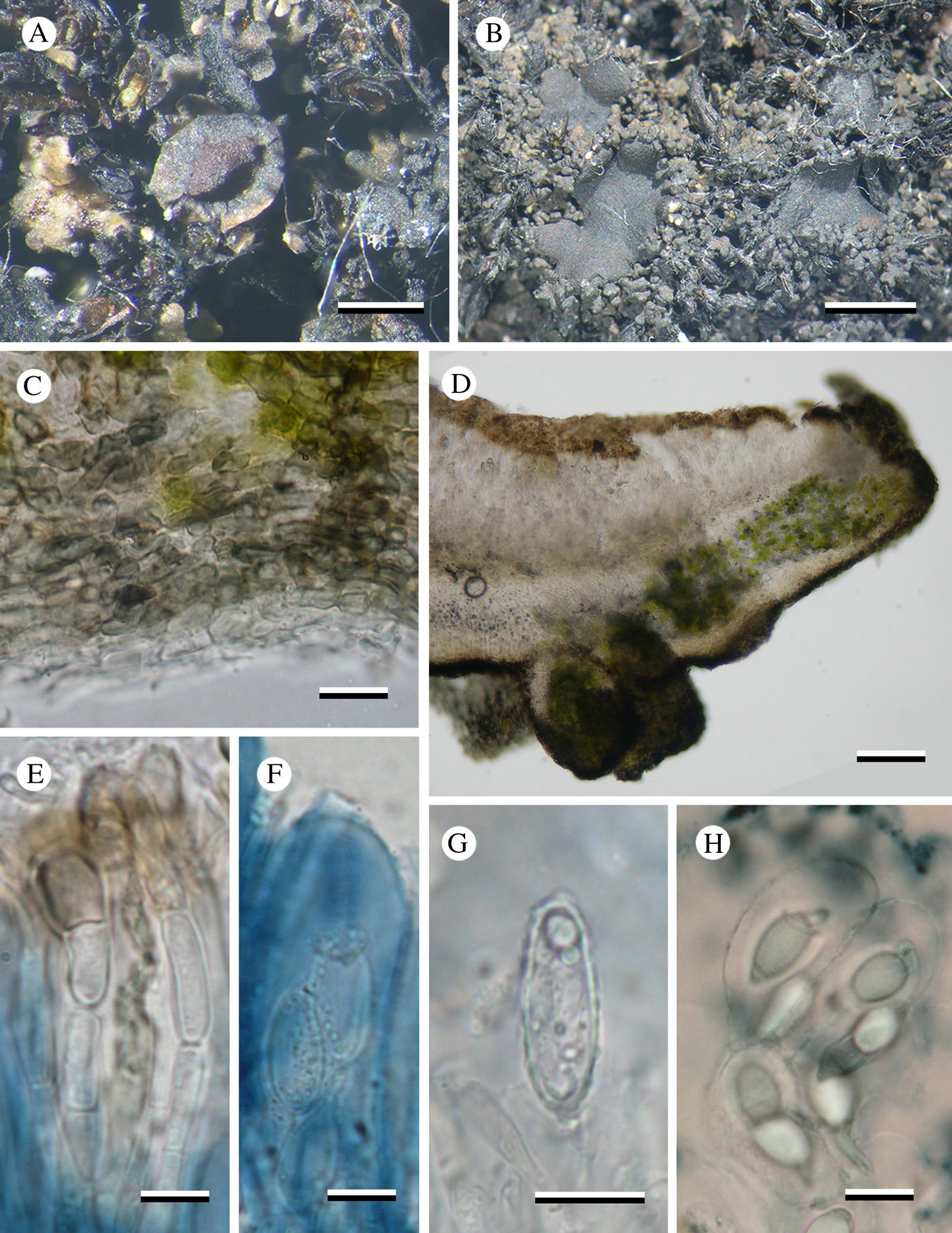
Psoroma nivale (holotype). A, immature apothecium; B, mature apothecia; C, lower cortex; D, section through mature apothecium; E, paraphyses; F, ascus in IKI; G, ascospore in water; H, ascospores in 70% nitric acid. Scales: A = 0·5 mm; B = 2·0 mm; C & F–H = 10 µm; D = 100 µm; E = 5 µm. In colour online.
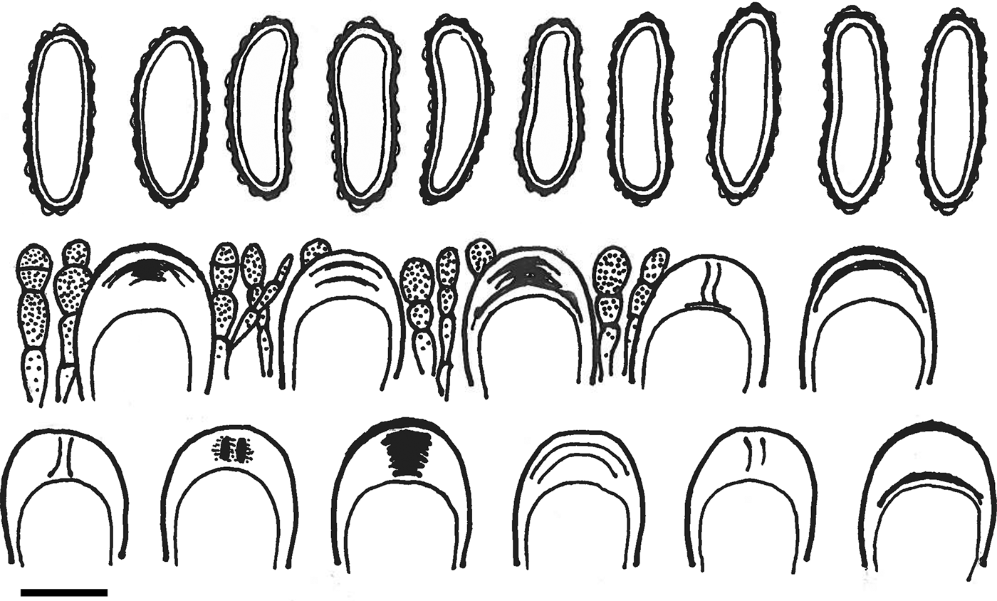
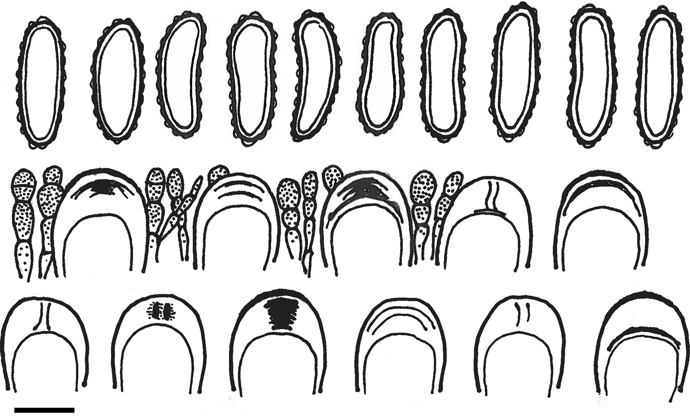
Ascospores, asci (showing the variation observed in IKI+ structures) and paraphyses of Psoroma nivale. Scale = 10 μm.
Apothecia common, lecanorine, sessile with a narrow base, arising from the upper surface of the squamules; initially flat, 0·4–0·8 mm diam., but up to 3 mm across with an irregular, often lobed outline and undulating surface when mature, covering much of the surface of the central part of the thallus; disc red when young becoming black when mature and turning dark red-brown when wet; thalline margin thick, raised, 0·10–0·15 mm wide, entire with shallow radial fissures when young, composed of proliferating ± digiform to coralloid, slightly flattened squamules similar to those of the thallus when mature. Exciple 150–175 µm wide, cupular and finely white-tomentose below, hyaline, composed of radiating hyphae 4–5 µm wide, cells 5–18 µm long, algal cells similar to those in the thallus; cortical cells dark brown pigmented (N+ red). Hymenium c. 150 µm tall, IKI+ strongly blue; paraphyses 3–4 µm wide, in mid hymenium, gradually widening to 5–7 µm wide at the apex with a brown-pigmented cap that sometimes forms a sheath around the terminal cell, simple, rarely branching above, septate, constricted at the septa, sometimes almost moniliform; epihymenium dilute brown. Hypothecium c. 50 µm deep, hyaline to dilute orange-brown. Asci cylindrical c. 100–120 × 15–20 µm, apical structure variable: usually with an IKI+ blue external cap and one or more IKI+ blue ±horizontal sheets in the upper tholus, but some asci have only the external cap and others have a weak but distinct IKI+ blue tube structure; proper ascospores hyaline, narrow elongate with rounded ends, mostly bacilliform, or weakly asymmetrical, 17–21 × 5·5–8·0 µm (l/w ratio: c. 2·5–3·0); perispores of the same shape, regularly low-verrucose, without or with modest pulvinate apical extensions, 18–22 × 6·0–8·5 µm, perispore gradually degenerating in K, but swelling to 4–5 µm thick in N.
Conidiomata not observed.
Chemistry
All spot tests negative; no substances detected by TLC.
Distribution and ecology
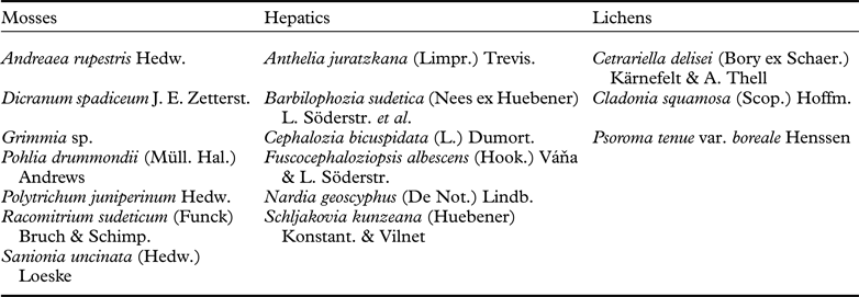
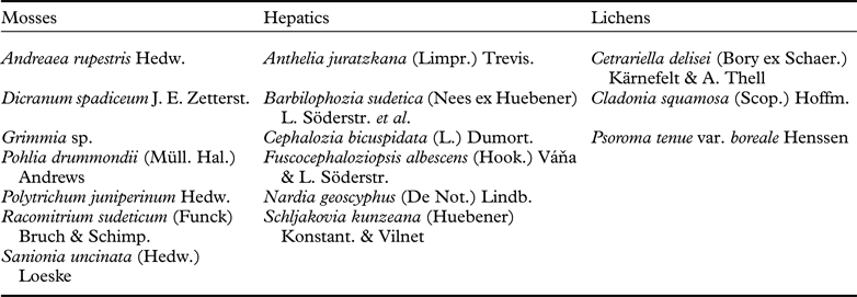
Psoroma nivale is known only from the type collection where it grew over Andreaea rupestris Hedw. on the upper surface of a low acidic boulder associated with glacial till at the lower edge of an area of late snow-lie (slope 15%; aspect 70°). Also present on the same boulder were Cladonia sp., Micarea incrassata Hedl., Peltigera sp. and Rhizocarpon sp. Other cryptogams recorded from the vicinity are listed in Table 1. The surrounding vegetation consisted of a dwarf shrub community dominated by Salix herbacea L. with Carex bigelowii Torr. ex Schwein. The Keglo Bay site is situated within the Arctic shrub tundra domain (Elvebakk et. al. Reference Elvebakk, Elven, Razzhivin, Nordal and Razzhivin1999; Ministère des Forêts, de la Faune et des Parcs 2019).
Cryptogams recorded from the vicinity of Psoroma nivale at Keglo Bay, Canada.
Remarks
The perispore of the ascospores of the new species expands dramatically in N, from c. 1 µm to 4–5 µm. We could find no mention of this phenomenon in the literature and so were unsure whether it was restricted to our new species or was common in other species of the genus, or even family. Consequently, we tested specimens of several further species of Psoroma and other Pannariaceae genera from the MSC herbarium (see ‘Additional collections examined’ above) and discovered that the perispore expanded in N, in all the studied species of Psoroma and Pannaria with a warted perispore, but not in the smooth-spored Fuscopannaria pacifica.
Systematic position
Initially, we thought that our collection might represent an extreme environmental modification of Psoroma hypnorum, but we soon dismissed this idea because of the characteristic elongate ascospores lacking large apical extensions (Figs 3G & 4), as well as the very thick paraphyses (4–7 µm; Fig. 3E), both of which are very different from those of P. hypnorum. The amyloid reaction of the ascus apex was also confusing. The asci of P. hypnorum have a well-defined IKI+ blue tube structure (Fig. 4C in Elvebakk et al. 2010), whereas the ascus structure of our new species appears variable. Most asci had an IKI+ blue external cap and one or more IKI+ blue ±horizontal sheets in the upper tholus (Fig. 4), whereas weak but distinct tube structures were also rarely observed (Figs 3F & 4). This is possibly due to differing degrees of maturity of the asci, with immature asci having only the external cap that later develops the internal horizontal sheets, with the tube-like structure developing only in fully mature asci. A similar process has been documented in the asci of Xenolecia Hertel, with immature asci having an internal cap (Lecidea-type) that develops a tube structure (Porpidia-type) when mature (Fryday & Thüs Reference Fryday and Thüs2017).
At first, the ascus characters of our new species appeared to exclude from consideration not only P. hypnorum but also the genus Psoroma in general. The shape and colour of the thallus squamules of our new species resemble those of a species of Fuscopannaria P. M. Jørg., and the apothecia have squamulose margins that are sunken below the squamules as in some species in that genus. Therefore, our next thought was that it might be a species of Fuscopannaria with a chlorococcoid photobiont. Although the photobiont in Fuscopannaria is usually a cyanobacterium, one species, Fuscopannaria viridescens P. M. Jørg. & Zhurb., does have a green chlorococcoid photobiont and occurs in the North American Arctic. However, that species has a fawn brown thallus with expanded marginal lobes (Jørgensen & Zhurbenko Reference Jørgensen and Zhurbenko2002; Nelson & Wheeler Reference Nelson and Wheeler2013) and because the ascospores of Fuscopannaria species have a smooth surface (James & Jørgensen Reference James, Jørgensen, Smith, Aptroot, Coppins, Fletcher, Gilbert, James and Wolseley2009) and their hymenia are IKI+ reddish brown, we excluded that genus from consideration for our species.
Based on the amyloid reaction of the ascus structure and the ascospores with a warted perispore, we next considered whether our collection could be accommodated in Protopannaria P. M. Jørg. However, all described species of Protopannaria have a cyanobacterium as photobiont and comparison with the type species, P. pezizoides (Weber) P. M. Jørg. & S. Ekman, revealed that our species had very little else in common with that genus. Protopannaria pezizoides differed in having a completely different thallus structure and apothecial anatomy with an adglutinated epihymenium of thin paraphyses, and asci with very distinct, external IKI+ blue apical caps. Some internal sheets were observed, but no tube structures. We also looked at several of the Southern Hemisphere species referred to Protopannaria, which differed widely from both P. pezizoides and each other. For example, Protopannaria hilaris (Zahlbr.) P. M. Jørg., described from the Juan Fernández Islands (Chile) had ascus IKI+ reactions different from those of P. pezizoides. The external caps were not so dominant, internal sheet-like structures were well developed, but many cases of true tube structures were also observed. However, unlike our new species, its ascospores were small and short-ellipsoid. Protopannaria, as presently understood, includes seven species but it appears to be heterogeneous and definitely in need of further study.
This brought us back to our original opinion, that the new species could only be accommodated in a widely circumscribed Psoroma as depicted by Ekman et al. (2014; clade 2c). However, as the paraphyses are different and the IKI+ tube structures less pronounced compared to those of P. hypnorum (the type species), we checked the phylogeny of Ekman et al. (Reference Ekman, Wedin, Lindblom and Jørgensen2014) for other taxa in their ‘clade 2c’ that might be congeneric with our species. Among these, Psorophorus species and Psoroma tenue have asci with well-developed IKI+ apical tube structures but no other similarities to our new species, except that P. tenue var. tenue has similar paraphyses. Austroparmeliella lacerata (P. M. Jørg.) P. M. Jørg. is also very different, including having smooth ascospores, and the two Austrella species do not have IKI+ blue hymenia and were recently found to be paraphyletic (Fryday et al. Reference Fryday, Ertz and Jørgensen2017). ‘Santessoniella’ arctophila is not a very useful reference for defining generic positions with the Pannariaceae as the type species of Santessoniella, S. polychidioides (Zahlbr.) Henssen, was recovered within the core Psoroma clade by Ekman et al. (Reference Ekman, Wedin, Lindblom and Jørgensen2014), leaving S. arctophila as an orphaned taxon. Our studied material of ‘S.’ arctophila may be heterogeneous; the specimen Elvebakk 11:021 has asci with a combination of IKI+ sheets and true tubes but the smooth ascospores are devoid of visible perispore structures, whereas Fryday 10221 and the type collection in UPS have ascospores with a warted perispore.
Within Psoroma, our new species is distinguished primarily by its unique ascospore shape (Figs 3G & 4), a character that is unknown in any previously described species of the genus: ascospores of Psoroma species are predominantly ellipsoid, with some ovoid and others weakly or strongly citriform. We have searched for Psoroma species with a similar morphology with regard to other characters, particularly in the Southern Hemisphere where the genus definitively has its evolutionary origin. Psoroma angustisectum Zahlbr., a rare species described from the Juan Fernandez Islands and also reported from southern mainland Chile and New Zealand, has long and dissected squamules but they have the brown melanin colour of P. hypnorum and its ascospores have been described as much larger (25–35(–38) × 10–15 µm) (Galloway Reference Galloway2007).
Most Psoroma species share the reddish brown hues of P. hypnorum or may even be cinnamon-coloured like P. tenue var. boreale. However, the species matching the colour of P. nivale most closely may be the panaustral species P. buchananii. It has a dark, blackish brown melanin colour, which can take on a more mustard yellow colour, as in the holotype of P. nivale, probably in shaded parts of the thallus. However, the squamules of P. buchananii are much less branched, it has conspicuous black pycnidia, and the ascospores are short-ellipsoid to subglobose with rather scattered, wide gibbae instead of regularly set verrucae.
We conclude that there are no known species from the Southern Hemisphere (or elsewhere) that resemble P. nivale in the combination of melanin colours and anatomical and macroscopic characters.
The new species will be included in extensive phylogenetic studies that are needed, both to challenge the present paraphyletic position of Psoroma shown by both Ekman et al. (Reference Ekman, Wedin, Lindblom and Jørgensen2014) and Park et al. (Reference Park, Hong and Elvebakk2018), and to define its nearest neighbouring genera. It is also our hope that the present paper can stimulate further searches in the field for this species, so far known only from its holotype specimen.
Key to species of Pannariaceae and similar families (Koerberiaceae, Massalongiaceae, Vahliellaceae) reported from arctic regions of North America
Geographically, the Arctic is the area of the earth north of the Arctic Circle but, biologically, this is of little relevance because it makes no allowance for altitude or continental position, the other main drivers of climatic conditions and the resulting vegetation. Elvebakk (Reference Elvebakk, Nordal and Razzhivin1999) proposed a bioclimatic delimitation of the ‘Arctic’ based on the presence of arctic vegetation that corresponds to that area north of the tree line. This has been refined by the CAVM Team (Walker et al. Reference Walker, Raynolds, Daniëls, Einarsson, Elvebakk, Gould, Katenin, Kholod, Markon and Melnikov2005) who have produced detailed maps of Arctic vegetation. The dominant vegetation of the Keglo Bay area is low shrub tundra, which is characteristic of the ‘Low Arctic’.
1 Primary photobiont a green chlorococcoid alga …………2
Primary photobiont a cyanobacterium …………6
2(1) Thallus placodioid, fawn brown with wide radiating marginal lobes
…………Fuscopannaria viridescens P. M. Jørg. & Zhurb.
Thallus composed of small squamules without a well-defined margin …………3
3(2) Apothecia lacking a thalline margin…………
………… Vahliella globigera (Fryday & P. M. Jørg.) P. M. Jørg.
Apothecia with a well-developed thalline margin …………4
4(3) Squamules elongate, much-branched, dark brownish black, ascospores bacilliform
………… Psoroma nivale Fryday et al.
Squamules rounded to granular, not branched, green to chestnut brown, ascospores wide ellipsoid to citriform …………5
5(4) Thallus of greenish brown to brown squamules; apothecial margin squamulose. Thallus lacking lichen substances …………Psoroma hypnorum (Vahl) Gray
Thallus of reddish brown, small and glossy granules; apothecial margin granular. Thallus containing porphyrilic acid methyl ester and pannaric acid………………
………… Psoroma tenue var. boreale Henssen
6(1) Thallus with soredia, sorediose isidia, isidia or coralloid granules …………7
Thallus lacking soredia, sorediose isidia and isidia, usually fertile …………11
7(6) Thallus with true isidia or coralloid granules …………8
Thallus with soralia or sorediose isidia (gymnidia) …………10
8(7) Squamules on a blue-black hypothallus, on rocks or trees; ascospores simple…………
………… Parmeliella triptophylla (Ach.) Müll. Arg.
Hypothallus absent …………9
9(8) Thallus of flat squamules, not adpressed to the surface; usually among bryophytes on rocks or on the ground; ascospores 1-septate…………
………… Massalongia carnosa (Dicks.) Körb.
Thallus of narrow radiating lobes, directly on rock; ascospores simple …………
………… Tingiopsidium isidiatum (Degel.) Hafellner & T. Sprib.
10(7) Thallus with enlarged, radiating marginal lobes; P– (fatty acids and terpenoids) …………
………… Fuscopannaria ahlneri (P. M. Jørg.) P. M. Jørg.
Thallus lacking enlarged, radiating marginal lobes; P+ red (pannarin)…………Pannaria conoplea (Ach.) Bory
11(6) Thallus composed of brown, ascending, frosted lobes; apothecia rare …………
………… Fuscopannaria praetermissa (Nyl.) P. M. Jørg.
Thallus otherwise; apothecia usually present …………12
12(11) Thallus subfruticose, composed of ascending greenish black lobes; apothecia greenish black …………Leciophysma finmarkicum Th. Fr.
Thallus crustose, squamulose or of radiating lobes …………13
13(12) Thallus ±crustose, in small patches; apothecia lacking a thalline margin …………14
Thallus squamulose or of radiating lobes; apothecia with a thalline margin …………15
14(13) Thallus olivaceous; apothecia dark brown, ascospores smooth …………
………… Leciophysma furfurascens (Nyl.) Gyeln.
Thallus brown; apothecia red-brown, ascospores with a warted perispore …………
………… ‘Santessoniella’ arctophila
15(13) Apothecia red-brown …………
…………Protopannaria pezizoides (Weber) P. M. Jørg. & S. Ekman
Apothecia black …………16
16(15) Thallus of narrow, fawn brown radiating lobes …………
………… Tingiopsidium elaeinum (Wahlenb.) Hafellner & T. Sprib.
Thallus squamulose or subfruticose 17
17(16) Thallus grey-white, placodioid to squamulose on a black hypothallus …………
………… Pannaria hookeri (Borrer) Nyl.
Thallus forming small, greenish brown cushions …………
………… Vahliella hookerioides (P. M. Jørg.) P. M. Jørg.
We thank the Kativik Regional Government for organizing and conducting botanical inventories in the Keglo Bay area on the east shore of Ungava Bay, Nunavik Québec, Canada, and Måns Svensson (UPS) for checking the ascospores of the type collection of Pannaria arctophila. Two specimens used in this study (Fryday 9715 & 9900) were collected from Glacier Bay NP as part of NPS project : R9815110518.