Introduction
There are many factors that determine the resultant development, viability and health of the preimplantation human embryo conceived through IVF. Several of these factors are external and therefore beyond the control of the IVF laboratory itself. These factors include patient age, genetics, aetiology of infertility, diet, smoking/drug habits and ovarian stimulation.Reference Gardner and Lane 1 With regards to ovarian stimulation, this approach overrides the dominant follicle, thereby creating a heterogeneous pool of oocytes, which increases the variability of gamete quality. Furthermore, the use of exogenous gonadotropins has been implicated in the induction of epigenetic alterations in the resultant oocytes and embryos,Reference Fortier, McGraw and Lopes 2 , Reference Huffman, Pak and Rivera 3 thereby contributing further variability and stress to the system, hence making it difficult to determine the precise effects of specific laboratory conditions.Reference Santos, Kuijk and Macklon 4
Within the IVF laboratory there reside numerous factors which are more controllable, and which can also impact subsequent development, viability and health of the embryo and resultant offspring. Such factors include media components and formulations, protein supplements, gas phase, embryo density, plasticware and oil overlay.Reference Wale and Gardner 5 To make this situation more complicated, many factors (both external and those within the laboratory) interact with each other, and once a gamete or embryo has been exposed to a specific stress, it is common for them to be more susceptible to a second, or even third, stress. Consequently, it is important to consider clinical IVF data with respect to external factors for a particular group of patients such as those with diabetes and/or who are obese, as their gametes are already significantly compromised before IVF, resulting in an altered embryo phenotype.Reference Finger, Harvey, Green and Gardner 6 , Reference Lane, Robker and Robertson 7 The term phenotype is not restricted to the morphological appearance of the embryo, but rather includes its physiological status, a key determinant of health and viability.
To make matters more convoluted, there are stage- and sex-specific sensitivities with regards to the impact of different stresses.Reference Wale and Gardner 5 , Reference Gardner and Lane 8 , Reference Gardner, Larman and Thouas 9 For example, the early stages of development from fertilization to compaction, are far more susceptible to stresses in their environment than the morula and blastocyst, attributed to both the transition from maternal to embryonic genetic control, and to the lack of a transporting epithelium and regulatory transport systems which are created at compaction.Reference Gardner and Lane 8 , Reference Lane and Gardner 10 Trauma manifest at the blastocyst stage is typically a result of a response to a stress during the cleavage stages, and embryos do not recover from early stress, even when this stress is relieved post-compaction.Reference Wale and Gardner 11 With regards to male and female embryos there exists a window period during the preimplantation period where both X-chromosomes are active in the female, resulting in an altered transcriptome, proteome and metabolism.Reference Gardner, Larman and Thouas 9 , Reference Picton, Elder and Houghton 12 – Reference Garcia-Herreros, Aparicio, Rath, Fair and Lonergan 16 Consequently, female human embryos respond differently to the in vitro environment than males, which can be manifest as altered growth kinetics and morphological score,Reference Alfarawati, Fragouli and Colls 17 , Reference Bodri, Kawachiya and Sugimoto 18 and in animals changes to culture conditions can result in altered sex ratio, secretome, transcriptome, apoptosis, and postimplantation viability and phenotype.Reference Bredbacka and Bredbacka 19 – Reference Siqueira and Hansen 22 Male and female human embryos could therefore be differentially vulnerable to certain stresses.Reference Kimura, Spate, Green and Roberts 23 – Reference Gardner and Kelley 25 Hence the multiple interactions between gamete quality, embryo sex, stage of development and other unknown factors can make it extremely challenging to understand the aetiology of any impaired embryo function in human IVF.
As the number of children conceived through IVF increases steadily each year, with over five million children born to date, more longitudinal studies are being undertaken to assess the health of ‘test-tube babies’. Although the birth weight of children born through IVF appears to have changed little over the past two decades, in spite of the introduction of several new technologies,Reference Maas, Galkina, Thornton, Penzias and Sakkas 26 there are concerns that the long-term health of children may be affected by the environment in vitro.Reference Scherrer, Rexhaj, Allemann, Sartori and Rimoldi 27 – Reference Feuer and Rinaudo 29 Therefore, here we consider the known impacts of specific components of the IVF laboratory on preimplantation embryo development, viability and health. Wherever possible, we have restricted our discussion to the preimplantation human embryo, but we have had to draw from the animal literature in a few cases. Consideration of the effects of the IVF laboratory on human prenatal and postnatal development has been considered by Chen and Heilbronn.Reference Chen and Heilbronn 30
The human IVF laboratory and the embryo culture system
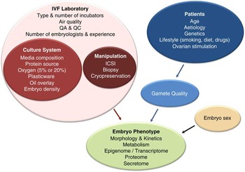
In order to support the development of a viable and healthy embryo, that is, one that is capable of both implanting and resulting in a healthy child, one needs to look far beyond the formulation of the culture media employed (although these are extremely important, as presented below). Figure 1 illustrates the specific components of an embryo culture system, and highlights how the embryo culture system is but one component of the IVF laboratory. Significantly, each of the individual components of the culture system and laboratory have the capacity to interact with each other, thereby confounding troubleshooting when poor embryo development is observed. Consequently, there is an absolute necessity for an effective quality management systemReference Mortimer 31 in order to ensure that variables such as equipment performance and changes in consumable lot numbers do not negatively impact embryo health.
Impact of the IVF laboratory environment on human embryo phenotype. The IVF laboratory is comprised of several key components, one of which is the embryo culture system, which in turn is made up from several components, only one of which is the embryo culture media. Independent of the IVF laboratory, patient age and overall health impact on the quality of gametes (both sexes), leading to an altered embryo phenotype. Furthermore, the use of exogenous gonadotropins to recruit follicles and induce ovulation has been implicated in the induction of epigenetic alterations in the resultant oocytes and embryos.Reference Fortier, McGraw and Lopes 2 , Reference Huffman, Pak and Rivera 3 Therefore, the use of hormones in human IVF may well be predisposing the resultant embryos to greater susceptibility to laboratory environment. Finally, the gender of the embryo could also determine its interactions with the components of the IVF laboratory. ICSI, intracytoplasmic sperm injection; IVF, in vitro fertilization; QC, quality control; QA, quality assurance.

Assessment of human embryo phenotype
Morphology and kinetics
How is the impact of IVF conditions on the resultant phenotype of the embryo evaluated? Since the inception of human IVF, analysis of embryo morphology has been the standard assessment.Reference Cummins, Breen and Harrison 32 – Reference Cohen, Trounson and Dawson 34 Through exhaustive analysis of key morphological traits, elegant grading systems have been developed to quantitate embryo development. These grading systems, from the pronucleate oocyte through to the blastocyst stage, reflect key features that are associated with subsequent viability post transfer.Reference Scott and Smith 35 – Reference Gardner and Balaban 39 Historically, embryo assessment was limited, requiring embryos to be removed from the incubator for analysis; hence it was only possible to obtain a snapshot of development. This approach has two inherent drawbacks: first, the removal of an embryo from the incubator for examination on an inverted microscope means that the embryo will be exposed to both pH and temperature shifts if the process takes more than just a few minutes;Reference Wale and Gardner 5 and second, not all of the key information regarding embryo development can be acquired as many events occur outside of working hours, and hence are not recorded. With the advent of time-lapse microscopy it is now possible to assess human embryo development almost as a continuum, with image acquisition taking place as frequently as every 10–15 min, while maintaining the embryo inside the incubation chamber.Reference Pribenszky, Matyas and Kovacs 40 , Reference Meseguer, Herrero and Tejera 41 Subsequently, through time-lapse analysis we are able to more accurately determine the effects of the IVF laboratory and the culture system on embryo phenotype. In addition, time-lapse allows the use of new parameters to assess embryo health that were previously impossible, such as reverse cleavage or the duration of cell cycles, and the development of algorithms to select the most viable embryos.Reference Gardner and Balaban 39 , Reference Liu, Chapple, Roberts and Matson 42 – Reference Petersen, Boel, Montag and Gardner 44
Metabolism
Through the continuous analysis of morphology over time, much can be gleaned about the development of the embryo, and indeed some aspects of physiology may be indirectly inferred by key events, e.g. the rate of blastocyst expansion as a reflection of metabolic activity. However, morphological features can only reveal so much about the physiology of the embryo,Reference Gardner and Balaban 39 and hence there have been considerable efforts to develop non-invasive means of assessing human embryo viability. One such approach has been the sampling of the surrounding culture medium for nutrient uptake and metabolite release into the medium, thereby creating a metabolic profile during development.Reference Leese and Barton 45 – Reference Houghton, Hawkhead and Humpherson 48 The significance of this approach has been realized in animal models, where the quantification of nutrient uptake and subsequent metabolism has been related to viability.Reference Renard, Philippon and Menezo 49 – Reference Lane and Gardner 53 It is now established that not only is the utilization of specific nutrients (including oxygen) related to the development of the human embryo in vitro,Reference Hardy, Hooper and Handyside 47 , Reference Houghton, Hawkhead and Humpherson 48 , Reference Gardner, Lane, Stevens and Schoolcraft 54 it is also related to pregnancy and live birth.Reference Gardner, Wale, Collins and Lane 13 , Reference Tejera, Herrero and Viloria 55 Hence alterations to the metabolic profile of the preimplantation human embryo can have significant consequence for its developmental potential, and therefore represents an important parameter to assess phenotype. Furthermore, it has recently been determined that metabolic functions, specifically the levels of key metabolites and co-factors such as nicotimamide adenine dinucleotide (NAD+), acetyl-CoA and lactate, are involved in signalling and can regulate cell function at the epigenetic level.Reference Gardner 56 – Reference Harvey, Rathjen and Gardner 58 Consequently, metabolism has taken on further significance in the analysis of the human embryo phenotype.
Secretome
In addition to metabolites, the proteins which the embryo releases into the surrounding medium (the secretome) can also somewhat reflect developmental ability.Reference Dominguez, Gadea and Mercader 59 , Reference Katz-Jaffe, Schoolcraft and Gardner 60 The earliest studies on the human embryo secretome revealed that conditioned media contained bioactive immunoregulatory molecules, and that this activity may be associated with implantation success.Reference Jones, Warnock, Urry, Edwin and Mitchell 61 – Reference Cocchiara, Di Trapani and Azzolina 64 One of these molecules was identified as PAF (1-o-alkyl-2-acetyl-sn-glycero-3-phosphocholine), which is one of the most well-characterized embryo-secreted autotrophic molecules, but is not used clinically as either a biomarker or a culture media additive.Reference O’Neill 65 Another immunologically active molecule that has been extensively investigated is soluble human leukocyte antigen G (sHLA-G), after early studies found that the presence of sHLA-G in conditioned media was predictive of clinical pregnancy.Reference Fuzzi, Rizzo and Criscuoli 66 – Reference Noci, Fuzzi and Rizzo 68 Despite some conflicting reports, the usefulness of sHLA-G as a biomarker was supported by the results of a multicentre retrospective studyReference Kotze, Kruger and Lombard 69 and a meta-analysis,Reference Vercammen, Verloes, Van de Velde and Haentjens 70 although it is not currently used routinely in IVF clinics.
Cytokines and growth factors have also been detected in human embryo culture media, such as transforming growth factor β, interleukins (IL-1α and IL-1β) and insulin-like growth factor 2 (IGF-2).Reference Thouas, Dominguez and Green 71 IL-1α and IL-1β have shown potential as viability markers,Reference Sheth, Roca and al-Sedairy 72 , Reference Sequeira, Espejel-Nunez, Vega-Hernandez, Molina-Hernandez and Grether-Gonzalez 73 but evidence is so far limited to a handful of studies.
More recently, proteomics methods have been employed to discover potential biomarkers, although there is little consistency in the results to date, which may be due to the use of a wide range of culture conditions, study designs and proteomics platforms. The first proteomics study on conditioned media found different protein profiles from blastocysts that implanted or did not, and ubiquitin was suggested as a candidate biomarker.Reference Katz-Jaffe, Gardner and Schoolcraft 74 Since this study, lipocalin-1,Reference McReynolds, Vanderlinden and Stevens 75 apolipoprotein A-1,Reference Nyalwidhe, Burch and Bocca 76 IL-6, stem cell factor,Reference Dominguez, Meseguer and Aparicio-Ruiz 77 IL-8,Reference Huang, Zhou and Wei 78 haptoglobin α-1Reference Montsko, Zrinyi and Janaky 79 and multiple other peptidesReference Cortezzi, Garcia and Ferreira 80 have been reported to correlate with embryo viability, but these potential biomarkers have not yet been validated in a large clinical trial.
In the last 3 years, there has been growing interest in embryo-secreted nucleic acids. Several studies have performed arrays for microRNAs to identify biomarkers; however, like the proteomics studies, there has been little consistency between study results.Reference Kropp, Salih and Khatib 81 – Reference Capalbo, Ubaldi and Cimadomo 84 Other nucleic acids such as genomic and mitochondrial DNA have also been suggested as indicators of embryo viabilityReference Stigliani, Anserini, Venturini and Scaruffi 85 or as a method of non-invasive embryo genotyping.Reference Galluzzi, Palini and Stefani 86 – Reference Xu, Fang and Chen 88 Although these new methods hold potential for clinical application, they have yet to be validated.
Combined technologies
Currently, time-lapse technology is being used in combination with analysis of metabolism, gene expression and the secretome in analysing the developmental potential and viability of mammalian embryos, including the human.Reference Dominguez, Meseguer and Aparicio-Ruiz 77 , Reference Lee, Thouas and Gardner 89 – Reference Milazzotto, Goissis and Chitwood 91 These important works are shedding light on how events during the first few cell cycles are manifest as physiological differences in the resultant blastocysts, such that embryos with different cleavage times result in blastocysts of different physiologies.
Transcriptome, proteome and karyotype
Beyond the non-invasive quantification of morphology, and the analysis of culture media, the technologies to assess either the embryonic transcriptome or proteome are invasive, requiring extraction of the tissue for subsequent analysis. Such approaches on donated human embryos have provided limited, but valuable, information and insight on how IVF conditions affect embryonic development. However, analysing embryonic tissue by taking samples for preimplantation genetic diagnosis (typically performed by removing several cells of the trophectoderm of the blastocyst),Reference Fragouli, Lenzi and Ross 92 is providing a means to determine how the IVF laboratory can impact the human transcriptome and chromosome stability. Using such an approach it has been documented that rates of chromosomal abnormalities are different between clinical laboratories, inferring that laboratory environment and/or hormonal stimulation affect the incidence of mosaicism.Reference Munne, Magli and Adler 93
Impact of the embryo culture system
Culture media
Perhaps of all components in the IVF laboratory, culture media have received the most attention with regards to their impact on embryo phenotype. Extensive data from animal models have clearly shown an effect of medium composition on the kinetics of embryo development and physiology.Reference Gardner and Lane 1 , Reference Bavister 94 – Reference Leese, Donnay and Thompson 96 The composition of current culture media used in human IVF are based around balanced salt solutions, a sodium bicarbonate/carbon dioxide buffer system, a protein source currently in the form of serum albumin (formerly in the form of serum), amino acids, vitamins and in some cases growth factors.Reference Bavister 94 , Reference Gardner 97 , Reference Gardner and Lane 98
Utilizing time-lapse microscopy, two studies have considered the effects of culture media on human embryo development. Basile et al.Reference Basile, Morbeck, Garcia-Velasco, Bronet and Meseguer 99 compared two commercial media in an randomized controlled trial on sibling oocytes and did not find an effect of medium formulation on any parameter up to day 3 of development. In contrast, in another study on sibling oocytes, Ciray et al.Reference Ciray, Aksoy, Goktas, Ozturk and Bahceci 100 did report an effect of culture medium on morphokinetics during the cleavage stages. Of note, this latter study employed two different media from the same company, thereby eliminating most variables other than the media formulations themselves. Of further significance, a recent study has reported that the culture conditions, that is medium formulation and/or pH, are associated with differences in the rate of human embryo mitotic aneuploidy.Reference Hickman, Wells and Lavery 101 What is known about the effects of individual medium components on embryo phenotype is considered below.
Amino acids and ammonium
Amino acids were once perceived simply as a nitrogen source for the embryo. However, amino acids are now known to be amongst the most important regulators of several intracellular functions during the preimplantation period, being involved in internal pH (pHi)Reference Edwards, Williams and Gardner 102 and osmotic regulation,Reference Lane 103 , Reference Dawson and Baltz 104 regulation of carbohydrate metabolismReference Gardner and Sakkas 51 , Reference Lane and Gardner 53 , Reference Lane and Gardner 105 and signalling.Reference Martin and Sutherland 106 , Reference Tan, Rathjen, Harvey, Gardner and Rathjen 107 Consequently, if amino acids are absent from the culture medium, for example human tubal fluid medium (a simple salt solution with three carbohydrates), the physiology and phenotype of the embryo will be significantly compromised.Reference Bavister 94 , Reference Gardner 108 , Reference Lane and Gardner 109 Devreker et al. showed that the inclusion of those amino acids known to stimulate mouse embryo development in vitro Reference Gardner and Lane 110 also improved human embryo development in vitro, resulting in a significant increase in blastocyst cell number.Reference Devreker, Hardy and Van den Bergh 111 Today, almost all culture media used for human IVF contain amino acids. Those media that do not contain amino acids will undoubtedly compromise the human embryo phenotype.
Although amino acids are required to maintain the health of the embryo in culture, a problem is created due to the fact that one is working in vitro and not within the female reproductive tract; when present in any medium and incubated at 37°C amino acids spontaneously deaminate and release ammonium, glutamine being the most labile.Reference Gardner and Lane 110 , Reference Heeneman, Deutz and Buurman 112 The levels of ammonium in a culture medium are further increased by the metabolism of amino acids by the embryos themselves.Reference Gardner, Lane, Stevens and Schoolcraft 54 , Reference Gardner and Lane 110 In animal models this accumulation of ammonium has been documented to inhibit embryo development,Reference Gardner and Lane 110 , Reference Gardner, Lane, Spitzer and Batt 113 alter metabolism,Reference Lane and Gardner 114 – Reference Lane and Gardner 116 gene expression,Reference Lane and Gardner 116 , Reference Gardner, Hamilton, McCallie, Schoolcraft and Katz-Jaffe 117 and ultimately compromises fetal development post transfer, and in some cases induces the neural tube defect exencephaly.Reference Lane and Gardner 118 The incidence of neural tube birth defects in mice as a result of ammonium exposure was initially found to be dose dependent.Reference Lane and Gardner 118 The incidence of this birth defect after exposure to ammonium accumulation in vitro was subsequently reported by one groupReference Sinawat, Hsaio and Flockhart 119 but not by another.Reference Biggers, McGinnis and Summers 120 The solution to this apparent paradox lies with the oxygen concentration used; at physiological oxygen concentration the embryo appears able to cope with ammonium,Reference Wale and Gardner 121 whereas at 20% oxygen the embryo cannot regulate its homoeostasis in the presence of an ammonium stress and exencephaly ensues. This is an example of how one stress can reduce the ability of the embryo to deal with a second trauma, something that is revisited below with regards to how oxygen predisposes embryos cultured individually to delayed growth.
Clinically, Virant-Klun et al.Reference Virant-Klun, Tomazevic, Vrtacnik-Bokal, Vogler, Krsnik and Meden-Vrtovec 122 determined that for human embryos, ammonium concentration in the medium was negatively correlated with human blastocyst formation. In a detailed analysis on the effects of ammonium on human embryo development, metabolism and transcription, it was determined that ammonium present in the culture medium as low as 150 μm delayed cleavage and was associated with a significant decrease in the uptake of pyruvate, the main nutrient of the cleavage-stage human embryo. Transcription analysis of the resultant human blastocysts revealed that ammonium significantly decreased the expression of 205 genes and increased the transcription of a further 185 genes. The affected genes were found to be associated with processes including metabolism, cell growth and/or maintenance, transcription, transport, cell communication and development. Given that a chronic exposure of the preimplantation mouse embryo to ammonium can result in neural tube defects,Reference Lane and Gardner 118 the observed changes in human embryo transcriptome are perhaps not too surprising, and highlight the significance of ensuring ammonium is not allowed to accumulate in a human embryo culture system.
Ammonium build up can be avoided by renewing or changing the culture media after a period of time, but this requires removing the embryos from the stable incubator environment and exposing them to fluctuations in temperature and pH. Balancing ammonium toxicity with the maintenance of a stable environment created through time-lapse microscopy is therefore an extremely important matter.Reference Hardarson, Bungum and Conaghan 123 As time-lapse and continuous culture are being adopted clinically, care must be taken to limit the presence of increasing ammonium in an embryo’s environment, and methods of toxicity alleviation such as partial medium renewal, or the addition of a second step medium to dilute out the ammonium, are being evaluated.
Carbohydrates
Within the fluids of the human female reproductive tract there exist gradients of carbohydrates. The oviduct is characterized by relatively high levels of the carboxylic acids pyruvate and lactate (0.32 and 10.5 mm, respectively), and low levels of glucose (0.5 mm). In contrast, uterine fluid contains much higher levels of glucose (3.15 mm), whereas the levels of pyruvate and lactate are much lower (0.1 and 5.87 mm, respectively).Reference Gardner, Lane, Calderon and Leeton 124 These changing levels of carbohydrates align with the dynamics of preimplantation embryo physiology, whereby cleavage stage embryos utilize carboxylic acids preferentially and are unable to utilize glucose as the sole substrate, followed by a switch in metabolic state around compaction, whereby glucose becomes the preferred and predominant nutrient of the blastocyst.Reference Biggers, Whittingham and Donahue 125 , Reference Gardner and Wale 126 Significantly, the levels of nutrients in a culture system are capable of changing the relative activities of specific metabolic pathways.Reference Gardner and Leese 127 , Reference Gardner 128 It is known from mouse embryo transfer studies that a lactate gradient impacts viability; with cleavage-stage embryos transferred on day 3 having a higher viability when cultured in a high lactate environment, whereas post-compaction embryo viability was higher when the lactate concentration was decreased.Reference Gardner and Sakkas 51 This effect on embryo function and health is plausibly modulated through the control of cytosolic redox at successive stages of development,Reference Lane and Gardner 129 as the relative ratio of pyruvate:lactate can affect the levels of intracellular NAD+:NADH.Reference Gardner and Harvey 57 NAD+ is known to be a key determinant of several intracellular functions, not the least of which is its regulation of several key metabolic enzyme activities.Reference Nikiforov, Kulikova and Ziegler 130 Furthermore, NAD+ levels can impact the epigenetic state of a cell though its regulation of NAD-dependent deacetylases (the sirtuins or SIRTs),Reference Ryall, Dell’Orso and Derfoul 131 , Reference Imai and Guarente 132 and its rate-limiting effect on adenosine diphosphate (ADP) ribosylation.Reference Quenet, El Ramy, Schreiber and Dantzer 133 , Reference Canto, Sauve and Bai 134
So do these gradients of carbohydrates within the female reproductive tract affect the preimplantation human embryo phenotype in vitro? It appears that the human embryo, similar to other mammalian species, cannot survive solely on glucose for the first 48 h of life, but rather utilizes pyruvate.Reference Hardy, Hooper and Handyside 47 , Reference Conaghan, Handyside, Winston and Leese 135 Furthermore, glucose present at a concentration of 1 mm (in the absence of amino acids) impaired the development of the cleavage-stage human embryo.Reference Conaghan, Handyside, Winston and Leese 135 However, there is a paucity of data regarding the effects of carbohydrates on human embryo development and physiology. Given the capacity of NAD+ levels to regulate the epigenomeReference Harvey, Rathjen and Gardner 58 and that NAD+ can be regulated by the level of lactate in the surroundings, studies are warranted on the role of nutrient gradients (particularly lactate) in regulating the human embryo phenotype.
Vitamins
Vitamins are key regulators of metabolic function and have been shown in the mouse model to regulate blastocyst metabolism and promote viability post transfer,Reference Lane and Gardner 53 and hence their inclusion in clinical IVF media. Given their role in one carbon metabolism,Reference Xu and Sinclair 136 further research on the human embryo’s requirement for vitamins warrants further evaluation.
Serum/albumin
Of all factors studied to date, it is the protein source which can have the most dramatic impact on embryo phenotype. The early years of human IVF were characterized by the presence of serum in culture media.Reference Gardner 97 , Reference Gardner and Lane 98 The most commonly used protein source in human IVF was patient’s serum, at a concentration of 5–20%. In some clinics fetal cord serum was used in preference.Reference Meldrum, Chetkowski and Steingold 137 Whole serum is not a pure protein preparation and contains many factors in varying amounts such as hormones, vitamins and fatty acids, as well as chelated metal ions and pyrogens.Reference Barnes and Sato 138 , Reference Barnes and Sato 139 The concentration of macromolecules and other components varies between patients and within the menstrual cycle, thereby exposing human embryos to diverse and uncontrolled environments. Furthermore, serum from several groups of patients such as those with endometriosis, polycystic ovary syndrome or unexplained infertility appears to be embryo toxic.Reference Damewood, Hesla and Schlaff 140 – Reference Deaton, Dempsey and Miller 144 From a physiological perspective, the mammalian embryo is never exposed to serum in vivo,Reference Menezo, Testart and Perrone 145 as the fluids of the female reproductive tract are not simple serum transudates,Reference Leese 146 but rather a specialized environment for the development of the embryo.Reference Gardner, Lane, Calderon and Leeton 124 It was extensive animal research however that revealed the need to eliminate serum from human embryo culture systems as it induces morphological, metabolic, epigenetic and ultrastructural changes in cultured blastocysts.Reference Gardner 97 , Reference Gardner, Lane, Spitzer and Batt 113 , Reference Khosla, Dean, Brown, Reik and Feil 147 , Reference Fernandez-Gonzalez, Moreira and Bilbao 148 The trophectoderm of blastocysts cultured in the presence of serum develops a vesicular appearance due to the sequestering of lipid in the blastomeres.Reference Thompson, Gardner, Pugh, McMillan and Tervit 149 , Reference Dorland, Gardner and Trounson 150 In Fig. 2a and 2b, the morphology of sibling sheep blastocysts, cultured with or without 20% human serum, can be seen to be extremely different, with those embryos cultured in the presence of serum exhibiting dark vacuoles, which were later shown to be lipid vesicles.Reference Thompson, Gardner, Pugh, McMillan and Tervit 149 These lipid vesicles were associated with mitochondria that exhibited abnormal folding of the cristae,Reference Thompson, Gardner, Pugh, McMillan and Tervit 149 , Reference Dorland, Gardner and Trounson 150 and loss of oxidative capacity.Reference Gardner, Lane, Spitzer and Batt 113 Of concern was that in the sheep model the inclusion of serum in the culture medium was associated with the birth of abnormally large lambs after the transfer of blastocysts to recipient ewes.Reference Thompson, Gardner, Pugh, McMillan and Tervit 149 , Reference Walker, Heard and Seamark 151 Although there have been no reports of abnormally large children born following culture with serum to the blastocyst stage, the morphology of human embryos cultured in the presence of serum does look remarkably like that of sheep embryos which have been exposed to human serumReference Bongso, Soon-Chye and Sathananthan 152 (Fig. 2c). In contrast, human blastocysts cultured in media in which the protein source is human albumin exhibit a morphology free of vesicular inclusions (Fig. 2d). Such dramatic differences in morphology plausibly reflect an accumulation of lipid by blastocysts cultured in the presence of serum, indicative of other alterations in human embryo phenotype.
Effect of protein source on blastocyst morphology. (a and b) Sibling sheep blastocyst cultured in the presence (a) or absence (b) of human serum.Reference Gardner 97 (c and d) The morphology of human blastocysts cultured in the presence (c) or absence (d) of human serum. The dark vesicles in (a) have been identified as lipid vesicles,Reference Thompson, Gardner, Pugh, McMillan and Tervit 149 resulting in compromised organelle structure and function.Reference Gardner, Lane, Spitzer and Batt 113 , Reference Dorland, Gardner and Trounson 150 (c) Taken from Bongso et al.Reference Bongso, Soon-Chye and Sathananthan 152 with permission.

Human serum albumin (HSA) has been used successfully for many years in the culture of human embryos,Reference Menezo, Testart and Perrone 145 , Reference Ashwood-Smith, Hollands and Edwards 153 – Reference Staessen, Van den Abbeel and Carle 155 and there exist several available products for serum replacement. Products include therapeutic albumin solutionsReference Ashwood-Smith, Hollands and Edwards 153 , Reference Staessen, Van den Abbeel and Carle 155 , Reference Huisman, Lo-A-Njoe and Alberda 156 and globulin-enriched albumin solutions such as plasmanate,Reference Adler, Reing, Bedford, Alikani and Cohen 157 plasmateinReference Pool and Martin 158 and synthetic serum substitute.Reference Weathersbee, Pool and Ord 159 – Reference Meintjes, Chantilis and Ward 162 However, there have been no prospective randomized trials evaluating the impact of such supplements on human embryo phenotype. Although serum albumin is a relatively pure fraction, it varies significantly with regards to fatty acids and other small moleculesReference Hanson and Ballard 163 as well as many components which are poorly characterized such as transition metals.Reference Dyrlund, Kirkegaard and Poulsen 164 , Reference Morbeck, Paczkowski and Fredrickson 165 These differences exist between both sources of serum albuminReference Batt, Gardner and Cameron 166 , Reference McKiernan and Bavister 167 and between batches from the same source.Reference Batt, Gardner and Cameron 166 , Reference Kane 168 The effects of such contaminants on human embryo phenotype have yet to be fully characterized.Reference Leonard, Charlesworth and Benson 169 New concerns with regards to the use of HSA have been raised since the revelation that serum albumin, added as the protein supplement, is the source of detectable levels of di(2-ethylhexyl)phthalate and mono(2-ethylhexyl)phthalate, as well as polybrominated diphenyl ethers in human embryo culture media.Reference Takatori, Akutsu and Kondo 170 , Reference Akutsu, Takatori, Nakazawa and Makino 171 Hence the inclusion of serum albumin in human embryo culture media warrants further reflection, and studies on alternatives such as recombinant albumin (which eliminate the problems inherent with using blood-derived products including lot-to-lot variability) would appear timely.
Incubation conditions
Oxygen
A key, and yet often overlooked, component of the embryo culture system is the gas phase employed. The level of oxygen employed varies dramatically between IVF laboratories around the world.Reference Christianson, Zhao and Shoham 172 Atmospheric oxygen is ~21%, which is significantly higher than the level of oxygen to which the human embryo is exposed to in vivo. Although oxygen levels in the human female reproductive tract have not been measured, levels in the oviduct of several mammalian species have been reported to be between 2 and 8%,Reference Fischer and Bavister 173 – Reference Ross and Graves 175 whereas the oxygen concentration in the uterus appears to be lower than in the oviduct, ranging from 5% in the hamster and rabbit to 1.5% in the rhesus monkey.Reference Fischer and Bavister 173 , Reference Maas, Storey and Mastroianni 176
The significance of differences between atmospheric and physiological oxygen levels is highlighted when one considers the effects of oxygen on mammalian embryo development. In all animal models studied it has been determined that embryo development and implantation post transfer are significantly decreased when embryos are cultured in the presence of atmospheric oxygen.Reference Batt, Gardner and Cameron 166 , Reference Quinn and Harlow 177 – Reference Thompson, Simpson, Pugh, Donnelly and Tervit 180 Indeed, exposure to atmospheric oxygen has been shown to alter embryo developmental kinetics,Reference Wale and Gardner 11 , Reference Kirkegaard, Hindkjaer and Ingerslev 181 transcription,Reference Gardner and Lane 8 , Reference Harvey, Kind, Pantaleon, Armstrong and Thompson 182 – Reference Rinaudo, Giritharan, Talbi, Dobson and Schultz 184 histone remodelling and methylation patterns,Reference Li, Goossens and Van Poucke 185 – Reference Ghosh, Coutifaris, Sapienza and Mainigi 187 the proteomeReference Katz-Jaffe, Linck, Schoolcraft and Gardner 188 and metabolic state.Reference Wale and Gardner 121 , Reference Khurana and Wales 189 , Reference Wale and Gardner 190
Analysis of human embryo development under different oxygen levels, facilitated through time-lapse, revealed that before compaction culture in atmospheric oxygen results in significantly slower embryo development compared with culture in 5% oxygen, with the timing of four to eight cells being retarded in 20% oxygen.Reference Kirkegaard, Hindkjaer and Ingerslev 181 Furthermore, the percentage of eight-cell embryos and early blastocysts was less in atmospheric oxygen, implying a damaging effect of high levels of oxygen on cleavage-stage human embryo development. These data are consistent with previous works on the human embryo cultured in atmospheric oxygen, which revealed a decrease in both blastocyst formation and resultant cell numberReference Dumoulin, Meijers and Bras 191 – Reference Kovacic and Vlaisavljevic 193 and an altered transcriptome.Reference Mantikou, Jonker and Wong 194 With regards to the long-term effects of oxygen on the preimplantation human embryo, both implantation and pregnancy rates are reduced following embryo culture in atmospheric oxygen,Reference Catt and Henman 195 , Reference Meintjes, Chantilis and Douglas 196 culminating in a decrease in the live birth rate.Reference Meintjes, Chantilis and Douglas 196 , Reference Gomes Sobrinho, Oliveira and Petersen 197 Furthermore, as discussed, exposure to atmospheric oxygen makes the mammalian embryo more susceptible to a second stressor (e.g. single embryo cultureReference Kelley and Gardner 198 , Reference Kelley and Gardner 199 or exposure to ammoniumReference Wale and Gardner 190 ). Therefore, it is essential to consider whether atmospheric oxygen was employed when considering reports inferring altered human embryo phenotype. Rather surprisingly, today around 75% of the world’s IVF clinics employ atmospheric oxygen for some or all of the culture period,Reference Christianson, Zhao and Shoham 172 making it difficult to determine the impact of specific components within the IVF laboratory on embryo phenotype.
Single culture
Historically, human embryos have typically been cultured in groups, but with advances in techniques for embryo assessment and selection, around half of clinics worldwide now culture embryos individually.Reference Christianson, Zhao and Shoham 172 This is of potential concern because data from animal models show that compared with group culture, single culture results in delayed cleavage divisions, fewer blastocysts, fewer cells per blastocyst and a reduction in inner cell mass (ICM), and more apoptotic cells.Reference Kelley and Gardner 198 – Reference Lane and Gardner 202 This is thought to be due to deprivation from paracrine signalling molecules.Reference Paria and Dey 201 In addition, single embryos are more susceptible to other stresses in the culture system such as atmospheric oxygen or peroxides from the oil overlay.Reference Kelley and Gardner 198 , Reference Hughes, Hudson and Walker 203 However, there is limited evidence to indicate if single culture is detrimental to human embryos. Two studies have found no difference between grouped and single human embryos,Reference Spyropoulou, Karamalegos and Bolton 204 , Reference Rijnders and Jansen 205 whereas an other four have observed a reduction in development rates, the number of quality blastocysts or pregnancy rate.Reference Almagor, Bejar, Kafka and Yaffe 206 – Reference Rebollar-Lazaro and Matson 209 All these studies used a variety of culture systems and study designs, and only one compared group and single culture for the entire period from day 1 to blastocyst, which resulted in poorer morphology scores on days 4 and 5, and a trend for a lower live birth rate for individually cultured embryos.Reference Ebner, Shebl and Moser 208 Together with animal data, these studies indicate that single culture may be detrimental to human embryos compared with group culture.
Embryo density
The volume of media per embryo (referred to as embryo density) varies widely between clinics,Reference Bolton, Cutting, Clarke and Brison 210 and its importance in human embryo development has not been determined. In other species, embryo density can influence embryo development and blastocyst cell numbers, but data from humans have been contradictory. One study found no effect of altering the media volume from day 3 to 5;Reference Rijnders and Jansen 205 however, a more recent study found that reducing the media volume for the entire duration of culture resulted in a higher blastocyst rate,Reference Minasi, Fabozzi and Casciani 211 and yet another study reported the reverse, that is reduced media volume resulted in a lower blastocyst rate.Reference De Munck, Santos-Ribeiro, Mateizel and Verheyen 212 These contradictory results are probably because optimum embryo density is dependent on other culture conditions, including oxygen,Reference Gardner 97 , Reference Kelley and Gardner 198 , Reference Kelley and Gardner 199 , Reference Kato and Tsunoda 213 – Reference Wiley, Yamami and Van Muyden 215 which suggests that individual clinics will need to optimize embryo density for their culture system.
Microwells
Along with new time-lapse technologies have come new culture dishes that contain microwells. In these dishes, an embryo is cultured at the bottom of a column of media, which can vary in volume and shape. Commercial microwell dishes come in two formats, with either each microwell totally separated from others in the dish, or with multiple microwells covered by a single media drop, referred to as well-of-the-well.Reference Vajta, Peura and Holm 216 Evidence from animal studies indicates that culture of single embryos in microwells is beneficial compared with standard culture, although this depends on the volume and shape of the microwells.Reference Pribenszky, Matyas and Kovacs 40 , Reference Kelley and Gardner 199 , Reference Vajta, Peura and Holm 216 , Reference Hoelker, Rings and Lund 217 Unfortunately, evidence on the benefits of microwells for human embryos is limited; two studies on custom-made microwell dishes have found an increase in blastocyst rates, morphology score and ICM cells compared with standard single culture,Reference Vajta, Korosi and Du 218 , Reference Hashimoto, Kato, Saeki and Morimoto 219 but other studies on commercially available microwell have used specialized incubators for the microwell dishes, and therefore have not controlled for differences in the incubator system between microwells and drops of medium in standard culture dishes. Given the growing prevalence of commercially available microwell dishes in human embryo culture, more research should be undertaken to determine their influence on human embryo development.
Undisturbed culture
With the recent development of time-lapse incubators, many clinics now culture embryos undisturbed to the blastocyst stage, rather than removing the dishes from the incubator for daily morphology checks, and are using single step rather than sequential media so that no media changeover is required. Undisturbed culture should cause less stress to the embryos because it eliminates fluctuations in temperature, oxygen and pH and therefore resulting in better development.Reference Wale and Gardner 5 An additional benefit of undisturbed culture may also be the establishment of an embryotrophic microenvironment via secretion of autocrine/paracrine factors. There is, however, the potential for nutrient depletion and accumulation of toxic waste products such as ammonium, although one study indicates this may not be a problem.Reference Hardarson, Bungum and Conaghan 123
In a comparison of time-lapse incubators (with no door openings, except one media change/renewal) compared with a standard incubator (with multiple daily door openings), two studies found that there were more good quality embryos after culture in the time-lapse incubator, and one study also found higher rates of implantation and lower early pregnancy loss.Reference Kahraman 220 , Reference Rubio, Galan and Larreategui 221 However, these studies used microwell dishes in the time-lapse systems and not in the conventional microdrops, which may influence embryo development as discussed above. Two other studies found that refreshing the medium on day 3 had no effect on morphokinetics, blastocyst development and morphology of human embryos or implantation and pregnancy rates compared with continuous culture,Reference Macklon, Pieters and Hassan 222 , Reference Costa-Borges, Belles and Meseguer 223 indicating that undisturbed culture may not have an obvious effect on embryo phenotype, although more subtle effects are possible. These results are in contrast to evidence from Zhang et al.,Reference Zhang, Li and Peng 224 who found that reducing the number of times dishes were removed from the incubator from six to four resulted in more good quality blastocysts, although there was no effect on implantation rate.
Air quality
The air in the laboratory and incubator can contain chemicals that are detrimental to embryo development. Filtering the air to remove particulate matter and harmful chemicals [in particular volatile organic compounds (VOCs)] has been shown to increase the number of embryos that are of high morphological quality, develop to the blastocyst stage and result in live birth.Reference Heitmann, Hill and James 225 – Reference Munch, Sparks, Duran and Van Voorhis 228 Because it would be unethical to intentionally expose human embryos to air pollutants to determine their effect, much of the existing evidence is from circumstantial retrospective studies, but animal studies have demonstrated a direct effect of VOCs on the embryo’s ability to develop into blastocyst.Reference Higdon, Blackhurst and Boone 229 , Reference Hall, Gilligan, Schimmel, Cecchi and Cohen 230
Oil
The choice of oil used to overlay the media can influence embryo phenotype. In a prospective randomized study, Sifer et al. Reference Sifer, Pont and Porcher 231 showed that some brands of commercially available mineral oil designed for embryo culture can result in fewer high-quality embryos on day 3 of culture. The exact reason for this difference is unknown, but human embryos can be damaged by peroxides in mineral oil, which may accumulate during inappropriate storage.Reference Otsuki, Nagai and Chiba 232 In addition, other contaminants have been identified in commercially available embryo culture oils, and these may also be damaging to embryos.Reference Morbeck, Khan, Barnidge and Walker 233 Washing or filtering oils before use can remove some contaminants and improve mouse embryo development, although the usefulness of washing oil has not been proven for human embryo culture, and may be unnecessary if high quality oil is selected and stored correctly.Reference Morbeck, Khan, Barnidge and Walker 233 , Reference Lee, Cho and Kim 234
Osmolality and dish preparation
Human embryos will grow in media with in a range of osmolality, but the optimum osmolality for human embryo culture media is not known. The osmolality of commercial embryo culture media ranges between 240 and 295 mOsm/kg,Reference Baltz 235 and this may be because optimal osmolality varies with media composition.Reference Baltz 235 In particular, the mammalian embryo’s tolerance for variations in osmolality is dependent on the presence of amino acids and other osmolytes.Reference Moravek, Fisseha and Swain 236
Osmolality is a key regulator of mammalian embryo development in vitro. High osmolality can reduce embryo size, increase apoptosis, activate stress response pathways, reduce cell division and completely block development.Reference Xie, Zhong and Wang 237 – Reference Hadi, Hammer, Algire, Richards and Baltz 239 Evidence regarding the effect of osmolality changes on human embryos is limited, but they have been found to respond to hypo-osmotic conditions (180–200 mOsm/kg) by releasing taurine, an organic osmolyte, and by activating swelling-activated anion channels.Reference Hammer, Kolajova, Leveille, Claman and Baltz 240 , Reference Dumoulin, van Wissen and Menheere 241 This shows that human embryos, like those from other mammalian species, are sensitive to changes in osmolality. Importantly, small changes in the laboratory can alter the osmolality of the culture media. Swain et al. Reference Swain, Cabrera, Xu and Smith 242 demonstrated that the method used for culture dish preparation can increase osmolality to levels that inhibit mouse embryo development, highlighting the importance of optimizing every aspect of the IVF laboratory.
pH
Like osmolality, human embryos can grow in media with a range of pH, the optimal pH will depend on other media components, and consequently the pH of commercial media varies.Reference Swain 243 Although the optimal pH of human embryo culture media is unknown, the pHi of human oocytes and embryos is reported to be 6.98–7.12Reference Phillips, Leveille, Claman and Baltz 244 or 7.4,Reference Dale, Menezo, Cohen, DiMatteo and Wilding 245 and the media should enable the embryo to maintain its pHi while using as few resources as possible and causing minimal stress. Furthermore, the optimal pH may even vary with stage of development.Reference Hentemann, Mousavi and Bertheussen 246
External pH (pHe) can fluctuate during handling or incubation, depending on incubator performance. Suboptimal pHe can perturb mouse and hamster embryo development, resulting in fewer blastocysts, reduced blastocyst cell number, altered gene expression, blastomere size, cytoskeletal and mitochondrial organization, and increased apoptosis.Reference Squirrell, Lane and Bavister 247 , Reference Zander-Fox, Mitchell, Thompson and Lane 248 It is not known if human embryos respond to changes in pH in the same way. Cleavage-stage human embryos can recover their pHi after exposure to alkaline or acidic environments, however, human oocytes cannot recover from acidosis,Reference Phillips, Leveille, Claman and Baltz 244 , Reference Dale, Menezo, Cohen, DiMatteo and Wilding 245 indicating that maintenance of optimal pH is particularly important during gamete handling and fertilization. Indeed, an acidic or alkaline environment during fertilization results in fewer embryos cleaving to two cells and a high degree of fragmentation.Reference Dale, Menezo, Cohen, DiMatteo and Wilding 245
Temperature
Human embryo culture is almost universally conducted at 37°C, but a 2008 retrospective study challenged this by reporting that culture at 36°C increased pregnancy rates.Reference Higdon, Blackhurst and Boone 229 This was later contradicted by a randomized-controlled trial which found that the conventional 37°C resulted in embryos with more cells on day 3, and a higher rate of blastocyst formation, with no difference in aneuploidy or pregnancy rate.Reference Hong, Lee, Forman, Upham and Scott 249 Human oocytes are also sensitive to temperature: fluctuations in temperature of only a few degrees for a few minutes can affect spindle stability and fertilization.Reference Almeida and Bolton 250 – Reference Wang, Meng, Hackett, Oldenbourg and Keefe 254 The choice of equipment and proper quality control is vital in maintaining temperature during culture and handling.Reference Cooke, Quinn and Kime 255 , Reference Swain 256 In particular, incubators vary in their recovery times after door openings, which may influence embryo morphological quality,Reference Fujiwara, Takahashi and Izuno 257 and temperature gradients can even exist within incubators.Reference Walker, Butler, Higdon and Boone 258
Light
Exposure of embryos or gametes to ambient light during handling is necessary, but animal studies show that light can be detrimental to development, resulting in fewer blastocysts, less proliferation, and increased reactive oxygen species (ROS) and apoptosis.Reference Daniel 259 – Reference Nakayama, Noda, Goto and Mori 265 This appears to be dependent on the wavelength of light, and longer wavelengths have less effect on stress response and development of animal embryos.Reference Oh, Gong, Lee, Lee and Lim 262 , Reference Li, Pedersen and Liu 266 , Reference Korhonen, Sjovall and Viitanen 267 Although there is no evidence regarding the effect of light on human embryos, the use of filters on microscopes has been suggested to reduce the potentially damaging effect of light in IVF clinics.Reference Ottosen, Hindkjaer and Ingerslev 268 The light used during time-lapse photography is usually red and therefore less likely to cause a stress response, and exposure is also very brief. Studies have found no detrimental effect of time-lapse on human embryos.Reference Nakahara, Iwase and Goto 269
Shear stress
Although it is unavoidable during IVF, pipetting and handling of embryos may influence their phenotype. A study by Xie et al. found that pipetting day 2.5 mouse embryos upregulates phosphorylated mitogen-activated protein kinase (MAPK) 8/9 and c-fos, which is a stress response.Reference Xie, Wang, Puscheck and Rappolee 270 No effect of this stress on embryos was observed on subsequent cell proliferation, but as discussed earlier it may cause embryos to be more susceptible to other stresses. The effect of pipetting and shear stress on human embryos is unknown, but such data reinforce the need to be gentle during gamete and embryo manipulations.
Embryo micromanipulations and cryopreservation
The focus of this discussion has been on the IVF laboratory environment. However, one must also acknowledge that there exist cases where the integrity of the oocyte or embryo are compromised, as in intracytoplasmic sperm injection (ICSI) for male-factor infertility, and embryo biopsy for preimplantation genetic analysis or aneuploidy screening. Although data on the possible consequences of ICSI are reassuring,Reference Palermo, Neri and Rosenwaks 271 the removal of a blastomere from the cleavage-stage embryo has been associated with impaired development.Reference Mastenbroek, Twisk and van Echten-Arends 272 Subsequently, it will be important to monitor closely the outcomes following blastocyst biopsy.
Oocyte and embryo cryopreservation were historically achieved using slow freezing. However, with the introduction of ultra-rapid vitrification, slow freezing is no longer the standard of care. Certainly, slow freezing can impart significant trauma to the embryo, and the cyroprotectants used in both types of cryopreservation procedures, such as propanediol and dimethyl sulfoxide (DMSO), are not inert,Reference Gardner, Sheehan, Rienzi, Katz-Jaffe and Larman 273 – Reference Thaler, Spitzer, Karlic, Klaushofer and Varga 277 and hence their impact on the human embryo phenotype warrants consideration. Although the initial data from the slow freezing have looked promising with regards to birth weights,Reference Shih, Rushford and Bourne 278 there are still some reports of large for gestational age following the transfer of frozen embryos.Reference Luke, Brown and Wantman 279 Consequently, monitoring the long-term health of children born following vitrification is paramount.
Asynchronous transfer ‘culture in vivo’
At first this may appear out of place in a discussion on IVF laboratory conditions, but we contend it is a logical endpoint for this discourse. Historically, the human embryo conceived through IVF was transferred asynchronously to the uterus on days 1–3, which is from the pronucleate oocyte to the eight-cell stage (stages which reside within the oviduct in vivo). The main rationale for this was that it was simply not feasible to culture the human embryo routinely to the blastocyst stage until the late 1990s.Reference Gardner and Lane 98 With the advent of blastocyst culture the discussion emerged as to whether it was better to replace the human embryo into the uterus as early as possible (thereby avoiding several of the issues raised herein associated with development in vitro) or transfer the embryo to the uterus at the stage and time it would normally reside there, that is the blastocyst, which is the most effective stage for transfer in animal models.Reference Barnes 280 , Reference Walker, Green and Gardner 281 After 20 years of clinical blastocyst transfer there is considerable evidence that blastocyst transfer can be more successful than cleavage stage transfer. However, findings have not been universal and this is likely due to the interactions between components of the culture system, the types of incubation system, training levels and number of embryologists, as well as investment in quality control. In a meta-analysis of prospective trials in which equal numbers of embryos were transferred, it was concluded that ‘The best available evidence suggests that the probability of live birth after fresh IVF is significantly higher after blastocyst-stage embryo transfer as compared with cleavage-stage embryo transfer when equal number of embryos are transferred…’.Reference Papanikolaou, Kolibianakis and Tournaye 282 In addition, the most recent Cochrane report on blastocyst transfer reported a significantly higher fresh live birth rate per couple favouring blastocyst transfer.Reference Glujovsky, Farquhar, Quinteiro Retamar, Alvarez Sedo and Blake 283 Evidently, the stress of asynchronous transfer of the cleavage-stage embryo to the uterus is greater than that imparted by the laboratory environment when effective culture systems are in place. Furthermore, initial concerns that extended culture of the human embryo would lead to an increase in epigenetic alterations do not appear valid, with the cleavage-stage embryo exhibiting the same frequency of methylation changes as the blastocyst,Reference Ghosh, Coutifaris, Sapienza and Mainigi 187 , Reference White, Denomme and Tekpetey 284 data consistent with the cleavage stages being more susceptible to their environment.Reference Gardner and Lane 8
Finally, it is important to consider that historically it has not been uncommon to transfer more than a single embryo, and that the transfer of multiple embryos represents one of the greatest stresses on pregnancy outcome and child health.Reference Adashi, Barri and Berkowitz 285 , Reference Bergh 286 Hence, it is essential that as well as heeding all the lessons we can from laboratory studies; we do not compromise the long-term health of the offspring by transferring multiple embryos.
Summary
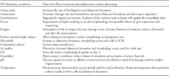
The IVF laboratory environment does affect the phenotype of the human embryo, and any stress present typically culminates in compromised transfer outcome, in the form of reduced implantation and pregnancy rates. Furthermore, the presence of two stressors can have more than a simple cumulative effect, and examples given of when oxygen is used at atmospheric levels reveal how potentially damaging this gas can be to the human embryo phenotype. Compared with animal models, there is a paucity of information on the human embryo regarding the direct effects of specific components of the laboratory and culture system. However, data to date on the human embryo are consistent with that from other mammalian species. Hence, while we cannot know for certain the effects of a specific variable, it would be prudent to extrapolate from animal studies. Table 1 summarizes some of the known effects of in vitro conditions on the human embryo phenotype. Although the field of human IVF has advanced significantly over the past four decades, culminating in the birth of over five million children worldwide, we still need ongoing research on the human embryo phenotype to ensure the optimization of IVF procedures and the birth of healthy children.
Observed effects of specific laboratory conditions on human embryo phenotype

ICM, inner cell mass.
Financial Support
D.K.G. is supported by the University of Melbourne; R.L.K. is supported by a postgraduate scholarship from the University of Melbourne.
Conflicts of Interest
D.K.G. receives research support from Vitrolife AB, Sweden.