In this malady the associations lose their continuity. Of the thousands of associative threads which guide our thinking, this disease seems to interrupt, quite haphazardly, sometimes such single threads, sometimes a whole group, and sometimes even large segments of them.
Eugen Bleuler (Reference Bleuler1950)From its earliest conceptualization, schizophrenia has been described as a disruption in the threads that bind ideational elements into coherent thought (Bleuler, Reference Bleuler1950). Modern neuroscience-based theories of schizophrenia also view the disorder as reflecting some form of disconnectivity (e.g., Bachman & Cannon, Reference Bachman and Cannon2005; Bullmore, Frangou, & Murray, Reference Bullmore, Frangou and Murray1997; Friston, Reference Friston1996, Reference Friston1997; Friston & Frith, Reference Friston and Frith1995; Weinberger, Aloia, Goldberg, & Berman, Reference Weinberger, Aloia, Goldberg and Berman1994; Weinberger, Berman, Suddath, & Torrey, Reference Weinberger, Berman, Suddath and Torrey1992). This idea is based on the understanding that the brain does not merely consist of a group of compartmentalized regions, but of a contiguous folded sheet of cortex drawn together from the inside by an elaborate web of connecting fibers. These fibers form an intricate network that allows for communication between cells throughout the cortex and that contributes to the infrastructure that makes complex cognitive functions possible. Given the level of cerebral “dexterity” necessary to facilitate interactions within such a complex structure, as well as the apparent lack of such coordination implicit in many of the symptoms of schizophrenia, it is not surprising that researchers have been intrigued by how connections both between and within particular brain regions may be disordered in patients. This theoretical approach has been supported in recent years by findings generated using modern imaging techniques, which allow us to observe structural and functional connectivity as they develop, mature, and deteriorate over the life span in healthy individuals as well as in patients with schizophrenia.
Several different models of disturbed connectivity in schizophrenia have been proposed. For example, Friston and Frith posit that schizophrenia is a result of disrupted communication between multiple regions (Friston & Frith, Reference Friston and Frith1995) and that this functional disconnectivity is based on alterations in synaptic connection strength rather than on gross anatomical changes (Friston, Reference Friston1998). In agreement with the basic theoretical notion of connective dysfunction in schizophrenia, but incorporating a structural component, Bullmore et al. (Reference Bullmore, Frangou and Murray1997) put forth the “dysplastic net” hypothesis, suggesting that schizophrenia involves a disruption of anatomical connectivity that arises at least in part during prenatal development. In support of this hypothesized time frame, it has been found that insults during the second trimester of gestation seem to carry a particularly high risk for later development of schizophrenia (Brown et al., Reference Brown, Hooton, Schaefer, Zhang, Petkova and Babulas2004; Mednick, Machon, Huttunen, & Bonett, Reference Mednick, Machon, Huttunen and Bonett1988). Moreover, the observed changes in patients with schizophrenia are subtle (Weinberger, Reference Weinberger1995), consistent with the idea that they are related not to gross stages of early development but later stages in which the neural circuitry is honed. A third model suggests that onset of schizophrenia is associated with aberrant synaptic pruning processes during late adolescence and early adulthood (McGlashan & Hoffman, Reference McGlashan and Hoffman2000), a view supported by findings of reduced dendritic complexity and spine and synapse density on cortical pyramidal neurons in schizophrenia patients compared with controls (Selemon, Rajkowska, & Goldman-Rakic, Reference Selemon, Rajkowska and Goldman-Rakic1995). The preceding examples are just three of the models viewing schizophrenia as a connective disorder; many other theorists have incorporated elements of functional and/or structural disconnectivity into their models of schizophrenia.
The goal of this paper is to evaluate the theoretical and empirical literatures bearing on the developmental course of disconnectivity in the pathophysiology of schizophrenia. We first consider work relevant to the timing of major risk factors for the disorder, followed by a review of the normal brain developmental mechanisms that underlie changes in connectivity during early (pre- and perinatal) as well as later (adolescent) maturational periods. We then consider the evidence bearing on whether there are changes in structural and functional connectivity associated with schizophrenia that can be linked to risk factors operative during these periods. We conclude with an analysis of specific molecular genetic mechanisms that have emerged from human and animal studies that may explain some of the developmental changes in neural connectivity that appear to underlie the onset and course of schizophrenia.
Developmental Course of Schizophrenia: Overview
Onset of schizophrenia tends to occur in late adolescence or early adulthood. Several lines of evidence indicate that schizophrenia involves risk factors impacting both early and late developmental brain changes (Cannon et al., Reference Cannon, van Erp, Bearden, Loewy, Thompson and Toga2003; McDonald & Murray, Reference McDonald and Murray2000). There are three prominent developmental periods that may influence disease risk and development: (a) conception, (b) early developmental, and (c) late developmental periods. First, the establishment of the genetic makeup at conception confers genetic liability for the disorder in terms of risk genes. Although this genetically mediated liability is instantiated at conception, it is not fixed; rather, different genes have differential relevance at different stages of development and under different environmental conditions. Heritability of schizophrenia is multifaceted and complex, and almost certainly involves multiple genes. Second, predisposing genotypes may then interact with both normal and disrupted developmental processes and environmental events that occur during specific phases of development. Two specific periods of influence have different potential impacts on functions related to connectivity. The “early” period includes brain development in utero and during the first few years after birth, and may involve factors such as obstetric complications. Evidence for involvement of risk factors during this period includes increased risk for schizophrenia among offspring of mothers who had infections during pregnancy (Buka, Cannon, Torrey, & Yolken, Reference Buka, Cannon, Torrey and Yolken2008) as well as evidence showing that some of the cortical and subcortical reductions in gray matter volume in patients with schizophrenia, in regions including the hippocampus, may arise from an interaction of an inherited genotype for schizophrenia and hypoxic complications in utero (Cannon, van Erp, et al., Reference van Erp, Saleh, Rosso, Huttunen, Lonnqvist and Pirkola2002; van Erp et al., Reference van Erp, Saleh, Rosso, Huttunen, Lonnqvist and Pirkola2002). These pre- and perinatal disturbances may interfere with some fundamental process in development of neural circuitry, such as cell migration, thus leaving the organism more vulnerable to later insult. The presence of a very early developmental change is consistent with the view that the neural changes associated with the development of schizophrenia are partly present from birth, and may therefore explain early neurocognitive and social deficits in many of the children who later develop schizophrenia (Bearden et al., Reference Bearden, Rosso, Hollister, Sanchez, Hadley and Cannon2000; Cannon, Caspi, et al., Reference Cannon, Caspi, Moffitt, Harrington, Taylor and Murray2002; Cannon et al., Reference Cannon, Rosso, Hollister, Bearden, Sanchez and Hadley2000; Jones, Rodgers, Murray, & Marmot, Reference Jones, Rodgers, Murray and Marmot1994; Rosso et al., Reference Rosso, Cannon, Huttunen, Huttunen, Lonnqvist and Gasperoni2000).
However, despite these early influences, and the presence of observable differences in childhood among some who later develop the disorder, formal diagnostic symptoms and signs of schizophrenia do not typically manifest until late adolescence or early adulthood. Thus, risk factors occurring during this later developmental period may play a role in disease onset. Late adolescence and early adulthood represents a period of continuing developmental brain changes. In particular, a natural pruning process takes place (Feinberg, Reference Feinberg1982; McGlashan & Hoffman, Reference McGlashan and Hoffman2000) reducing extraneous excitatory synapses and honing more mature circuitry. Findings of progressive anatomical deterioration in schizophrenia (Gur et al., Reference Gur, Cowell, Turetsky, Gallacher, Cannon and Bilker1998; Thompson, 2001) suggest that additional factors active during the late developmental period and more proximal to the onset of schizophrenia, in addition to early-acting genetic and perinatal risk factors, may also play a role in the etiology and pathophysiology of schizophrenia.
Given that complex cognitive functions arise from assemblies of interacting brain regions, and considering that disruptions in complex cognitive functions in schizophrenia occur in a developmental context, it is paramount to understand the normal development of neural connectivity before considering how such processes may go awry in the developmental course of schizophrenia.
Early and Late Maturational Mechanisms Affecting Neural Connectivity
The structural elements supporting neural connectivity include cell number, dendritic arborization, and density of synaptic contacts, which are gray matter components supporting both intra- and interregional connectivity, as well as axons, which are a white matter component supporting primarily interregional connectivity. During development, after the initial cell proliferation period occurs, neuronal migration begins at around 8 weeks gestation (Monk, Webb, & Nelson, Reference Monk, Webb and Nelson2001). The basic organizational structure of axonal connections is laid down as soon as cells have migrated and begin to form synapses. It is generally assumed that “the main lines of cortical circuitry are connected during the rapid phase of synaptogenesis” (Krasnegor, Lyon, & Goldman-Rakic, Reference Krasnegor, Lyon and Goldman-Rakic1997). Once the migrating cells have reached their target regions, they send out processes that begin to form connections with other neurons, which may be directly adjacent or quite a distance away, ultimately resulting in connections between neighboring neurons, between neighboring sulci (arcuate fibers), between different regions of the same hemisphere (association fibers), or between the two hemispheres (commissural fibers). As the axons extend, their chemosensitive growth cones guide them to the cells with which they will connect. The initial connection is via a “pioneer axon,” which uses cues from the epithelium to find its path (Patterson, Reference Patterson and Hall1992), whereas later arriving axons extend along the bundles of axons that have grown around this pioneer. For subsequent axons the challenge is to ensure that they stay on the appropriate path, avoiding crossing fibers. To facilitate this, each tract appears to have a distinct molecular label that can be sensed by the growth cones and that serves as a guide to the migrating axon when it reaches a potential intersection point. Once a group of axons are laid in a tract, membrane proteins, in particular, those from the cadherin family, aid in fasciculation by causing this grouped set of axons to bind to each other. A disturbance in this highly orchestrated process could easily result in misguided or otherwise unsuccessful formation of connections.
Once the growth cones have reached their target, synaptic connections are formed, and then over time they are strengthened. The newly migrated axons must form synapses with appropriate partners, a task they accomplish through cues such as a local chemical gradient and cell location (Patterson, Reference Patterson and Hall1992). Although this process is generally successful, in the presence of extenuating circumstances such as an excess of space in which to maneuver, axons may improperly connect. In addition, if they are unable to locate an appropriate connection point, they will connect with whatever else they do find. These observations appear to offer a mechanism by which proper migration and extension of a neuron may still not result in a proper connection.
Once the basic connections have been made, the second phase of white matter maturation begins as the resulting axons begin to undergo myelination. This phase may play a role in not only early but also later developmental changes. The myelination of the cortical tracts occurs on a different time frame than their initial development. The first myelination occurs around 10 weeks in the spinal cord, but other brain structures are not fully myelinated until much later. At birth, the pons and cerebellar peduncles are myelinated, followed by the internal capsule, splenium of the corpus callosum, anterior limb of the internal capsule, and genu, and, by ~1 year after birth, parts of the frontal, parietal, and temporal lobes (Paus et al., Reference Paus, Collins, Evans, Leonard, Pike and Zijdenbos2001). The hippocampus and frontal lobes undergo the majority of their myelination during adolescence and do not finish until early adulthood (Arnold & Rioux, Reference Arnold and Rioux2001). In fact, normally during early adolescence, the gray matter volume is decreasing because of increased synaptic pruning and decreased neuropils, but the white matter volume actually undergoes a simultaneous increase (Paus et al., Reference Paus, Collins, Evans, Leonard, Pike and Zijdenbos2001).
In addition to the long connecting fibers between cortical regions that develop early on, the local synaptic connections enabling neuronal communication are the other important aspect of structural connectivity. However, in the context of schizophrenia, this level of connectivity may be more affected by events that occur during “late” adolescent development. Generally, two types of neurodevelopmental events contribute to increases and decreases in brain volume during postnatal development: progressive phenomena, such as cell proliferation, dendritic arborization, myelination, and regressive phenomena, such as apoptosis, synaptic and axonal pruning, and atrophic processes (Pfefferbaum et al., Reference Pfefferbaum, Mathalon, Sullivan, Rawles, Zipursky and Lim1994). Cell growth, arborization, and synaptogenesis are likely to contribute to notable increases in cortical gray matter volume during the first 5 to 10 years of life. Development of synapses occurs around the 27th week of gestation, and although synaptic density peaks in the visual cortex at 3 months of age, the frontal cortex (middle frontal gyrus) is delayed and the peak does not occur until around 3.5 years (Huttenlocher & Dabholkar, Reference Huttenlocher and Dabholkar1997). Synapses are initially over produced, and there is an accompanying overproduction of axons, dendrites, and dendritic spines (Webb, Monk, & Nelson, Reference Webb, Monk and Nelson2001).
Although synapses develop early and the human brain reaches 75% of its adult weight by age 2 (Carmichael, Reference Carmichael1990), considerable changes in brain structure continue to occur until at least the second decade of life (Blows, Reference Blows2003; Giedd et al., Reference Giedd, Snell, Lange, Rajapakse, Casey and Kozuch1996; Huttenlocher, Reference Huttenlocher1979). As a part of this later development, a normal aspect of brain maturation during adolescence and early adulthood is the reduction of gray matter volume, which is attributed largely to the process of synaptic pruning (Thompson et al., Reference Thompson, Giedd, Woods, MacDonald, Evans and Toga2000). This process results in the normal elimination of about 40% of cortical synapses over the course of development (Huttenlocher, Reference Huttenlocher1979). In humans, different regions of cortex undergo synaptic pruning at different times and rates. For example, pruning occurs in the visual cortex between the ages of 1 and 12, yet in Layer 3 of the prefrontal cortex it occurs from the ages of ~5 to 16 years (Pfefferbaum et al., Reference Pfefferbaum, Mathalon, Sullivan, Rawles, Zipursky and Lim1994), and an adult level of synaptic density is reached by age 16 (Huttenlocher, Reference Huttenlocher1990). Reduction of dendritic branching can occur through either axonal retraction, in which the integrity of the cellular material is retained and recycled, or axonal degeneration, in which the entire axon is destroyed and then taken up by local phagocytes (Saxena & Caroni, Reference Saxena and Caroni2007). Developmental pruning occurs through a highly regulated process of axonal degeneration, which is mechanistically separate from either axonal retraction or from lesion-induced degeneration. The processes that initiate it may include changes in levels and distribution of excitatory and inhibitory neurotransmitters, and changes in availability of neurotrophic factors (Webb et al., Reference Webb, Monk and Nelson2001). Overall, the process of pruning in prefrontal cortical regions in conjunction with the growth of corticocortical connections is thought to increase the overall efficiency of the brain by improving the effectiveness of neural transmission, which allows for increased executive control of cognitive functions (Feinberg, Reference Feinberg1982).
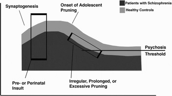
In the developmental pruning hypothesis of schizophrenia, it is proposed that pruning is disrupted and occurs to an excessive degree in patients, placing them below a threshold for the presentation of psychotic symptoms (e.g., Hoffman & McGlashan, Reference Hoffman and McGlashan1997; see Figure 1). This disrupted pruning might take a variety of forms. For example, there could be a paucity of synapses beginning in early development, possibly as a result of pre- or perinatal insult or of genetic disruptions in early development; in this case even a normally functioning pruning process would still result in a critically low number of synapses. It is also possible that the initial period of synaptogenesis is intact, but that the pruning process is either prolonged or excessive, again resulting in too few functional synapses. Of course, it is also possible that there is a combination of early and late factors at play. Data indicating that patients with schizophrenia when compared with controls have a reduction in cortical thickness and a reduction in dendritic spine density on prefrontal cortical pyramidal neurons (Glantz & Lewis, Reference Glantz and Lewis2000), but without an accompanying reduction in the number of neurons (Selemon et al., Reference Selemon, Rajkowska and Goldman-Rakic1995), are consistent with theories of reduced neuropil via disrupted synaptic pruning.
A schematic diagram of synaptic pruning as related to the onset of schizophrenia.

With the improvements in structural and functional magnetic resonance imaging (sMRI and fMRI) and the advent of diffusion tensor imaging (DTI), we now have the ability to investigate the course of brain development in ways not previously possible in human subjects. In a longitudinal MRI study with 145 subjects ages 4–22 years scanned once or multiple times, Giedd and colleagues (Reference Giedd, Blumenthal, Jeffries, Castellanos, Liu and Zijdenbos1999) reported age-related brain lobar gray and white matter changes. Although white matter volumes showed a linear increase, cortical gray matter followed nonlinear lobar-specific patterns. In particular, gray matter volumes in the frontal, parietal, and temporal lobes followed an inverted U-shaped curve with age, declining after achieving their peak during adolescence. These findings are consistent with several other studies (for review, see Durston et al., Reference Durston, Hulshoff Pol, Casey, Giedd, Buitelaar and van Engeland2001, including one large cross-sectional study of normal development and aging across the life span (Sowell et al., Reference Sowell, Peterson, Thompson, Welcome, Henkenius and Toga2003). In this latter study, gray matter density along the dorsal surfaces of the frontal and parietal lobes showed rapid decline before middle age. Further, Gogtay and colleagues (1999) showed detailed dynamic mapping of cortical changes in a multiple time-point follow-up study. Although the primary sensorimotor cortices underwent early changes, the thinning of cerebral cortex was particularly delayed in association areas including the prefrontal regions (Gogtay et al., Reference Gogtay, Giedd, Lusk, Hayashi, Greenstein and Vaituzis2004). Overall, the pattern of cortical developmental trajectory in adolescence and early adulthood is consistent with the proposed synaptic elimination profile, featured by prolonged maturation of higher order association cortices, especially the prefrontal cortex.
In addition, a small number of studies have used DTI to examine developmental changes in white matter tracts longitudinally. An increase in anisotropy and decrease in diffusion over time has been shown in studies of infant development (Morriss, Zimmerman, Bilaniuk, Hunter, & Haselgrove, Reference Morriss, Zimmerman, Bilaniuk, Hunter and Haselgrove1999; Mukherjee et al., Reference Mukherjee, Miller, Shimony, Philip, Nehra and Snyder2002). Although the changes are maximal during the first 2 years of life, studies including older samples suggest that changes in diffusion (Paus et al., Reference Paus, Zijdenbos, Worsley, Collins, Blumenthal and Giedd1999; Schmithorst, Wilke, Dardzinski, & Holland, Reference Schmithorst, Wilke, Dardzinski and Holland2002) and anisotropy (Klingberg, Vaidya, Gabrieli, Moseley, & Hedehus, Reference Klingberg, Vaidya, Gabrieli, Moseley and Hedehus1999; Mukherjee et al., Reference Mukherjee, Miller, Shimony, Philip, Nehra and Snyder2002; Schmithorst et al., Reference Schmithorst, Wilke, Dardzinski and Holland2002) are likely to persist into late adolescence or early adulthood. For instance, fractional anisotropy (FA) increases have been observed in the corpus callosum, corona radiata, caudate, and putamen during childhood (Snook, Paulson, Roy, Phillips, & Beaulieu, Reference Snook, Paulson, Roy, Phillips and Beaulieu2005), and in the arcuate fasciculus, internal capsule, prefrontal gyrus, temporal gyrus, and corpus callosum during adolescence (Ashtari et al., Reference Ashtari, Cervellione, Hasan, Wu, McIlree and Kester2007). It is important to note that although myelination continues until the second decade of life, particularly in the hippocampus (Benes, Reference Benes1989) and prefrontal cortex (Yakovlev & Lecours, Reference Yakovlev and Lecours1967), these changes in diffusion and anisotropy are not necessarily entirely myelin dependent. Previous work has shown that anisotropy can exist even in unmyelinated tracts (Beaulieu & Allen, Reference Beaulieu and Allen1994), possibly reflecting other aspects of the cellular infrastructure, such as tissue hydration, cell packing density, axonal fiber diameter, and directional coherence (Schmithorst et al., Reference Schmithorst, Wilke, Dardzinski and Holland2002). The changes may be partially related to myelination, however, especially as increased myelination is one cause of increased axonal size. One way in which to dissociate the roles of myelination and other influences is through the use of a coherence measure. Klingberg and colleagues (1999) found that after covarying for coherence (the degree to which the direction of one voxel's primary eigenvector is the same as those of neighboring voxels), the remaining anisotropy differences between children and adults are likely to be linked to myelination. Regardless, changes during development that occur with the timing of disease onset may prove particularly interesting for understanding development in schizophrenia.
White Matter Findings in Schizophrenia
The white matter tracts that are laid down early during development and that mature throughout adolescence are the basis for large-scale intercortical connectivity, and as such, they have been an important focus of research on disconnectivity in schizophrenia. Previous studies have found that white matter abnormalities in patients with schizophrenia measured by histology, MRI, and DTI are not associated with age (Buchsbaum et al., Reference Buchsbaum, Tang, Peled, Gudbjartsson, Lu and Hazlett1998; Foong et al., Reference Foong, Maier, Barker, Brocklehurst, Miller and Ron2000; Highley, Walker, Esiri, Crow, & Harrison, Reference Highley, Walker, Esiri, Crow and Harrison2002) medication history (Agartz, Andersson, & Skare, Reference Agartz, Andersson and Skare2001; Buchsbaum et al., Reference Buchsbaum, Tang, Peled, Gudbjartsson, Lu and Hazlett1998; Foong et al., Reference Foong, Maier, Barker, Brocklehurst, Miller and Ron2000; Kubicki et al., Reference Kubicki, Westin, Maier, Frumin, Nestor and Salisbury2002; Suzuki et al., Reference Suzuki, Nohara, Hagino, Kurokawa, Yotsutsuji and Kawasaki2002), or duration of illness (Foong et al., Reference Foong, Maier, Barker, Brocklehurst, Miller and Ron2000; Okugawa, Sedvall, & Agartz, Reference Okugawa, Sedvall and Agartz2002; Suzuki et al., Reference Suzuki, Nohara, Hagino, Kurokawa, Yotsutsuji and Kawasaki2002), suggesting that these changes are more likely due to disturbed developmental processes rather than degenerative processes associated with the disease or its treatment. Moreover, developmental disorders of the white matter, such as metachromatic leukodystrophy (MLD), can result in schizophrenia-like behavior. MLD results in a demyelination of prefrontal white matter, and can cause psychotic symptoms in patients whose disease onset occurs during adolescence. The specific nature of its psychosis, which includes primarily auditory hallucinations, disorganization of thought, and bizarre behavior, makes this disorder a good neurological model for schizophrenia (Hyde, Ziegler, & Weinberger, Reference Hyde, Ziegler and Weinberger1992). In addition to MLD, patients with developmental disturbances of white matter tracts connecting the prefrontal cortex with other regions show schizophrenia-like deficits in inhibitory control and executive functions (Christ, White, Brunstrom, & Abrams, Reference Christ, White, Brunstrom and Abrams2003). Taken together, the support for the hypothesis that schizophrenia is a syndrome that involves disconnectivity is bidirectional; not only do patients with schizophrenia potentially show disrupted functional and structural connectivity, patients with developmentally disrupted structural connectivity show schizophrenia-like symptoms.
Overall white matter changes: Histology and MRI
Initial attempts to quantify white matter changes were based on anatomical dissection and standard structural MRI. Historically, imaging techniques have not allowed detailed analysis of white matter in vivo, particularly at a cellular level, and so investigations have been predominately postmortem. However, postmortem studies carry with them a variety of methodological issues, such as the influence of postmortem interval and cause of death on the tissue, as well as the necessity of relying on previous diagnoses, that can seriously complicate interpretation of the data collected (Lewis, Reference Lewis2002). One finding from postmortem work has been a change in distribution of interstitial cells of the white matter (Akbarian, Reference Akbarian, Kim, Potkin, Hetrick, Bunney and Jones1996; Kirkpatrick, Reference Kirkpatrick, Conley, Kakoyannis, Reep and Roberts1999, Reference Kirkpatrick, Messias, Conley and Roberts2003). Interstitial neurons develop from the cortical subplate, which is a transitory structure present only during the second and third trimester and which plays a role in guiding the formation of cortical connections, possibly by dictating each cell's ultimate location. Normally the cells from the subplate undergo programmed cell death at the end of the third trimester and shortly after birth. The findings of changes in interstitial neuron distribution may indicate an imbalance in the factors initiating the cell death. Because of its critical role in migratory processes, any disturbance of the functioning of the subplate could lead to a disruption in the appropriate formation of cortical connections. Thus, this finding offers a potential mechanism for disruption of structural connectivity in the absence of gliosis or large-scale gross pathology.
As an alternative to histological and dissection studies, neuroimaging techniques have been applied to the investigation of white matter in living schizophrenic patients. However, although traditional MRI does allow a gross overview of the white matter, any abnormalities other than the overall size of the area are beyond the scope of this methodology. Traditional MRI cannot assess whether cells are in place but not properly connected, whether axons are disorganized, or whether normal functional cells have been replaced with other cells; in each case white matter volume may remain unchanged while functional connectivity may be fundamentally altered.
Many structural MRI studies have examined whole brain or lobar white matter volumes in patients with schizophrenia compared with controls. Breier and colleagues (1992) reported reduced bilateral prefrontal white matter volumes in patients with schizophrenia compared with controls. In addition, they reported that variation in prefrontal white matter volume correlated with right (but not left) amygdala and hippocampal volumes in the patients but not in the controls, which may be consistent with the idea of early hippocampus and amygdala damage, resulting in abnormal connectivity with the frontal lobe in patients with schizophrenia. This interpretation is also consistent with the finding that greater hippocampal pathology in schizophrenia is correlated with decreased prefrontal activity assessed using positron emission tomography (PET; Weinberger et al., Reference Weinberger, Berman, Suddath and Torrey1992). Similarly, Hulshoff Pol and colleagues (Reference Hulshoff Pol, Schnack, Bertens, van Haren, van der Tweel and Staal2002) found reduced white matter volume only in the prefrontal region in schizophrenia patients; there were no case–control differences in whole-brain white matter volumes. Specific tracts have also been investigated; for instance, decreases in the volume of the anterior limb of the internal capsule (connecting the medial dorsal thalamus and the frontal lobes) have been shown (Suzuki et al., Reference Suzuki, Nohara, Hagino, Kurokawa, Yotsutsuji and Kawasaki2002; Zhou et al., Reference Zhou, Suzuki, Hagino, Takahashi, Kawasaki and Nohara2003).
The relationship between gray and white matter volumes has also been investigated (Bartzokis et al., Reference Bartzokis, Nuechterlein, Lu, Gitlin, Rogers and Mintz2003). It was noted that in normal development across the life span (from late adolescence until midadulthood) there is a significant reduction in the gray/white matter ratio in the brain (Bartzokis et al., Reference Bartzokis, Beckson, Lu, Nuechterlein, Edwards and Mintz2001). In adolescence there is a period of substantial loss of gray matter (pruning), and then throughout adulthood, until the age of approximately 47, there is a normal increase in white matter. The combination of these events leads to a decrease in the gray/white matter ratio over time. In a cross-sectional study of schizophrenics and controls, it was found that the patients do not demonstrate this decrease in the way that normal subjects do; rather, the patients appear to fail to increase their white matter with age in the manner of the controls, and the deficit becomes more pronounced over time, making the gray matter deficit more apparent in younger subjects, whereas the white matter deficit becomes relatively larger with age (Bartzokis et al., Reference Bartzokis, Nuechterlein, Lu, Gitlin, Rogers and Mintz2003).
DTI assessments of white matter changes
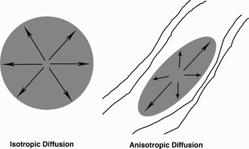
A relatively new MRI technique, DTI, allows for a more detailed analysis of the integrity of the white matter tracts. The data collected in DTI are based on Brownian motion, the normal motion driven by thermal energy, of water molecules (Peled, Gudbjartsson, Westin, Kikinis, & Jolesz, Reference Peled, Gudbjartsson, Westin, Kikinis and Jolesz1998). If unobstructed, that motion has an equal likelihood of going in any direction (i.e., isotropic diffusion). Water molecules in the brain, however, are moving in an environment full of cell membranes, fibers, and other tissue components that limit their motion. In white matter tracts, the motion of the molecules is restricted by the myelinated axons and tends to move more easily in the direction along the axon than perpendicular to it, creating motion in an ellipsoid shape rather than a spherical one (i.e., anisotropic diffusion; see Figure 2). The shape of this ellipsoid can give information such as the degree of myelination, the average fiber diameter, and the similarity in the direction of the fibers within the tract. An index known as FA characterizes the eccentricity of the ellipsoid, and is interpreted as a measure of white matter integrity. The direction of the ellipsoid can give information about the orientation of fibers in that region, and it is possible to use tractography to follow fiber tracts through adjacent voxels with a common principle direction (Catani, Howard, Pajevic, & Jones, Reference Catani, Howard, Pajevic and Jones2002; Klingberg et al., Reference Klingberg, Vaidya, Gabrieli, Moseley and Hedehus1999). This procedure allows the tracing of anatomical connections between regions of interest, or the determination of whether the tracts themselves are appropriately placed and connected. With these capabilities, coupled with the ability to perform the technique in a standard MRI scanner over relatively short scan periods, DTI has become a very practical way to investigate structural integrity of fiber tracts in living subjects.
Isotropic and anisotropic diffusion, which are the basis of FA calculations used to calculate white matter integrity using DTI.

Initially, many DTI studies in schizophrenia assessed changes at the level of entire lobes or large cortical regions. These approaches have shown decreased FA in frontal (Buchsbaum et al., Reference Buchsbaum, Tang, Peled, Gudbjartsson, Lu and Hazlett1998; Hao et al., Reference Hao, Liu, Jiang, Gong, Liu and Tan2006; Kitamura et al., Reference Kitamura, Matsuzawa, Shioiri, Someya, Kwee and Nakada2005; Kumra et al., Reference Kumra, Ashtari, McMeniman, Vogel, Augustin and Becker2004; Lim et al., Reference Lim, Hedehus, Moseley, de Crespigny, Sullivan and Pfefferbaum1999; Minami et al., Reference Minami, Nobuhara, Okugawa, Takase, Yoshida and Sawada2003; Szeszko, Ardekani et al., Reference Szeszko, Ardekani, Ashtari, Kumra, Robinson and Sevy2005) and parietal lobes (Ardekani, Nierenberg, Hoptman, Javitt, & Lim, Reference Ardekani, Nierenberg, Hoptman, Javitt and Lim2003; Kyriakopoulos, Vyas, Barker, Chitnis, & Frangou, Reference Kyriakopoulos, Vyas, Barker, Chitnis and Frangou2008; Minami et al., Reference Minami, Nobuhara, Okugawa, Takase, Yoshida and Sawada2003). However, the affected subregions vary between studies, with some studies finding no patient–control differences (Foong et al., Reference Foong, Symms, Barker, Maier, Miller and Ron2002; Price, Bagary, Cercignani, Altmann, & Ron, Reference Price, Bagary, Cercignani, Altmann and Ron2005). Some of these discrepancies may be related to differences in registration techniques, as accurate registration is an ongoing issue for DTI studies. As new methods have emerged, it has been possible to obtain more subtle information about not only specific tracts or small regions implicated, but also about the relationship of changes in those regions with changes in cognition, symptomatology, and outcome.
One region of particular interest is the uncinate fasciculus, a tract providing connections between the temporal pole near the amygdala, and the orbitofrontal/ventrolateral prefrontal cortex (Petrides & Pandya, Reference Petrides, Pandya, Stuss and Knight2002). FA in the uncinate fasciculus has been shown to be reduced in patients with schizophrenia (Burns et al., Reference Burns, Job, Bastin, Whalley, Macgillivray and Johnstone2003; Kubicki et al., Reference Kubicki, Westin, Maier, Frumin, Nestor and Salisbury2002; Nakamura et al., Reference Nakamura, McCarley, Kubicki, Dickey, Niznikiewicz and Voglmaier2005; Nestor et al., Reference Nestor, Kubicki, Gurrera, Niznikiewicz, Frumin and McCarley2004; Price et al., Reference Price, Cercignani, Parker, Altmann, Barnes and Barker2008; Szeszko et al., Reference Szeszko, Robinson, Ashtari, Vogel, Betensky and Sevy2008) and schizotypal personality disorder (Nakamura et al., Reference Nakamura, McCarley, Kubicki, Dickey, Niznikiewicz and Voglmaier2005), a genetically related syndrome. Moreover, because of the regions it connects, this tract is of particular interest to emotional processing, and efforts have been made to determine the behavioral correlates of white matter changes in this tract. For example, decreased FA in this tract is associated with negative symptoms (Szeszko et al., Reference Szeszko, Robinson, Ashtari, Vogel, Betensky and Sevy2008) and with reduced executive functions (Kubicki et al., Reference Kubicki, Westin, Maier, Frumin, Nestor and Salisbury2002), verbal learning (Nakamura et al., Reference Nakamura, McCarley, Kubicki, Dickey, Niznikiewicz and Voglmaier2005; Szeszko et al., Reference Szeszko, Robinson, Ashtari, Vogel, Betensky and Sevy2008), and episodic memory (Nestor et al., Reference Nestor, Kubicki, Gurrera, Niznikiewicz, Frumin and McCarley2004), all cognitive constructs that would be expected to rely on coordinated communication between frontal and temporal lobes.
The cingulate gyrus is another region of interest based on functional imaging studies of patients with schizophrenia. It serves as a primary connection to the frontal lobes, which are compromised in schizophrenia, and is associated with a number of cognitive and affective processes that may be impaired in schizophrenia. FA is decreased in the anterior and posterior cingulate in schizophrenia (Fujiwara, Hirao, et al., Reference Fujiwara, Hirao, Namiki, Yamada, Shimizu and Fukuyama2007; Fujiwara, Namiki, et al., Reference Fujiwara, Namiki, Hirao, Miyata, Shimizu and Fukuyama2007; Kubicki et al., Reference Kubicki, Westin, Nestor, Wible, Frumin and Maier2003; Sun et al., Reference Sun, Wang, Cui, Breeze, Du and Wang2003; Wang et al., Reference Wang, Sun, Cui, Du, Wang and Zhang2004). Changes in FA in this region have been associated with positive symptom scores (Fujiwara, Namiki, et al., Reference Fujiwara, Namiki, Hirao, Miyata, Shimizu and Fukuyama2007), performance on the Wisconsin Card Sort Test, an index of executive function (Kubicki et al., Reference Kubicki, Westin, Nestor, Wible, Frumin and Maier2003) and performance on an attention task (Lim et al., Reference Lim, Ardekani, Nierenberg, Butler, Javitt and Hoptman2006).
Patients with schizophrenia have decreased hippocampal volumes (Nelson, Saykin, Flashman, & Riordan, Reference Nelson, Saykin, Flashman and Riordan1998) as well as reduced overall temporal lobe gray matter volumes (Gur et al., Reference Gur, Turetsky, Cowell, Finkelman, Maany and Grossman2000) and impairments in cognitive functions mediated by the medial temporal lobe such as episodic memory (Saykin et al., Reference Saykin, Gur, Gur, Mozley, Mozley and Resnick1991). FA reductions in patients with schizophrenia have been observed in the hippocampus (White et al., Reference White, Kendi, Lehericy, Kendi, Karatekin and Guimaraes2007) and the temporal lobes (Schlosser et al., Reference Schlosser, Nenadic, Wagner, Gullmar, von Consbruch and Kohler2007), although negative findings exist (Begre et al., Reference Begre, Federspiel, Kiefer, Schroth, Dierks and Strik2003). Further, associations between decreased FA in the hippocampus with decreased verbal declarative memory performance (Lim et al., Reference Lim, Ardekani, Nierenberg, Butler, Javitt and Hoptman2006), and decreased FA in the fornix with decreased episodic memory performance (Nestor et al., Reference Nestor, Kubicki, Kuroki, Gurrera, Niznikiewicz and Shenton2007), have been reported. White matter changes have been shown to be present even in the first episode of schizophrenia (Begre et al., Reference Begre, Federspiel, Kiefer, Schroth, Dierks and Strik2003; Federspiel et al., Reference Federspiel, Begre, Kiefer, Schroth, Strik and Dierks2006; Hao et al., Reference Hao, Liu, Jiang, Gong, Liu and Tan2006; Price et al., Reference Price, Bagary, Cercignani, Altmann and Ron2005; Szeszko, Ardekani, et al., Reference Szeszko, Ardekani, Ashtari, Kumra, Robinson and Sevy2005), suggesting that they occur during development and are not secondary to the disease or its treatment. Recently, our group has begun to assess changes in DTI measures prior to the onset of schizophrenia, in an ultra high-risk (UHR) population. In a cross-sectional investigation, the hippocampus and temporal tracts have shown a very interesting pattern across adolescence and young adulthood in which healthy controls increase FA with age, while high-risk subjects fail to show the same increase. This pattern may indicate that the differences in some white matter tracts that are observable in adult schizophrenia patients arise gradually as a part of an abnormal process of white matter development in adolescence (Karlsgodt, Niendam, et al., Reference Karlsgodt, Niendam, van Erp, Poldrack, Nuechterlein and Cannon2008).
One white matter tract emerging as a point of particular interest given its role in supporting working memory is the superior longitudinal fasciculus (SLF), which is a primary connection linking the frontal and parietal lobes (Petrides & Pandya, Reference Petrides, Pandya, Stuss and Knight2002). The frontal and parietal cortices are critical for working memory (Chafee & Goldman-Rakic, Reference Chafee and Goldman-Rakic2000; Paulesu, Frith, & Frackowiak, Reference Paulesu, Frith and Frackowiak1993), and cellular activity in these regions during working memory processing has been shown to be coordinated (Chafee & Goldman-Rakic, Reference Chafee and Goldman-Rakic1998), suggesting that these regions function as a working memory network. Patients with schizophrenia show impairments in working memory (Lee & Park, Reference Lee and Park2005), and working memory deficits have been proposed to be a core feature of the illness, as they are associated with other cognitive processes (Silver, Feldman, Bilker, & Gur, Reference Silver, Feldman, Bilker and Gur2003), symptomatology (Goldman-Rakic, Reference Goldman-Rakic1994), and functional outcomes (Green, Reference Green1996). Moreover, the pattern of maturation of the white matter tracts between the frontal and parietal lobe regions correlates both with the pattern of maturation of working memory capacity and functional activation (Klingberg, Reference Klingberg2006), linking intact working memory performance to integrity of the entire circuit. Finally, FA in the SLF has been shown to be decreased in patients with schizophrenia (Federspiel et al., Reference Federspiel, Begre, Kiefer, Schroth, Strik and Dierks2006; Karlsgodt, van Erp, et al., Reference Karlsgodt, van Erp, Poldrack, Bearden, Nuechterlein and Cannon2008; Szeszko et al., Reference Szeszko, Robinson, Ashtari, Vogel, Betensky and Sevy2008).
This relatively new body of work using DTI as a measure to investigate abnormalities in schizophrenia has been growing rapidly. It is an important area to expand upon, given DTI's ability to investigate white matter microstructure in vivo longitudinally, and given that this safe, repeatable procedure enables study of the functional behavioral correlates of changes in connectivity during normal and abnormal brain development.
Gray Matter Findings in Schizophrenia
Gray matter, including dendritic branches supporting synaptic connectivity, constitutes a primary component of local intra- and interregional connectivity. Postmortem findings support the theory of continuous synaptic/neurite elimination (Huttenlocher, Reference Huttenlocher1979; Huttenlocher & Dabholkar, Reference Huttenlocher and Dabholkar1997) during the late development stage. However, although these findings are in line with those of animal studies (Bourgeois, Goldman-Rakic, & Rakic, Reference Bourgeois, Goldman-Rakic and Rakic1994), given the paucity of available studies and the sparse sampling of human brain specimens in the studies to date, the anatomical profiles and the dynamics of brain changes during development need further study. With the advent of modern brain imaging and analysis techniques, changes that may arise from these developmental processes are observable (albeit indirectly) in adult patients with schizophrenia. sMRI is one of the most widely used imaging modalities for investigating brain changes during normal brain development and during the development of schizophrenia. Because of noninvasiveness of MRI techniques, brain images can be acquired in vivo, making well-controlled, large-scale studies possible. Moreover, compared to postmortem studies that can assess only the terminal state of the brain, the possibility of following the same subjects and tracking brain changes over time makes MRI one of the best techniques for studying progressive changes in normal and disordered development.
MRI of disrupted development in schizophrenia
Modern imaging findings have well established that schizophrenia is associated with structural brain abnormalities, with the most consistent findings being enlarged ventricles and reduced medial temporal, superior temporal, and prefrontal gray matter volumes (Pantelis et al., Reference Pantelis, Yucel, Wood, Velakoulis, Sun and Berger2005; Shenton, Dickey, Frumin, & McCarley, Reference Shenton, Dickey, Frumin and McCarley2001). Cross-sectional neuroimaging studies have shown gray matter volume deficits not only in established but also in prodromal (Borgwardt et al., Reference Borgwardt, McGuire, Aston, Berger, Dazzan and Gschwandtner2007; Pantelis et al., Reference Pantelis, Velakoulis, McGorry, Wood, Suckling and Phillips2003) and early stages of schizophrenia (Shenton et al., Reference Shenton, Dickey, Frumin and McCarley2001), suggesting that structural abnormalities exist at least in part before the onset of illness. Furthermore, abnormal brain asymmetry and cortical folding, signs of relatively early developmental deviations, have been found in schizophrenia patients (Nakamura, Nestor, et al., Reference Nakamura, Nestor, McCarley, Levitt, Hsu and Kawashima2007; Yucel et al., Reference Yucel, Stuart, Maruff, Velakoulis, Crowe and Savage2001). These findings are in accordance with the early neurodevelopmental model of schizophrenia (Weinberger, Reference Weinberger1995). At the same time, accumulating longitudinal neuroimaging data suggest that brain volume changes in schizophrenia are not restricted to early developmental stages, but may be progressive around the time of onset of frank symptoms (for a review, see Pantelis et al., Reference Pantelis, Yucel, Wood, Velakoulis, Sun and Berger2005), in line with the late developmental hypothesis of the illness.
Recent longitudinal studies using high-resolution MRI and advanced image analysis techniques have demonstrated progressive volume loss in various brain regions in schizophrenia patients. In a series of first-episode schizophrenia follow-up studies, DeLisi, Sakuma, Maurizio, Relja, and Hoff (Reference DeLisi, Sakuma, Maurizio, Relja and Hoff2004) and colleagues found accelerated lateral ventricular expansion over a 10-year period. Accelerated progressive ventricular expansion in patients at various stages of illness has also been reported by other researchers (Garver, Nair, Christensen, Holcomb, & Kingsbury, Reference Garver, Nair, Christensen, Holcomb and Kingsbury2000; Nair et al., Reference Nair, Christensen, Kingsbury, Kumar, Terry and Garver1997; Nakamura, Salisbury, et al., Reference Nakamura, Salisbury, Hirayasu, Bouix, Pohl and Yoshida2007). Progressive frontal volume loss was reported initially by Gur and colleagues (1998) in first-episode/chronic patients and subsequently replicated in several other studies (Ho et al., Reference Ho, Andreasen, Nopoulos, Arndt, Magnotta and Flaum2003; Lieberman et al., Reference Lieberman, Tollefson, Charles, Zipursky, Sharma and Kahn2005; Mathalon, Sullivan, Lim, & Pfefferbaum, Reference Mathalon, Sullivan, Lim and Pfefferbaum2001; Nakamura, Salisbury, et al., Reference Nakamura, Salisbury, Hirayasu, Bouix, Pohl and Yoshida2007; Rapoport et al., Reference Rapoport, Giedd, Blumenthal, Hamburger, Jeffries and Fernandez1999; Sun et al., in press; Thompson et al., Reference Thompson, Vidal, Giedd, Gochman, Blumenthal and Nicolson2001; van Haren et al., Reference van Haren, Pol, Schnack, Cahn, Brans and Carati2008). Excessive volume loss in the whole temporal lobe has been reported in several studies (Nakamura, Salisbury, et al., Reference Nakamura, Salisbury, Hirayasu, Bouix, Pohl and Yoshida2007; Rapoport et al., Reference Rapoport, Giedd, Blumenthal, Hamburger, Jeffries and Fernandez1999), and this finding seems largely attributable to changes in the superior temporal gyrus (Kasai, Shenton, Salisbury, Hirayasu, Lee, et al., Reference Kasai, Shenton, Salisbury, Hirayasu, Lee and Ciszewski2003; Kasai, Shenton, Salisbury, Hirayasu, Onitsuka, et al., Reference Kasai, Shenton, Salisbury, Hirayasu, Onitsuka and Spencer2003; Thompson et al., Reference Thompson, Vidal, Giedd, Gochman, Blumenthal and Nicolson2001).
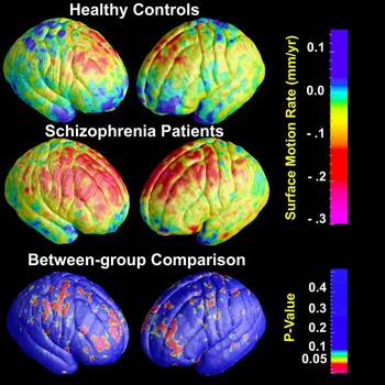
The profile of brain volume decline in schizophrenia relative to the normal developmental profile was recently examined in a longitudinal study by our group in collaboration with the Melbourne group (Sun, Stuart, et al., in press). We applied cortical pattern matching (Thompson et al., Reference Thompson, Hayashi, de Zubicaray, Janke, Rose and Semple2003) in combination with fMRI of the brain structural image evaluation using normalization of atrophy (SIENA) to repeated MRI scans in first-episode schizophrenia patients and healthy controls, mapping local brain surface contractions throughout the lateral cortical surface. Significantly greater prefrontal surface contraction was found in patients compared with controls, consistent with previous findings of frontal progression mentioned above. More interestingly, patients and healthy controls demonstrated very similar anatomical patterns of brain surface contractions, both showing prominent prefrontal contraction, with that of the former group exaggerated in both magnitude and spatial extent (see Figure 3). Further analyses demonstrated that the surface contraction observed in prefrontal regions was driven by changes in gray matter volume. The similarity of surface contraction patterns between normal and diseased cohorts nicely fits the prediction of the hypothesis of synaptic overpruning in schizophrenia (Feinberg, Reference Feinberg1982; McGlashan & Hoffman, Reference McGlashan and Hoffman2000), indicating the involvement of late developmental deviations in the neuropathophysiology of schizophrenia.
Maps of the average brain surface contraction rates and p-maps for comparison of first-episode schizophrenia patients versus healthy controls.

MRI of disease onset: Prodromal states
Despite the mentioned findings, the major difficulties in interpreting the longitudinal brain changes as primary pathological processes in schizophrenia are the inevitable existence of confounding factors secondary to the illness, which include long-term illness, medications, or fluctuation of symptoms. Even in longitudinal studies following first-episode patients, the reported duration of illness in the majority of studies were over 1 year at intake (Pantelis et al., Reference Pantelis, Yucel, Wood, Velakoulis, Sun and Berger2005), and antipsychotics were generally administered to control psychotic symptoms. All these factors are likely to bring changes to brain structures, and for ethical and practical reasons, these factors cannot be controlled in patients.
One strategy to address these questions is to identify individuals who are at heightened risk for developing psychosis because of mental state change and/or family history of psychosis, and to track brain changes from the premorbid stage to the onset of illness (Cannon et al., Reference Cannon, van Erp, Bearden, Loewy, Thompson and Toga2003; Johnstone et al., Reference Johnstone, Abukmeil, Byrne, Clafferty, Grant and Hodges2000; McGorry, Yung, & Phillips, Reference McGorry, Yung and Phillips2003). When some of these individuals experience a frank onset during follow-up, brain changes occurring around the time of onset can be assessed. This approach aims to observe brain changes at a very early prodromal stage, before full-blown psychosis emerges, thereby avoiding confounding factors related to long-term treatment and illness chronicity (Cannon et al., Reference Cannon, van Erp, Bearden, Loewy, Thompson and Toga2003). Indeed, if exaggerated brain volume loss is found during the prodromal phase of the illness, these changes may be involved in the onset of the disorder.
Although only a few longitudinal studies of high-risk individuals have appeared, their results corroborate the notion of preonset brain volume decline. In a 1-year follow-up study, Pantelis and colleagues (2003) examined brain changes in a cohort at UHR of psychosis determined by both state and trait criteria. They found progressive gray matter loss in left inferior frontal, medial temporal, and cerebellar regions and left and right cingulate regions in the high-risk individuals who later converted to psychosis (converters). In a 2-year follow-up study on individuals at high risk of schizophrenia for familial reasons, Job Whalley, Johnstone, and Lawrie (Reference Job, Whalley, Johnstone and Lawrie2005) reported gray matter loss in the temporal lobes, the right frontal lobe, and right parietal lobe in the high-risk subjects (converters and nonconverters), and left temporal and right cerebellar gray matter loss in the converters. However, no significant differences in brain volume changes were observed in either study in the comparisons of converters versus nonconverters. Furthermore, factors shared by the groups that could cause brain changes (e.g., transient psychotic states, drug use, and normal developmental factors) were not controlled, and therefore the question of whether progressive changes differ among converters, nonconverters, and healthy controls was not answered conclusively.
We recently reported on brain surface contraction in an expanded sample of the UHR cohort examined by Pantelis and colleagues (2003), using cortical pattern matching (Thompson et al., Reference Thompson, Hayashi, de Zubicaray, Janke, Rose and Semple2003) in combination with SIENA (Sun, Phillips, et al., in press). In a direct between-group comparison, converters showed a significantly greater surface contraction rate in right prefrontal regions when directly compared to nonconverters. This result largely excludes the possibility that progressive brain volume changes observed in schizophrenia/psychosis are due to long-term medication or illness chronicity, and strongly suggests that progression is a primary pathological process in psychosis and schizophrenia that possibly leads to the onset of full-blown symptoms. In addition, the brain surface contractions in the converters resembled the patterns found in healthy controls and first-episode schizophrenia patients in the above-mentioned study from our group (Sun, Stuart, et al., in press), indicating the continuous involvement of deviations of normal brain development in the pathology of schizophrenia from prodromal stages to early years of illness.
The high anatomical correlation between differential rates of surface contraction in homologous brain regions between healthy individuals, UHR converters, and schizophrenia patients suggests that more pronounced synaptic pruning occurs in regions that experience greater normal developmental changes in the late adolescent/early adult period. This finding may provide an explanation of the apparent differential involvement of prefrontal cortical changes in schizophrenia. The prefrontal region experiences the most pronounced developmental changes during late adolescence and early adulthood, and it is the region where anatomical and physiological changes have been most consistently observed in schizophrenia. In addition, this brain region plays a central role in working memory, executive function and other higher order cognitive functions, all of which are impaired in schizophrenia. It is also the prefrontal region that shows signs of reduced neuronal connectivity in postmortem studies (Glantz & Lewis, Reference Glantz and Lewis2000; Selemon et al., Reference Selemon, Rajkowska and Goldman-Rakic1995; Selemon, Rajkowska, & Goldman-Rakic, Reference Selemon, Rajkowska and Goldman-Rakic1998). Cortical mapping in twins discordant for schizophrenia shows both genetic and disease-specific influences on gray matter deficits in the prefrontal region (Cannon, Thompson, et al., Reference Cannon, Thompson, van Erp, Toga, Poutanen and Huttunen2002), and several susceptibility genes for schizophrenia have been associated with prefrontal structural and functional abnormalities (Cannon et al., Reference Cannon, Hennah, van Erp, Thompson, Lonnqvist and Huttunen2005; Egan et al., Reference Egan, Goldberg, Kolachana, Callicott, Mazzanti and Straub2001). It is conceivable that excessive synaptic pruning in schizophrenia results from inherited variations in genes that are involved in the normal pruning process, suggesting the testable hypothesis that progressive brain changes are associated with schizophrenia-related gene variations. In this regard, the disrupted in schizophrenia 1 (DISC1) gene is a promising candidate, as it has been linked to prefrontal gray matter deficit in schizophrenia (Cannon et al., Reference Cannon, Hennah, van Erp, Thompson, Lonnqvist and Huttunen2005).
Disruption of Functions Reliant on Connectivity: Emotional Processing
Emotion and symptoms of schizophrenia
We know that complex structural changes occur across development and with the gradual onset of schizophrenia. The ramifications of these impairments in neural connectivity can be more easily conceptualized by considering the effects on complex functions that are known to rely on interconnected and communicating regions. For example, socioemotional information processing is a complex cognitive function that is reliant on a network of regions, which is disrupted in schizophrenia. Blunted emotional expression and avolition (diminished interest in work, social, or other goal-directed activities), are included among the negative symptoms, and are central characteristics of schizophrenia (American Psychiatric Association, 2000). Such symptoms tend to be pervasive and stable over time (Fenton & McGlashan, Reference Fenton and McGlashan1991) and are strongly associated with social or occupational functional impairment (Green, Kern, Braff, & Mintz, Reference Green, Kern, Braff and Mintz2000; Sayers, Curran, & Mueser, Reference Sayers, Curran and Mueser1996). Emotion processing is integrally related to social behavior as it facilitates both relationships and reciprocal communication. The basis of the social impairments associated with schizophrenia may be due to core social information processing deficits and, by extension, deficits in processing of emotion information (Penn, Corrigan, Bentall, Racenstein, & Newman, Reference Penn, Corrigan, Bentall, Racenstein and Newman1997).
The developmental emergence of emotional processing deficits is of particular interest, as emotional and social skills, particularly those involving self-regulation and expressivity, continue to develop into adolescence (Campos, Frankel, & Camras, Reference Campos, Frankel and Camras2004; Casey, Giedd, & Thomas, Reference Casey, Giedd and Thomas2000). The ability to regulate emotional reactivity is likely supported by maturation of frontal lobe regions such as the anterior cingulate and medial prefrontal cortex (Fuster, Reference Fuster2002; Posner & Rothbart, Reference Posner and Rothbart1998). This regulatory capacity may have important ramifications; regulatory skills in adolescence have been associated with increased resilience to adverse events (Buckner, Mezzacappa, & Beardslee, Reference Buckner, Mezzacappa and Beardslee2003), a finding that may inform investigations about different effects of environmental factors on at risk adolescents. It is possible that the excessive pruning or impaired myelination believed to occur in schizophrenia might interfere with the normal development of these emotional and social functions. The reliance of emotional processing on a widespread network of interactive regions makes it particularly vulnerable to even subtle insult in any implicated region or the connections between them. Consistent with this vulnerability, poor social role functioning, deficient social skills, and inadequate support networks are among the earliest reliable indicators of the disorder, consistently reported in pre-illness, prodromal, and recent-onset cases (Horan, Subotnik, Snyder, & Nuechterlein, Reference Horan, Subotnik, Snyder and Nuechterlein2006; Pinkham, Penn, Perkins, Graham, & Siegel, Reference Pinkham, Penn, Perkins, Graham and Siegel2007). Accordingly, social and role functioning have increasingly become high priority targets for assessment and treatment, especially in at-risk individuals and at early stages of the disorder (e.g., Cornblatt et al., Reference Cornblatt, Auther, Niendam, Smith, Zinberg and Bearden2007; Mueser & Bond, Reference Mueser and Bond2000).
Disruption of emotional processing in schizophrenia
Emotion processing is complex and involves several stages (e.g., Phillips, Drevets, Rauch, & Lane, Reference Phillips, Drevets, Rauch and Lane2003): appraisal, the identification of a stimulus as salient or self-significant (Campos et al., Reference Campos, Frankel and Camras2004; LeDoux, Reference LeDoux2000); experience, the production of a specific affective state (i.e., “feeling”) in response to an emotional stimulus (Phillips et al., Reference Phillips, Drevets, Rauch and Lane2003); expression, the conveyance (either verbal or nonverbal) of information regarding our affective state to others; and regulation, modulation and/or inhibition of this set of processes that is important for the contextual appropriateness of our affective states and subsequent behavior (e.g., Ochsner & Gross, Reference Ochsner and Gross2005). The impairments in perception of emotional and social stimuli observed in schizophrenia could implicate deficits in any of these areas. For example, individuals with schizophrenia have difficulty identifying emotional faces and affective prosody (Edwards, Pattison, Jackson, & Wales, Reference Edwards, Pattison, Jackson and Wales2001; Kohler, Reference Kohler2004). Likewise, flat affect or blunted emotional expressivity is a hallmark of the disorder (Shtasel et al., Reference Shtasel, Gur, Gallacher, Heimberg, Cannon and Gur1992), although impaired emotional expression is not necessarily concordant with reduced emotional experience (Berenbaum & Oltmanns, Reference Berenbaum and Oltmanns1992; Kring, Kerr, Smith, & Neale, Reference Kring, Kerr, Smith and Neale1993).
It is important that deficits in basic emotion perception may easily extend to impaired perception of social signals in general (e.g., Aghevli, Blanchard, & Horan, Reference Aghevli, Blanchard and Horan2003; Hooker & Park, Reference Hooker and Park2002). Deficits in theory of mind, the ability to appreciate the mental states of others, and empathy are commonly reported in patients (Lee, Farrow, Spence, & Woodruff, Reference Lee, Farrow, Spence and Woodruff2004). This coupling of impaired social cognition and perception may be critical to the interpersonal problems associated with schizophrenia (Brune, Reference Brune2005; Sergi et al., Reference Sergi, Rassovsky, Widmark, Reist, Erhart and Braff2007). Behavioral findings to date indicate that the full picture of socioemotional processing deficits in schizophrenia is anything but simple. Elucidating the complex neurobiological mechanisms and interacting brain regions implementing such processes is critical to a complete understanding of these deficits.
Neural bases of emotional processing
Extant literature on the neural correlates of emotion processing consistently implicates a network of subcortical brain regions as the seat of the emotional response (e.g., Burgdorf & Panksepp, Reference Burgdorf and Panksepp2006). It is this reliance on a group of interacting regions that make this domain an interesting probe of disrupted connectivity. Specifically, the amygdaloid complex is critical for automatic evaluation of incoming stimuli from primary sensory processing regions (via the thalamus) as well as cortical sensory association areas with which it has bidirectional connections (Aggleton, Reference Aggleton1993). The amygdala has been implicated as a site of primary appraisal of highly salient (e.g., fearful or threatening) stimuli in particular, but may also be influenced by and exert influence on perceptual, attentional, and memory functions of the cortex (for a review, see LeDoux, Reference LeDoux2000) and play a key role in the coordination of behavioral, neuroendocrine, and autonomic nervous system responses to threatening stimuli via efferent projections to the hypothalamus and brainstem (Holland & Gallagher, Reference Holland and Gallagher1999; Posner, Reference Posner2001). In line with the complexity of its reciprocal connections, research suggests that the amygdala may also play a role in processing predictive social signals, and as such, aids in resolving broad-spectrum biologically relevant associative ambiguities (Whalen et al., Reference Whalen, Shin, McInerney, Fischer, Wright and Rauch2001).
In addition to the fear response, positive emotional systems have also become an established area of inquiry (Seligman & Csikszentmihalyi, Reference Seligman and Csikszentmihalyi2000), and these are thought to rely on a complex network as well. Burgdorf and Panksepp (Reference Burgdorf and Panksepp2006) characterize two distinct classes of positive affect subserved by partially overlapping neuroanatomical networks as revealed by animal models and PET studies in humans: an appetitive (e.g., reward seeking) system involving the ventral striatal dopamine system and a consummatory system (e.g., hedonic tastes) involving the opiate and GABA system in the ventral striatum and orbital frontal cortex. Given the well-established role of dopamine dysregulation in schizophrenia (Davis, Kahn, Ko, & Davidson, Reference Davis, Kahn, Ko and Davidson1991), these data suggest important avenues for further research. PET and single-photon emission computerized tomography studies may be particularly suited for providing valuable information about various catecholamine and opioid neurotransmitter (NT) receptor characteristics and synaptic NT concentrations (Posner, Reference Posner2001). For example, several PET studies with converging evidence from rodent and nonhuman primate work implicate interactions between the amygdala and orbitofrontal cortex in reward expectancy and appetitive learning (see Holland & Gallagher, Reference Holland and Gallagher2004, for a review).
Moving toward a cohesive structural network model of emotion, several researchers have theorized that conscious perception of emotion may involve the recognition in working memory (implicating dorsolateral prefrontal cortex, orbitofrontal cortex, and anterior cingulate) of an immediately present stimulus that triggers both long-term explicit memories (involving diffuse cortical regions) and amygdala activation (LeDoux, Reference LeDoux1989, Reference LeDoux2000). Similarly, emotion regulation strategies such as attentional control and cognitive reappraisal are clearly processes of cognition–emotion interactions that may depend upon distinct neural systems acting in concert (e.g., Ochsner & Gross, Reference Ochsner and Gross2005; see also Lazarus, Reference Lazarus1991). Specifically, prefrontal and cingulate regions may exert inhibitory influence upon subcortical structures (Green & Malhi, Reference Green and Malhi2006), possibly mediated by medial prefrontal cortex (Lane et al., Reference Lane, Reiman, Bradley, Lang, Ahern and Davidson1997; Ochsner, Bunge, Gross, & Gabrieli, Reference Ochsner, Bunge, Gross and Gabrieli2002). Several neuroimaging studies have found decreased amygdalar activation with a corresponding increased right hemispheric ventrolateral prefrontal activation during affective labeling (Hariri, Bookheimer, & Mazziotta, Reference Hariri, Bookheimer and Mazziotta2000; Hariri, Mattay, Tessitore, Fera, & Weinberger, Reference Hariri, Mattay, Tessitore, Fera and Weinberger2003; Lieberman et al., Reference Lieberman, Eisenberger, Crockett, Tom, Pfeifer and Way2007), suggesting a potential brain mechanism for emotion regulation involving multiple interacting systems (see also Keightley et al., Reference Keightley, Winocur, Graham, Mayberg, Hevenor and Grady2003). Given this interactive inhibitory relationship, disruption in either of these regions or the white matter tracts connecting them could give rise to emotional processing deficits.
Our limited understanding of the neural networks involved in emotion processing clearly highlights an area that requires further research, and this need is especially evident as we try to translate our knowledge into explication of the possible neural underpinnings of deficits in social cognition in schizophrenia. As outlined previously, structural analyses of schizophrenia patients consistently reveal overall reductions in brain volume and cortical gray matter (Lawrie & Abukmeil, Reference Lawrie and Abukmeil1998; Mueser & McGurk, Reference Mueser and McGurk2004). These regions include areas implicated in the socioemotional circuitry, including the frontal lobes, amygdala, hippocampus, and thalamus (Andreasen et al., Reference Andreasen, Flashman, Flaum, Arndt, Swayze and O'Leary1994; Byne et al., Reference Byne, Buchsbaum, Mattiace, Hazlett, Kemether and Elhakem2002; Wright et al., Reference Wright, Rabe-Hesketh, Woodruff, David, Murray and Bullmore2000). In addition to gray matter changes specific to areas relevant to emotional processing, there is evidence for impaired connectivity between these regions. First, structural imaging provides evidence that decreases in prefrontal white matter volumes correlates with amygdala and hippocampal volumes in patients (Breier et al., Reference Breier, Buchanan, Elkashef, Munson, Kirkpatrick and Gellad1992) as well as findings that greater hippocampal pathology correlates with decreased prefrontal activity (Weinberger et al., Reference Weinberger, Berman, Suddath and Torrey1992). Second, there are DTI findings of decreased FA in schizophrenia patients in several relevant brain regions, including the prefrontal cortex (Buchsbaum et al., Reference Buchsbaum, Tang, Peled, Gudbjartsson, Lu and Hazlett1998; Hao et al., Reference Hao, Liu, Jiang, Gong, Liu and Tan2006; Kitamura et al., Reference Kitamura, Matsuzawa, Shioiri, Someya, Kwee and Nakada2005; Kumra et al., Reference Kumra, Ashtari, McMeniman, Vogel, Augustin and Becker2004; Lim et al., Reference Lim, Hedehus, Moseley, de Crespigny, Sullivan and Pfefferbaum1999; Minami et al., Reference Minami, Nobuhara, Okugawa, Takase, Yoshida and Sawada2003; Szeszko, Ardekani, et al., Reference Szeszko, Ardekani, Ashtari, Kumra, Robinson and Sevy2005), temporal lobe regions (Schlosser et al., Reference Schlosser, Nenadic, Wagner, Gullmar, von Consbruch and Kohler2007; White et al., Reference White, Kendi, Lehericy, Kendi, Karatekin and Guimaraes2007), and frontotemporal tracts such as the uncinate fasciculus (Burns et al., Reference Burns, Job, Bastin, Whalley, Macgillivray and Johnstone2003; Kubicki et al., Reference Kubicki, Westin, Maier, Frumin, Nestor and Salisbury2002; Nakamura et al., Reference Nakamura, McCarley, Kubicki, Dickey, Niznikiewicz and Voglmaier2005; Nestor et al., Reference Nestor, Kubicki, Gurrera, Niznikiewicz, Frumin and McCarley2004; Price et al., Reference Price, Cercignani, Parker, Altmann, Barnes and Barker2008; Szeszko et al., Reference Szeszko, Robinson, Ashtari, Vogel, Betensky and Sevy2008). Third, given the relationship of negative symptoms and socioemotional functioning, the correlation of decreased FA in the uncinate fasciculus to negative symptoms (Szeszko et al., Reference Szeszko, Robinson, Ashtari, Vogel, Betensky and Sevy2008) indicates that associations between emotional processing and uncinate integrity may be of interest.
Functional neuroimaging has been used as a probe of emotional processing deficits in patients; however, the existing literature is sparse and has yet to be organized into a coherent framework. Several fMRI studies have shown abnormal amygdalar activation in response to emotional faces in individuals with schizophrenia (Gur et al., Reference Gur, Loughead, Kohler, Elliott, Lesko and Ruparel2007; Kosaka et al., Reference Kosaka, Omori, Murata, Iidaka, Yamada and Okada2002). Williams and colleagues (Reference Williams, Das, Liddell, Olivieri, Peduto and David2007) found that facial expressions of fear, anger, and disgust abnormally increased autonomic arousal in patients (assessed via skin conductance response) but concurrent fMRI activity of the amygdala, insula, and anterior cingulate cortex were decreased compared to healthy controls. Taylor Phan, Britton, and Liberzon (Reference Taylor, Phan, Britton and Liberzon2005) found reduced fMRI activity in the amygdala and ventral striatum in response to both positive and aversive stimuli in patients with schizophrenia versus controls, although their prediction that these differences may be related to increased “tonic” activity in these regions in patients was not wholly supported. Future studies explicitly addressing the basis of socioemotional processing deficits by assessing functional connectivity of brain regions associated with each of the areas of emotion perception, expression, and regulation are needed, as are studies directly addressing white matter connections as related to such deficits.
Genetic Influences on Connectivity and Development
Many of the early neurodevelopmental processes that are believed to be disrupted in schizophrenia are likely to be mediated by genetic mechanisms. Familial aggregation of schizophrenia is well documented, and overall estimates of heritability for schizophrenia and schizophrenia-spectrum disorders have been placed around 80–85% (Cardno & Gottesman, Reference Cardno and Gottesman2000). Examinations of the heritability of specific neurological and neurocognitive markers of disturbance have also suggested that aspects of brain structure, organization, and function are all under genetic influence in patients. High estimates of heritability have led to the search for individual genes that might confer risk for schizophrenia. Potential candidate genes include catechol-O-methyltransferase (COMT); neuregulin 1 (NRG1), and the gene for its receptor (ERBB4); G72, regulator of G-protein signaling 4 (RGS4); dystrobrevin binding protein 1 (DTNBP1 or dysbindin); and DISC1 (Chowdari et al., Reference Chowdari, Mirnics, Semwal, Wood, Lawrence and Bhatia2002; Chumakov et al., Reference Chumakov, Blumenfeld, Guerassimenko, Cavarec, Palicio and Abderrahim2002; Harrison & Weinberger, Reference Harrison and Weinberger2005, image 45; Hennah et al., Reference Hennah, Varilo, Kestila, Paunio, Arajarvi and Haukka2003; Morris et al., Reference Morris, Rodgers, McGhee, Schwaiger, Scully and Quinn2004; Schwab et al., Reference Schwab, Knapp, Mondabon, Hallmayer, Borrmann-Hassenbach and Albus2003; Stefansson et al., Reference Stefansson, Sigurdsson, Steinthorsdottir, Bjornsdottir, Sigmundsson and Ghosh2002, Reference Stefansson, Sarginson, Kong, Yates, Steinthorsdottir and Gudfinnsson2003; Straub et al., Reference Straub, Jiang, MacLean, Ma, Webb and Myakishev2002; Thomson et al., Reference Thomson, Wray, Millar, Evans, Hellard and Condie2005; Tunbridge, Harrison, & Weinberger, Reference Tunbridge, Harrison and Weinberger2006; Wang et al., Reference Wang, He, Gu, Yang, Tang and Chen2004; Williams et al., Reference Williams, Preece, Spurlock, Norton, Williams and McCreadie2004). Although many other genes have been investigated because of their proposed biological roles, linkage and association studies have only supported the role of a handful of genes as risk markers. The exact mechanism by which many of these genes may affect neuronal development and connectivity is still under investigation. Some of these genes may not be broadly involved in the neurodevelopmental process, but are believed to interact with specific neural pathways involved in symptom presentation and functional deficit. However, many of the potential risk genes identified thus far are able to exert a myriad of effects on the processes of neuronal migration, myelination, neuronal integrity and intracellular transport, all of which are highly implicated in the developmental path of the disorder (Arnold, Talbot, & Hahn, Reference Arnold, Talbot and Hahn2005; Lang, Puls, Muller, Strutz-Seebohm, & Gallinat, Reference Lang, Puls, Muller, Strutz-Seebohm and Gallinat2007; Ozeki et al., Reference Ozeki, Tomoda, Kleiderlein, Kamiya, Bord and Fujii2003; Sawamura & Sawa, Reference Sawamura and Sawa2006; Stefansson, Steinthorsdottir, Thorgeirsson, Gulcher, & Stefansson, Reference Stefansson, Steinthorsdottir, Thorgeirsson, Gulcher and Stefansson2004).
Neuroimaging investigations of candidate genes
A number of risk genes for schizophrenia have been investigated using neuroimaging techniques to examine their effects on neural developmental processes, and their relationship to phenotypes associated with schizophrenia. Here we will briefly review a subset of these genes that have been investigated with respect to neuroimaging, development, and connectivity.
Brain-derived neurotrophic factor (BDNF)
The gene for BDNF was one of the earliest candidate genes considered because of the well-known effects of BDNF on neurodevelopment and neuronal activity. BDNF, like other neurotrophins, promotes the growth of cells and is therefore highly influential in the development, regeneration and survival of neurons (Lang et al., Reference Lang, Puls, Muller, Strutz-Seebohm and Gallinat2007). However, BDNF also specifically influences the development of the mesolimbic dopaminergic pathway, and therefore stands to directly alter some of the main neural signaling pathways implicated in the cardinal symptoms of schizophrenia (Gourion et al., Reference Gourion, Goldberger, Leroy, Bourdel, Olie and Krebs2005). Furthermore, BDNF is one of the neurotrophic factors that has been implicated in both dendritic arborization and the later process of pruning in adolescence (Saxena & Caroni, Reference Saxena and Caroni2007; Webb et al., Reference Webb, Monk and Nelson2001). BDNF protein levels have also been shown to differ in patients and controls in response to fetal hypoxia (Cannon, Yolken, Buka, & Torrey, in press), implicating it in the early developmental course of schizophrenia. Despite some conflicting findings in linkage studies, BDNF remains a gene of interest in schizophrenia and likely plays a role in the disrupted neurodevelopmental processes occurring in schizophrenic patients.
BDNF is one of the most heavily assessed genes with regard to structural imaging phenotypes, and has yielded remarkably stable results across studies. Genetic variation in BDNF has most commonly been assessed with regard to the val66met genotype, and met allele carries have been found to have reduced volume in the parahippocampus, frontal lobe, temporal lobe, and occipital lobe (Agartz et al., Reference Agartz, Sedvall, Terenius, Kulle, Frigessi and Hall2006; Bueller et al., Reference Bueller, Aftab, Sen, Gomez-Hassan, Burmeister and Zubieta2006; Ho, Andreasen, Dawson, & Wassink, Reference Ho, Andreasen, Dawson and Wassink2007; Ho et al., Reference Ho, Milev, O'Leary, Librant, Andreasen and Wassink2006; Nemoto et al., Reference Nemoto, Ohnishi, Mori, Moriguchi, Hashimoto and Asada2006; Szeszko, Lipsky, et al., Reference Szeszko, Lipsky, Mentschel, Robinson, Gunduz-Bruce and Sevy2005) and increased white matter intensities (Taylor et al., Reference Taylor, Zuchner, McQuoid, Payne, MacFall and Steffens2008). Although these effects are also found in healthy controls, they are more pronounced in patients (Agartz et al., Reference Agartz, Sedvall, Terenius, Kulle, Frigessi and Hall2006; Szeszko, Lipsky, et al., Reference Szeszko, Lipsky, Mentschel, Robinson, Gunduz-Bruce and Sevy2005). Moreover, there may be an interaction of genotype with developmental aging changes during early adulthood with carriers of the met allele having greater age related reductions in volume (Ho et al., Reference Ho, Andreasen, Dawson and Wassink2007; Nemoto et al., Reference Nemoto, Ohnishi, Mori, Moriguchi, Hashimoto and Asada2006). This finding may indicate that BDNF has an ongoing role in developmental changes associated with schizophrenia not only during early brain formation, but also across the entire life span.
COMT
The COMT gene is one of the most thoroughly explored schizophrenia susceptibility genes due to its mechanistic influence on dopamine metabolism and the critical role of dopamine dysfunction in schizophrenia. Although linkage and association studies have yielded discrepant results (Williams, Owen, & O'Donovan, Reference Williams, Owen and O'Donovan2007), there does appear to be evidence that it influences neural activation as measured by fMRI as well as functional connectivity. There has been a large body of work assessing how allelic variance (i.e., methionine or valine allele) affects brain structure and function. For example, it has been shown that carrying the valine allele is associated with ventricular volume (Crespo-Facorro et al., Reference Crespo-Facorro, Roiz-Santianez, Pelayo-Teran, Perez-Iglesias, Carrasco-Marin and Mata2007), volume of temporal lobe structures (Taylor et al., Reference Taylor, Zuchner, Payne, Messer, Doty and MacFall2007), and gray matter density in the cingulate (McIntosh, Baig, et al., Reference McIntosh, Baig, Hall, Job, Whalley and Lymer2007). Functionally the COMT genotype has been shown to influence functional activation as measured by fMRI (Caldu et al., Reference Caldu, Vendrell, Bartres-Faz, Clemente, Bargallo and Jurado2007; Ettinger et al., in press; Smolka et al., Reference Smolka, Schumann, Wrase, Grusser, Flor and Mann2005), as well as being implicated in functional connectivity between frontal and temporal structures (Bertolino et al., Reference Bertolino, Rubino, Sambataro, Blasi, Latorre and Fazio2006). It is of interest that recent evidence has indicated that many of these effects may be modulated by epistasis with other risk related genes such as dopamine transporter (DAT, particularly 3′ variable number tandem repeats (Bertolino et al., Reference Bertolino, Di Giorgio, Blasi, Sambataro, Caforio and Sinibaldi2008), metabotropic glutamate receptor mgluR3 (GRM3; Tan, Chen, et al., Reference Tan, Chen, Sust, Buckholtz, Meyers and Egan2007), glutamic acid decarboxylase (GAD1; Straub et al., Reference Straub, Lipska, Egan, Goldberg, Callicott and Mayhew2007), and RGS4 (Buckholtz et al., Reference Buckholtz, Sust, Tan, Mattay, Straub and Meyer-Lindenberg2007). Investigations into such epistatic relationships may lead us to further understanding of how these genes interact to confer the complex changes observed in schizophrenia.
DISC1
The DISC1 gene is a promising target gene based on several independent studies using both cytogenetic and linkage-based evidence (Ekelund et al., Reference Ekelund, Hovatta, Parker, Paunio, Varilo and Martin2001; Gasperoni et al., Reference Gasperoni, Ekelund, Huttunen, Palmer, Tuulio-Henriksson and Lonnqvist2003; Jones et al., Reference Jones, Catani, Pierpaoli, Reeves, Shergill and O'Sullivan2006; Kanaan et al., Reference Kanaan, Shergill, Barker, Catani, Ng and Howard2006; Millar et al., Reference Millar, Wilson-Annan, Anderson, Christie, Taylor and Semple2000). DISC1 is thought to be developmentally regulated in the hippocampus because it is expressed during all stages of hippocampal development in the mouse brain (Austin, Ky, Ma, Morris, & Shughrue, Reference Austin, Ky, Ma, Morris and Shughrue2004). In addition, the DISC1 protein forms a functional complex with the developmentally regulated proteins Nudel and Lis1 (Brandon et al., Reference Brandon, Handford, Schurov, Rain, Pelling and Duran-Jimeniz2004; Morris, Kandpal, Ma, & Austin, Reference Morris, Kandpal, Ma and Austin2003; Ozeki et al., Reference Ozeki, Tomoda, Kleiderlein, Kamiya, Bord and Fujii2003) and is involved in cell migration, neurite outgrowth, and synaptogenesis (Kamiya et al., Reference Kamiya, Kubo, Tomoda, Takaki, Youn and Ozeki2005; Millar, Christie, & Porteous, Reference Millar, Christie and Porteous2003; Ozeki et al., Reference Ozeki, Tomoda, Kleiderlein, Kamiya, Bord and Fujii2003). These lines of evidence support the potential role of DISC1 in brain development, and therefore, possible involvement in the neurodevelopmental abnormalities thought to contribute to schizophrenia (Schurov, Handford, Brandon, & Whiting, Reference Schurov, Handford, Brandon and Whiting2004). Molecular biology research has produced evidence that DISC1 may act as a “hub gene” (or hub protein) in multiple genetically mediated neurodevelopmental pathways in schizophrenia. This view is supported by the findings that, in addition to having unique effects on neurodevelopment, DISC1 also interacts with the potential functioning and expression of other target genes such as neuregulin and dysbindin (Camargo et al., Reference Camargo, Collura, Rain, Mizuguchi, Hermjakob and Kerrien2007). To support a developmental role for DISC1, using an inducible transgenic mouse model, Li and colleagues (2007) showed that induction of a mutant C-terminal fragment of the DISC1 protein early in postnatal development, but not during adulthood, results in a number of phenotypic changes, including decreased dendritic complexity in the hippocampus, impaired spatial working memory, sociability and depressive-like traits. Other variations of DISC1 animal models also support the finding of neurocognitive impairments associated with the gene (Clapcote et al., Reference Clapcote, Lipina, Millar, Mackie, Christie and Ogawa2007; Hikida et al., Reference Hikida, Jaaro-Peled, Seshadri, Oishi, Hookway and Kong2007; Kvajo et al., Reference Kvajo, McKellar, Arguello, Drew, Moore and MacDermott2008). DISC1 allelic variation was also shown to predict greater deficits in sociability and spatial working memory within families that have a history of schizophrenia (Cannon et al., Reference Cannon, Hennah, van Erp, Thompson, Lonnqvist and Huttunen2005; Hennah et al., Reference Hennah, Tuulio-Henriksson, Paunio, Ekelund, Varilo and Partonen2005; Li et al., Reference Li, Zhou, Jentsch, Brown, Tian and Ehninger2007), deficits that have been implicated in genetic risk for schizophrenia through earlier twin studies.
Neuroimaging studies have explored the effects of DISC1 genetic variation in human samples as well. In patients, variation in DISC1 genotype has been shown to predict reduced gray matter density in the prefrontal cortex and hippocampus as well as decreased gray matter volume in the superior frontal gyrus and cingulate (Callicott et al., Reference Callicott, Straub, Pezawas, Egan, Mattay and Hariri2005; Cannon et al., Reference Cannon, Hennah, van Erp, Thompson, Lonnqvist and Huttunen2005; Szeszko, Robinson, Sevy et al., Reference Szeszko, Robinson, Sevy, Kumra, Rupp and Betensky2007). With regard to white matter integrity, DTI investigations have indicated decreased frontal lobe FA associated with the risk allele (Hashimoto et al., Reference Hashimoto, Numakawa, Ohnishi, Kumamaru, Yagasaki and Ishimoto2006). Finally, small animal imaging studies indicate that DISC1 transgenic mice may have increases in ventricular volume, similar to that found in patients with schizophrenia (Hikida et al., Reference Hikida, Jaaro-Peled, Seshadri, Oishi, Hookway and Kong2007; Pletnikov et al., Reference Pletnikov, Ayhan, Nikolskaia, Xu, Ovanesov and Huang2008).
DTNBP1
Another recently identified risk gene, DTNBP1, binds to β-dystrobrevin, a member of the dystrophin protein complex (DPC) found in postsynaptic densities (Benson, Newey, Martin-Rendon, Hawkes, & Blake, Reference Benson, Newey, Martin-Rendon, Hawkes and Blake2001; Blake, Hawkes, Benson, & Beesley, Reference Blake, Hawkes, Benson and Beesley1999; Li et al., Reference Li, Zhang, Oiso, Novak, Gautam and O'Brien2003). Alternate forms of DTNBP1 may disrupt DPC function and consequently influence synaptic signaling and plasticity (Numakawa et al., Reference Numakawa, Yagasaki, Ishimoto, Okada, Suzuki and Iwata2004). In addition, dysbindin is involved in glutamate release associated with upregulation of presynaptic proteins (SNAP-25 and synapsin I), neurotrophic effects through the PI3–kinase–Akt signaling pathway, and vesicle docking and fusion through interactions with the BLOC-1 complex (Numakawa et al., Reference Numakawa, Yagasaki, Ishimoto, Okada, Suzuki and Iwata2004).
DTNBP1 has been associated with schizophrenia in a number of populations (e.g., Funke et al., Reference Funke, Finn, Plocik, Lake, DeRosse and Kane2004; Hovatta et al., Reference Hovatta, Lichtermann, Juvonen, Suvisaari, Terwilliger and Arajarvi1998; Hwu et al., Reference Hwu, Lin, Lee, Lee, Ou-Yang and Liu2000; Schwab et al., Reference Schwab, Hallmayer, Albus, Lerer, Eckstein and Borrmann2000; Straub et al., Reference Straub, MacLean, O'Neill, Burke, Murphy and Duke1995), with certain haplotypes being over transmitted in family members of schizophrenia patients (Schwab et al., Reference Schwab, Knapp, Mondabon, Hallmayer, Borrmann-Hassenbach and Albus2003; Straub et al., Reference Straub, Jiang, MacLean, Ma, Webb and Myakishev2002; Tang et al., Reference Tang, Zhou, Fan, Li, Shi and Gu2003; Williams et al., Reference Williams, Preece, Spurlock, Norton, Williams and McCreadie2004). DTNBP1 variants have been associated with cognitive decline in schizophrenia (Burdick et al., Reference Burdick, Goldberg, Funke, Bates, Lencz and Kucherlapati2007) and has also been found to mediate deficits in spatial working memory in patient populations (Donohoe et al., Reference Donohoe, Morris, Clarke, McGhee, Schwaiger and Nangle2007), a function heavily associated with the glutamatatergic and dopaminergic circuitry known to be impaired in schizophrenia. In addition to association with cognition, DTNBP1 variants have been associated with negative symptoms (DeRosse et al., Reference DeRosse, Funke, Burdick, Lencz, Ekholm and Kane2006; Fanous et al., Reference Fanous, van den Oord, Riley, Aggen, Neale and O'Neill2005; Tosato et al., Reference Tosato, Ruggeri, Bonetto, Bertani, Marrella and Lasalvia2007). Altered levels of DTNBP1 in adult patients with schizophrenia suggest that the effects of this gene on symptoms and functioning may not merely occur during early stages of development.
Dysbindin mutant mice (“sandy” or sdy mice) have been previously investigated as a model of Hermansky-Pudlak syndrome, and are currently under investigation as models of schizophrenia. These mice may have increased dopamine turnover in cortex and hippocampus (Murotani et al., Reference Murotani, Ishizuka, Hattori, Hashimoto, Matsuzaki and Yamatodani2007) and behavioral differences are being explored. Although the effect of variation in dysbindin has not yet been explored using MRI, a recent EEG study found that variations in the DTNBP1 gene may affect the functioning of the prefrontal cortex in healthy controls (Fallgatter et al., Reference Fallgatter, Herrmann, Hohoff, Ehlis, Jarczok and Freitag2006). This finding lends further support to the idea that DTNBP1 may be effecting cognitive functioning in patients and controls by modulating the organization and efficiency of prefrontal cortical circuits.
NRG1
NRG1 has known effects on neural development that are of particular interest with regard to connectivity, and has been implicated as a schizophrenia risk gene in linkage (Stefansson et al., Reference Stefansson, Sigurdsson, Steinthorsdottir, Bjornsdottir, Sigmundsson and Ghosh2002, Reference Stefansson, Sarginson, Kong, Yates, Steinthorsdottir and Gudfinnsson2003) and association studes (see Harrison & Law, Reference Harrison and Law2006). Its role in development includes facilitation of neuronal migration, neurite formation, and outgrowth via protein kinase C and mitogen-activated protein kinase pathways, and myelination (Gamett et al., Reference Gamett, Greene, Wagreich, Kim, Koland and Cerione1995; Vaskovsky, Lupowitz, Erlich, & Pinkas-Kramarski, Reference Vaskovsky, Lupowitz, Erlich and Pinkas-Kramarski2000; Villegas et al., Reference Villegas, Villegas, Longart, Hernandez, Maqueira and Buonanno2000). Recent research suggests that neuregulin may also affect symptoms and brain development in schizophrenia through its effects on cell adhesion (Kanakry, Li, Nakai, Sei, & Weinberger, Reference Kanakry, Li, Nakai, Sei and Weinberger2007), and that it may be involved in the direct internalization of N-methyl-d-aspartate (NMDA) receptors as well as indirect inhibition of NMDA activity in the prefrontal cortex (Lang et al., Reference Lang, Puls, Muller, Strutz-Seebohm and Gallinat2007). In addition, NRG1 is crucial for oligodendrocyte development and proliferation (Liu, Ford, Mann, & Fischbach, Reference Liu, Ford, Mann and Fischbach2001; Marchionni et al., Reference Marchionni, Goodearl, Chen, Bermingham-McDonogh, Kirk and Hendricks1993; Vartanian, Fischbach, & Miller, Reference Vartanian, Fischbach and Miller1999), thus altered expression of NRG1 or other myelin-related genes may result in abnormal myelination or oligodendrocyte function. In support of the importance of neuregulin in the early developmental period, it has been shown that expression of the Type IV isoform may be particularly increased in the fetal brain, and that there are splice variants that are uniquely observed in fetal and not adult brains (Tan, Wang, et al., Reference Tan, Wang, Gold, Chen, Dean and Harrison2007). In addition to pre- and perinatal development, it may be involved in later development as well. In an investigation assessing change in gene expression across development, both NGR1 and ErbB3 were found to be part of a group of genes that had a different expression pattern during the risk period for schizophrenia onset than it did during later adulthood, which might indicate a role in disrupted neural maturation proximal to disease onset (Colantuoni et al., in press).
Consistent with this role in white matter development, recent neuroimaging work has shown that individuals with the schizophrenia risk-associated NRG1 genotype have white matter abnormalities in frontothalamic connections as assessed by MRI and DTI (McIntosh, Moorhead, et al., Reference McIntosh, Moorhead, Job, Lymer, Munoz Maniega and McKirdy2007) as well as decreased hippocampal volume (Gruber et al., in press). fMRI investigations have further shown the risk allele to be associated with lower frontal and temporal activation (Hall et al., Reference Hall, Whalley, Job, Baig, McIntosh and Evans2006). In an effort to use imaging to explore the developmental component of neuregulin, Addington et al. (Reference Addington, Gornick, Shaw, Seal, Gogtay and Greenstein2007) demonstrated that carrying a risk allele can affect development differently in patients and healthy controls. Risk allele carriers who had childhood-onset schizophrenia showed a steeper decline in white and gray matter volume from childhood to adolescence than nonrisk allele carriers; unaffected controls with the risk allele showed a similar but attenuated effect indicating a possible interaction of disease state and genotype.
Closely linked to NRG1, and also with important implications for development and connectivity, is the family of ErbB tyrosine kinase receptors. Association studies in human populations have yielded conflicting results for the ErbB3 gene itself and schizophrenia (Kanazawa et al., Reference Kanazawa, Glatt, Tsutsumi, Kikuyama, Koh and Yoneda2007; Watanabe et al., Reference Watanabe, Fukui, Nunokawa, Muratake, Kaneko and Kitamura2007). However, recent evidence indicates that risk may actually be conveyed via interactions with other related genes from the ErbB and NRG families (Benzel et al., Reference Benzel, Bansal, Browning, Galwey, Maycox and McGinnis2007). Translational work indicates that mice mutant for NRG1 and ErbB4 exhibit behavioral alterations related to cognitive deficits of schizophrenia (O'Tuathaigh et al., Reference O'Tuathaigh, Babovic, O'Meara, Clifford, Croke and Waddington2007). Furthermore, blocking of ErbB signaling in transgenic mice leads to disruption in oligodendrocyte structure and reduced myelin (Roy et al., Reference Roy, Murtie, El-Khodor, Edgar, Sardi and Hooks2007). These gene families are excellent candidates for further investigation of connectivity and white matter integrity.
RGS4
RGS4 (regulator of G-protein signaling 4) modulates g-protein pathways affecting postsynaptic signal transduction in a number of neurotransmitter systems associated with schizophrenia, including dopamine, glutamate, and serotonin, making it a plausible candidate gene. Although association findings have been somewhat mixed, variation in RGS4 has been associated with differences in clinical presentation in schizophrenia (Bakker et al., Reference Bakker, Hoogendoorn, Hendriks, Verzijlbergen, Caron and Verduijn2007; Lane et al., Reference Lane, Liu, Huang, Chang, Wu and Huang2008; So et al., Reference So, Chen, Chen, Cheung, Li and Sham2008) However, RGS4 has also been implicated in neurodevelopmental processes and functional connectivity. Particularly, the RGS family may play an important role in early and continuing nervous system development and plasticity, including axonal growth and synapse formation, because of their modulatory roles in cellular signaling (Ingi & Aoki, Reference Ingi and Aoki2002). Moreover, findings indicate that it is one of four proteins that continually increase their expression frontopolar cortex across adulthood (Colantuoni et al., in press), and may have a role in development across the life span. Recent imaging genetics studies have shown that RGS4 affects gray and white matter density as well as frontotemporal and frontoparietal connectivity in healthy individuals (Buckholtz et al., Reference Buckholtz, Sust, Tan, Mattay, Straub and Meyer-Lindenberg2007). Of interest, the effect of RGS4 on functional activation may be modulated by an interaction with the COMT genotype, indicating epistasis between RGS4 and COMT (Buckholtz et al., Reference Buckholtz, Sust, Tan, Mattay, Straub and Meyer-Lindenberg2007).
Genetic and environmental interaction
It is generally accepted that the effects of variations in any single gene are not likely to be highly influential in the disease process. Rather, genetic influences in schizophrenia and all other psychiatric illnesses do not directly alter the phenotype in a simple Mendelian manner, but are complex (multigenetic) in nature, such that a large network of small genetic variations may add and interact to produce the disruption of neural architecture that is behaviorally expressed as the schizophrenia syndrome.
Some of the developmental disruptions observed in patients with schizophrenia appear influenced by environmental rather than genetic factors. The most consistently reported environmental risk factors affecting early developmental processes in schizophrenia are obstetric complications (Clarke, Harley, & Cannon, Reference Clarke, Harley and Cannon2006). For example, hypoxic events appear to be among some of the most influential obstetric complications in the development of schizophrenia (Cannon, van Erp, et al., Reference Cannon, van Erp, Rosso, Huttunen, Lonnqvist and Pirkola2002). Fetal hypoxia has been found in several studies to both confer risk for schizophrenia and contribute to the brain abnormalities commonly associated with the early neurodevelopmental disturbances seen in probands (Buka, Tsuang, & Lipsitt, Reference Buka, Tsuang and Lipsitt1993; Dalman et al., Reference Dalman, Thomas, David, Gentz, Lewis and Allebeck2001; Murray et al., Reference Murray, Sham, Van Os, Zanelli, Cannon and McDonald2004; van Erp et al., Reference van Erp, Saleh, Rosso, Huttunen, Lonnqvist and Pirkola2002; Zornberg, Buka, & Tsuang, Reference Zornberg, Buka and Tsuang2000). The hippocampus in particular appears to be most influenced by environmental factors in schizophrenia and is highly vulnerable to hypoxic events (van Erp et al., Reference van Erp, Saleh, Rosso, Huttunen, Lonnqvist and Pirkola2002). Family studies have supported the idea of a gene–environment interaction, indicating that obstetric complications are most predictive of schizophrenia later in life, only in individuals who have a strong family history of psychosis (Cannon et al., Reference Cannon, Rosso, Hollister, Bearden, Sanchez and Hadley2000). In addition, many of the genes that have been implicated in the developmental process in schizophrenia, including BDNF, COMT, DTNBP1, NRG1, and RGS4, are also highly influenced by hypoxic events (Schmidt-Kastner, van Os, Steinbusch, & Schmitz, Reference Schmidt-Kastner, van Os, Steinbusch and Schmitz2006).
Later development is susceptible to environmental factors as well. There are a number of environmental risk factors that may not have clearly defined mechanisms, but that have been shown to contribute to risk for psychosis. These include life stress, immigrant status, living in an urban environment, and substance abuse (McDonald & Murray, Reference McDonald and Murray2000). One example getting much recent attention is cannabis use, which often begins during adolescence, and which may increase risk for schizophrenia (Arseneault, Cannon, Witton, & Murray, Reference Arseneault, Cannon, Witton and Murray2004; Veling, Mackenbach, van Os, & Hoek, Reference Veling, Mackenbach, van Os and Hoek2008). Gene–environment interactions with cannabis are beginning to be explored, but it is not yet clear the degree to which an interaction exists or with what particular genes cannabis may interact (Caspi et al., Reference Caspi, Moffitt, Cannon, McClay, Murray and Harrington2005; Veling et al., Reference Veling, Mackenbach, van Os and Hoek2008; Zammit et al., Reference Zammit, Spurlock, Williams, Norton, Williams and O'Donovan2007).
Taken together, these considerations suggest that for some, the genetic susceptibility to schizophrenia may only “come on-line” and result in the accompanying developmental disturbances when an individual is exposed to specific environmental influences. The exploration of how the environmental and genetic factors combine to influence normal developmental processes is an important direction of future research. Also important is exploring the differences between people who have similar risk factors but have differences in clinical or functional outcome. There may be differences in resilience to early insults based on either baseline differences that make the brain more robust or differences in plasticity, enabling it to better withstand insults (Cicchetti & Blender, Reference Cicchetti and Blender2006). Exploring these issues may bring us closer to understanding the complex interplay of events that unfold across development to lead to schizophrenia.
Conclusions
Schizophrenia has long been viewed as a disorder of disrupted connectivity. Modern imaging techniques provide us with powerful tools to gather empirical evidence of this disruption in humans as well as animal models. Findings provided by these methods include changes in gray matter structure indicative of connectivity deficits at the level of synaptic connections and neuropil, white matter changes indicative of large-scale deficits in connections between cortical regions, and deficits in cognitive and socioemotional functions indicative of abnormalities in the interaction of connected regions. More recent investigations have enabled the exploration of the influences of genetic variance on neural changes associated with schizophrenia. Given that neuroimaging provides the ability of longitudinal assessments in both humans and in animal models, these technological advances provide us with a promising avenue for investigating the influences of genetic and environmental risk factors on the development of neural disconnectivity in schizophrenia.