Introduction
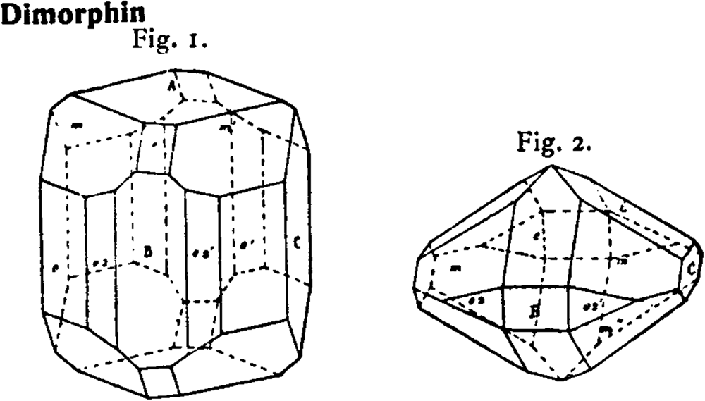
In his ‘Memorie geologiche sulla Campania’, a detailed report of the mineralogical phases occurring in the fumaroles of the Campi Flegrei, near Napoli, Italy, Arcangelo Scacchi (Reference Scacchi1850) observed the presence of an arsenic sulfide, with probable composition As4S3, displaying two distinct crystalline forms. From accurate goniometric measurements, he found two distinct morphologies and axial ratios, corresponding to two possibly distinct phases, a more abundant form I with an a:b:c ratio of 1:1.287:1.153 and a form II with the a:b:c ratio 1:1.658:1.508. (Fig. 1). Nevertheless, Scacchi considered his own analyses indecisive for establishing a definitive chemical formula and the existence of two different mineral species, because of the small quantity of material used in the analysis and because the method used involved quantitative analysis only for sulfur. For these reasons the name ‘dimorfina’ or later dimorphite (from the Greek), with reference to the two forms in which it was thought to exist, was used with no distinction for the two minerals.
Morphology of the two As4S3 forms observed by Scacchi (Reference Scacchi1850) and redrawn in the ‘Atlas der Kristallformen’ by Goldschmidt (Reference Goldschmidt1913). Form I (paradimorphite) left, form II (dimorphite) right.

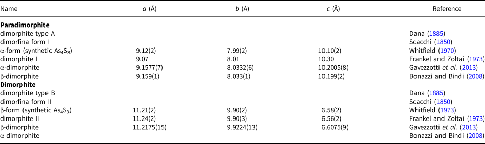
After Scacchi's discovery, there has been considerable discussion about whether dimorphite is indeed a distinct mineral or instead a morphologically unusual orpiment, As2S3, not recognised by Scacchi (Kenngott, Reference Kenngott1870; Dana, Reference Dana1885; Palache et al., Reference Palache, Berman and Frondel1944). Schuller (Reference Schuller1894) and Krenner (Reference Krenner1907) showed As4S3 can be synthesised by direct combination of the elements mixed in stoichiometric proportion, and their work allowed the existence of the synthetic counterparts of one of the two dimorphites to be demonstrated. In fact Krenner (Reference Krenner1907) showed that the forms and angles of the second type of dimorphite discovered by Scacchi (form II) agree closely with those of synthetic As4S3 prepared by Schuller (Reference Schuller1894) and that Scacchi's second type of dimorphite should be considered a valid mineral species. In the Commission on New Minerals, Nomenclature and Classification of the International Mineralogical Association (IMA–CNMNC) list of mineral species updated January 2022 (Pasero, Reference Pasero2022), dimorphite is indeed considered a valid mineral and corresponds to Sacchi's form II, the polymorph, stable at room temperature, with a = 11.21(2), b = 9.90(2), c = 6.58(1) Å, space group Pnma, whose structure was determined by Whitfield (Reference Whitfield1973) on the synthetic phase. Whitfield called this phase β-form, despite this polymorph being stable at room temperature. The structure of the other polymorph with a = 9.12(2), b= 7.99(2), c = 10.10(2) Å and space group Pnma, had been reported earlier (Whitfield, Reference Whitfield1970) by the same author, who called this phase α-form. The existence of the two distinct natural polymorphs was confirmed, using powder X-ray diffraction and single-crystal precession data by Frankel and Zoltai (Reference Frankel and Zoltai1973), working on samples of dimorphite from Vesuvius, obtained by the Swedish Natural History Museum. They also observed that in some crystals, having the morphology of form I, both polymorphs were present, and they concluded that the existence of dimorphite II crystals as pseudomorphs after dimorphite I suggests that dimorphite I is unstable under conditions to which it was exposed after crystallisation, and transforms to dimorphite II. The contemporary presence of both polymorphs was also observed by us in some specimens. An accurate structure refinement on both phases of natural origin was later carried out by some of us (Gavezzotti et al., Reference Gavezzotti, Demartin, Castellano and Campostrini2013) together with a theoretical evaluation of the stability of both polymorphs at room temperature. In a crystallographic review of arsenic sulfides by Bonazzi and Bindi (Reference Bonazzi and Bindi2008) the two forms are labelled according to their field of stability, i.e. α-dimorphite the phase stable at room temperature and β-dimorphite the phase stable above 130°C.
It can be seen that much confusion exists in the literature for the use of α- and β-descriptors of the As4S3 polymorphs (see Table 1). Following the rules in the nomenclature of temperature-depending phase transitions, the low-temperature and the high-temperature forms should be labelled as α- and β-form, respectively, as reported by Bonazzi and Bindi (Reference Bonazzi and Bindi2008).
History of the different naming of the two As4S3 polymorphs.

As a part of our studies on fumarolic minerals (Russo et al. Reference Russo, Campostrini and Demartin2017; Campostrini and Demartin, Reference Campostrini and Demartin2021; Campostrini et al., Reference Campostrini, Demartin and Russo2019a, Reference Campostrini, Demartin and Scavini2019b; Demartin et al., Reference Demartin, Campostrini, Castellano and Russo2014), we have investigated some ‘dimorphite’ crystals from Solfatara di Pozzuoli and from Vesuvius crater and the results of a crystallographic study on both natural polymorphs of As4S3 have been reported in Gavezzotti et al. (Reference Gavezzotti, Demartin, Castellano and Campostrini2013). A recent complete characterisation of the high-temperature phase, not yet recognised as a valid mineral species, prompted us to submit a proposal to the IMA–CNMNC, to include this phase in the list of the mineral species (IMA2020-101, Campostrini et al., Reference Campostrini, Castellano, Demartin, Rocchetti, Vignola and Russo2022). On April 2021 we received a communication from the chairman of the Commission reporting that the proposed new mineral had major YES votes but, as the CNMNC was going to consider revision of the guidelines for the nomenclature of polymorphs and polysomes, the voting result for the approval was suspended. According to the new rules established by IMA-CNMNC only recently, the mineral name was finally approved as paradimorphite, by analogy with the low temperature polymorph dimorphite. This decision overcomes the confusion arisen using the α- and β-descriptors. The approved mineral name abbreviation is Pdim (Warr, Reference Warr2021).
The holotype (from Solfatara di Pozzuoli) and cotype (from Vesuvius) specimens of paradimorphite are deposited in the Reference Collection of the Dipartimento di Chimica, Università degli Studi di Milano, catalogue numbers 2020-03/6121 and 2020-04/4226, respectively.
In Strunz classification paradimorphite is in 2.FA.10 (Dimorphite group) 2: SULFIDES and SULFOSALTS (sulfides, selenides, tellurides; arsenides, antimonides, bismuthides; sulfarsenites, sulfantimonites, sulfbismuthites, etc.) F: Sulfides of arsenic, alkalies; sulfides with halide, oxide, hydroxide, H2O, A: With As, (Sb), S. In the Dana classification paradimorphite belongs to the 2.6.1.1 class, 2: SULFIDES 6: AmBnXp, with (m+n):p = 4:3.
Occurrence
The Solfatara di Pozzuoli is an explosion volcano formed 4285 years ago, located in the central part of the Campi Flegrei caldera, Napoli, Campania, Italy (40°49’41”N, 14°08’30”E). The deposits of the Solfatara consist of pyroclastic products dispersed over an area of about one square kilometre. Inside the crater the deposits are strongly altered by the intense fumarolic activity. The morphological characteristics of the volcano represent a unicum for the Campi Flegrei area as it is a maar-diatreme. The holotype specimen of paradimorphite was collected here at the Bocca Grande fumarole. Associated minerals are realgar, salammoniac, mascagnite, alacránite, adranosite and russoite (Russo et al., Reference Russo, Campostrini and Demartin2017).
The Somma–Vesuvius is a strato-volcano, whose oldest part is represented by Monte Somma in whose interior the ‘Gran Cono’ of Vesuvius was formed. The volcano's explosive activity took place in some ‘Plinian’ eruptions (e.g. Pomici di Avellino eruption, ~4000 years ago and Pompei eruption, 79 A.D.); subsequently the formation of the Vesuvius cone began. After the great eruption of 1631, Vesuvius entered a state of ‘open conduit’ activity with frequent eruptions, on average one every seven years. The most important eruptions of the last century were those of 1906 and the last one in March 1944. Currently, Vesuvius is in state of quiescence with some seismic and fumarolic evidence. The cotype specimen of paradimorphite was collected from an active fumarole after the 1906 eruption and was found among old specimens belonging to the, now dispersed, collection of the Istituto Geomineralogico Italiano (Campostrini and Russo, Reference Campostrini and Russo2012). Associated minerals in the cotype specimen are anhydrite and sassolite. Realgar, lafossaite, anhydrite, bonazziite and an unknown arsenic thallium chloride, probably related to lucabindiite are associated with paradimorphite in other specimens. In both localities the mineral is a fumarolic sublimate.
Physical and optical properties
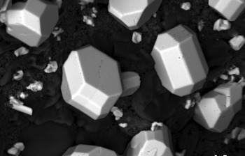
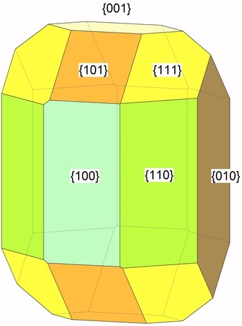
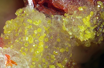
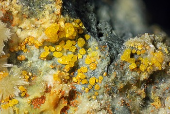
Crystals of paradimorphite are orange yellow, transparent or semitransparent, with adamantine lustre. Habit is prismatic and observed forms are {110}, {101}, {111}, {100}, {010} and {001} (Figs 2–5). Tenacity is brittle, no distinct cleavage is observed and fracture is conchoidal. The mineral does not fluoresce in long- or shortwave ultraviolet light. No twinning is apparent. The streak is saffron yellow. Hardness (Mohs) = 1–2. Vickers hardness (micro-indentation): VHN25 = 70 (range 59–80 kg/mm2) (obtained using a Shimadzu type-M microhardness tester; average of five indentation measurements). The density, measured by flotation in a thallium malonate/formate solution (Clerici solution) for the Solfatara sample (holotype), is 3.510(3) g/cm3. The calculated density is 3.500 g/cm3 (Solfatara), 3.520 g/cm3 (Vesuvius), using the empirical formula and single-crystal cell data.
Back-scatter electron image of paradimorphite from Solfatara di Pozzuoli (Field of view 120 μm, specimen #EDS-5955).

Idealised drawing of a paradimorphite crystal similar to that on the right of Fig. 2. Using Kristall 2000 (Klaus Schilling, http://www.kristall2000.de/).

Paradimorphite crystals on mascagnite with realgar from Solfatara di Pozzuoli (Field of view 3 mm).

Paradimorphite crystals with anhydrite, cotype sample (#2020-04/4226), from Vesuvius crater (Field of view 3 mm).

Refractive indices were not measured conventionally, because they were found to be higher than available reference liquids (>1.9). The mineral is biaxial (+), dispersion is weak to very weak, and r > v. Pleochroism is barely noticeable.
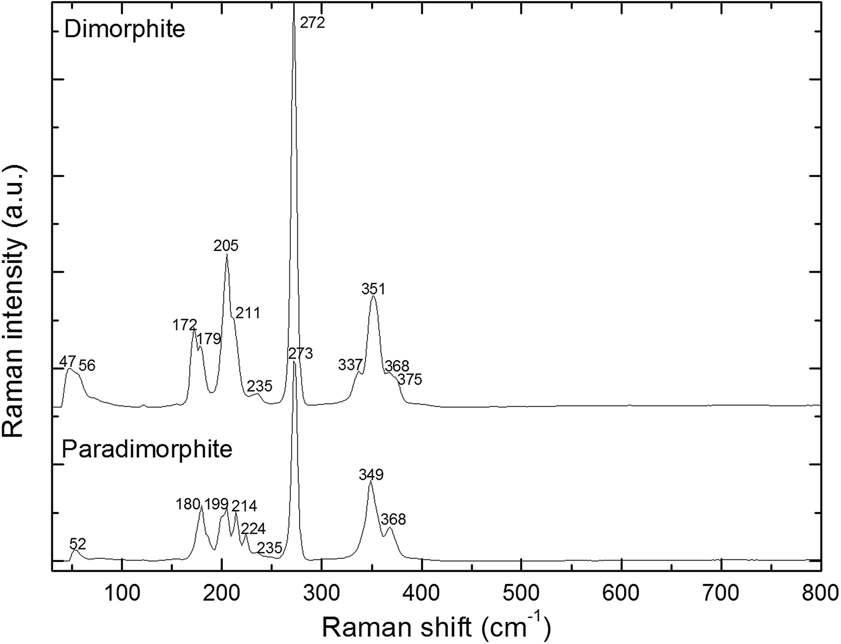
The Raman spectrum was obtained with an ANDOR 303 spectrometer equipped with a CCD camera iDus DV420A-OE and using the 532 nm line of an OXXIUS solid state laser for excitation. Figure 6 shows a comparison of the Raman spectrum of paradimorphite with that of a dimorphite collected recently at Vesuvius. Due to the similarity in the packing of the molecules in the two polymorphs (see below), frequencies related to lattice modes should essentially occur at very similar values. A broad band at ~52 cm–1, is indeed observed in paradimorphite whereas the corresponding band of dimorphite appears at 47 cm–1 with a shoulder at 56 cm–1. All the other bands fall in the range of vibrational frequencies (125–386 cm–1) calculated by Gavezzotti et al. (Reference Gavezzotti, Demartin, Castellano and Campostrini2013) for the As4S3 molecule in the gas phase. It should be pointed out the lack of a shoulder at 337 cm–1 in paradimorphite with respect to dimorphite and the presence of a band at 224 cm–1.
Raman spectra of paradimorphite from Solfatara di Pozzuoli and dimorphite from Vesuvius.

Chemical analysis
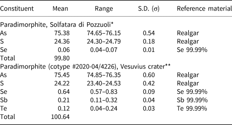
Quantitative chemical analyses (6) were carried out in wavelength dispersive spectroscopy (WDS) mode using a JEOL JXA–8200 WDS electron microprobe (15 kV excitation voltage, 5 nA beam current and 5 μm beam diameter). The following mineral and pure elements served as standards: realgar (As and S), pure elements (99.99% for Se, Sb and Te). X-ray intensities were converted to wt.% by ZAF quantitative analysis software. Chemical data for the Solfatara and Vesuvius samples are reported in Table 2. The empirical formula, calculated on the basis of 7 atoms per formula unit, for the Solfatara sample is: As3.986(S3.011Se0.003), that of the Vesuvius crater sample is: (As3.975Sb0.007)(S2.982Se0.032Te0.004). The simplified formula is: As4S3.
Analytical data (in wt.%) for paradimorphite (average of 6 analyses).

*The empirical formula calculated on the basis of 7 atoms per formula unit is: As3.986(S3.011Se0.003)
**The empirical formula calculated on the basis of 7 atoms per formula unit is: (As3.975Sb0.007)(S2.982Se0.032Te0.004)
S.D. – standard deviation
The ideal formula is As4S3 which requires: As 75.70, S 24.30 wt.%, total 100 wt.%.
X-ray crystallography and crystal structure determination
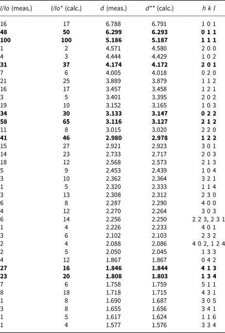
Powder X-ray diffraction data were collected for the Solfatara specimen with a Rigaku DMAX II powder diffractometer with graphite monochromatised CuKα radiation. Data (in Å for CuKα) are listed in Table 3. Unit cell parameters refined from the powder data using UNITCELL (Holland and Redfern, Reference Holland and Redfern1997) are a = 9.1596(9), b = 8.0365(9), c = 10.1195(10) Å and V = 744.92(9) Å3. None of the intense reflections corresponding to dimorphite were observed in the powder pattern of this sample.
Powder X-ray diffraction data for paradimorphite.

*Calculated from the refined structure; **calculated from the unit cell a = 9.1596(9), b = 8.0365(9), c = 10.1195(10) Å and V = 744.92(9) Å3 obtained from least-squares refinement of the above data using the program UNITCELL (Holland and Redfern, Reference Holland and Redfern1997). The strongest lines are given in bold.
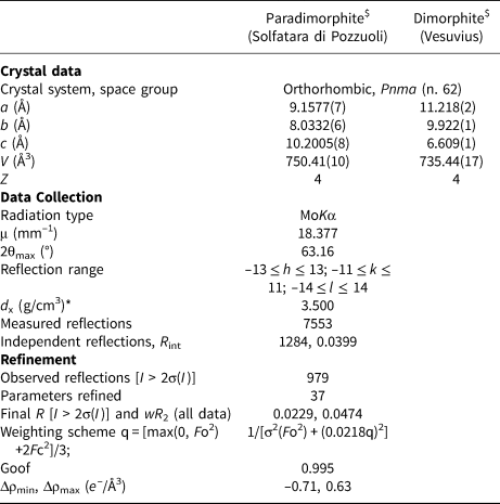
Single crystal data for paradimorphite were obtained from a crystal fragment of the Solfatara sample. Details about the data collection and refinement are summarised in Table 4, whereas the final atom coordinates and anisotropic displacement parameters and selected interatomic distances have already been reported by Gavezzotti et al. (Reference Gavezzotti, Demartin, Castellano and Campostrini2013). Single-crystal data of the Vesuvius sample gave the following unit-cell parameters: a = 9.155(3), b = 8.026(2), c = 10.201(6) Å and V = 749.55(20) Å3. The a:b:c ratio calculated from the unit-cell parameters is 1.1400:1:1.2698 (single-crystal data, Solfatara sample), 1.1407:1:1.2710 (single-crystal data, Vesuvius sample). The crystallographic information file has been deposited with the Principal Editor of Mineralogical Magazine and is available as Supplementary material (see below).
Crystal data of the two As4S3 polymorphs, data collection and structure refinement details for paradimorphite.

$ Data from Gavezzotti et al. (Reference Gavezzotti, Demartin, Castellano and Campostrini2013); *calculated using the empirical formula and single-crystal cell data.
Notes: R = Σ||F o|–|F c||/ Σ|F o|; wR 2 = {Σ[w(F o2–F c2)2] / Σ[w(F o2)2]}½; Goof ={Σ[w(F o2–F c2)]/(n–p)}½ where n is the number of reflections and p is the number of refined parameters.
Description of the crystal structure and discussion
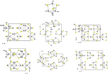
A comparison of the structures of the two polymorphs, paradimorphite and dimorphite, is shown in Fig. 7. Crystals of both phases contain cage-like As4S3 molecules of C 3v idealised symmetry located about a crystallographic mirror and packed together by weak van der Waals interactions. Other cage-like covalently bonded As4Sn (n = 4 and 5) molecules are also the building blocks of some other arsenic sulfides (see Bonazzi and Bindi, Reference Bonazzi and Bindi2008 for a general review), such as the As4S4 molecules in realgar (Mullen and Nowacki, Reference Mullen and Nowacki1972), pararealgar (Bonazzi et al., Reference Bonazzi, Menchetti and Pratesi1995) and bonazziite (Bindi et al., Reference Bindi, Pratesi, Muniz-Miranda, Zoppi, Chelazzi, Lepore and Menchetti2015); the As4S5 molecules in uzonite (Bindi et al., Reference Bindi, Popova and Bonazzi2003); and those in alacránite, As8S9 (Bonazzi et al., Reference Bonazzi, Bindi, Popova, Pratesi and Menchetti2003) that contains both As4S4 and As4S5 molecules. In the As4S3 molecules of paradimorphite and dimorphite, the four arsenic atoms are arranged with a triangular pyramidal geometry, and the sulfur atoms bridge the three apical edges of the pyramid. Molecular dimensions and conformation are within standard uncertainties the same for the molecules in the two polymorphs. These are also very similar to those observed in the tetrakis(tris(μ2-sulfido)-tetra-arsenic)-tetraphenylphosphonium chloride complex (Siewert and Müller, Reference Siewert and Müller1992) where the As–As bonds are in the range 2.464(3)–2.494(3) Å and the As–S bonds in the range 2.229(6)–2.243(6) Å. For paradimorphite, the As–As bonds of the triangular base are in the range 2.4673(4)–2.4855(6) Å and are on average significantly shorter than those observed for the As4S4 molecules in realgar (2.566(1)–2.571(1) Å), bonazziite (2.579(1) Å), pararealgar (2.484(4)–2.534(4) Å) and alacránite (2.579(5) Å), and those in the As4S5 molecules in uzonite (2.527(1) Å) and alacránite (2.566(6) Å). The As–S bonds fall in the range 2.2155(7)–2.2360(10) Å and are again on the average shorter than those observed in realgar (2.228(2)–2.247(2) Å), pararealgar (2.228(10)–2.261(8) Å) and uzonite (2.237(1)–2.261(1) Å), but closer to those found in alacránite (2.205(9)–2.238(7) Å) and bonazziite (2.222(1)–2.234(1) Å). These geometrical differences can be ascribed to the different bonding pattern in the As4Sn molecules. For n = 4 and 5, instead of being arranged in a pyramidal fashion as in As4S3, the As atoms are located at the vertices of a more-or-less regular disphenoid where there are (6 – n) disphenoidic edges corresponding to As–As bonds and the sulfur atoms bridge n among the six disphenoidic edges. Thus, the number of As–S bonds formed by each As atom, combined with the steric hindrance of the additional S atom, determines the geometry of the As4 framework.
The As4S3 molecule and comparison of the structures of paradimorphite (up) and dimorphite (down); drawn with DIAMOND software (Crystal Impact GbR. Bonn, Germany).

From a comparison of the projections of the structure of both polymorphs reported in Fig. 7 it seems that there are no substantial differences in the molecular packing and orientation of the As4S3 molecules. A detailed study of the intermolecular interactions, carried out by Gavezzotti et al. (Reference Gavezzotti, Demartin, Castellano and Campostrini2013), showed that slight, but significant differences in the packing of the As4S3 molecules could be inferred from radial distribution curves of the centres of mass of the molecules, that are essentially rigid cages. In dimorphite the As4S3 molecules are oriented with their pseudo threefold axis along [102] and [10$\bar{2}$ ] whereas in paradimorphite the threefold axes are along [101] and [10$\bar{1}$
 ], but these directions form similar angles with the crystallographic axes. These features can account for the fact that the space group is maintained during the transition from one polymorph to the other, due to slight deformation of the lattice. The larger unit-cell volume of paradimorphite with respect of that of the low-temperature form dimorphite (see Table 4) is in line with its stability at higher temperatures.
The lack of a distinct cleavage and the conchoidal fracture in paradimorphite are in keeping with the absence of molecular layers that are instead present in other molecular arsenic sulfides such as realgar.
Acknowledgements
Valuable suggestions and constructing comments for improving this paper have been given by the Principal Editor Dr. Stuart Mills, Prof. Peter Leverett, Dr. Sergey M. Aksenov and by an anonymous referee.
Supplementary material
To view supplementary material for this article, please visit https://doi.org/10.1180/mgm.2022.47
Competing interests
The authors declare none.