Introduction
Diethylstilbestrol (DES) is a synthetic estrogen that was prescribed to over 2 million pregnant women from the 1940s through the early 1970s. It was later discovered to be associated with the occurrence of vaginal clear cell adenocarcinoma,Reference Herbst, Ulfelder and Poskanzer 1 anatomic abnormalities of the reproductive tractReference Stillman 2 and poor reproductive outcomes in prenatally exposed daughters.Reference Beral and Colwell 3 – Reference Hatch, Troisi and Wise 6
Prenatal DES exposure is hypothesized to exert long-term effects on female endocrine function via dysregulation of the hypothalamic–pituitary–ovarian axis and alterations in hormone biosynthesis.Reference Hatch, Troisi and Wise 6 – Reference Wu, Mangan, Burtnett and Mikhail 9 Two animal studiesReference Haney, Newbold and McLachlan 10 , Reference Fuller, Yates, Helton and Hobson 11 and two human studiesReference Peress, Tsai, Mathur and Williamson 8 , Reference Wu, Mangan, Burtnett and Mikhail 9 have assessed the effects of DES on endogenous sex steroid hormones or gonadotropins, but results were inconclusive. Exposure was documented by medical record in both human studies,Reference Peress, Tsai, Mathur and Williamson 8 , Reference Wu, Mangan, Burtnett and Mikhail 9 but neither timed blood collection to a particular phase of the menstrual cycle.
In the Harvard Study of Moods and Cycles (HSMC), a prospective cohort study of late reproductive-aged women, we evaluated the associations of self-reported prenatal DES exposure with early follicular phase concentrations of estradiol, follicle-stimulating hormone (FSH), luteinizing hormone (LH) and inhibin B.
Method
Study population
Data for the present analysis were derived from the HSMC (1995–1999), a prospective cohort study designed to investigate the influence of depression on ovarian function. Methods for this study are described in greater detail elsewhere.Reference Harlow, Cohen, Otto, Spiegelman and Cramer 12 , Reference Harlow, Cohen and Otto 13 In 1995, study investigators identified women between the ages of 36 and 45 years from seven communities in the Greater Boston area using Massachusetts Town Books (annual publications that list residents by name, age and address according to voter precincts). Self-administered screening questionnaires eliciting data on reproductive, menstrual and medical history were mailed to 6228 women with a verified address and telephone number; 73.5% responded to this questionnaire. The questionnaire also assessed past diagnosis with or treatment for depression, and included the Center for Epidemiologic Studies Depression Scale to measure current depressive symptoms.Reference Radloff 14
Based on response to the initial survey, study investigators invited women to participate in a 3-year study that included structured clinical psychiatric interviews, baseline and semi-annual medical questionnaires and blood collection. Women were deemed ineligible for enrollment due to pregnancy, postmenopausal status or loss to follow-up after the initial screening assessment. The remaining women provided blood specimens and participated in follow-up in-person interviews every 6 months for a total of 3 years. The study was approved by the Institutional Review Board at Brigham and Women’s Hospital. All participants provided written informed consent.
Assessment of DES and covariates
On the baseline questionnaire, women were asked ‘Did your mother use the drug DES when she was pregnant with you?’ with the following response options: ‘No,’ ‘Yes,’ or ‘Don’t know.’ Information on demographics, lifestyle, anthropometry, medical history, smoking and other predictors of reproductive hormones were collected on the baseline questionnaire. Caffeine and alcohol consumption were assessed separately by means of the validated Willett food frequency questionnaire.Reference Lucero, Harlow, Barbieri, Sluss and Cramer 15 Body weight and height were used to calculate body mass index (BMI, kg/m2). Medical questionnaires assessed menstrual history including age at menarche, average cycle length, pregnancy and breastfeeding history, and oral contraceptive (OC) or other hormone use. History of infertility was assessed as follows: ‘Did you ever try for more than 2 years to get pregnant or have problems carrying a pregnancy?’
Assessment of hormone concentrations
To measure hormone concentrations, blood was drawn on one of the first 5 days of the menstrual cycle with ‘day 1’ defined as the 1st day of menstrual bleeding. The specimen was drawn during the first menstrual period after study enrollment and was scheduled every 6 months over 36 months of follow-up. Early follicular-phase concentrations of FSH, LH and estradiol were measured through 36 months of follow-up. Inhibin B concentrations were measured among subjects that had provided specimens at baseline and at least twice over the first 18 months of follow-up.Reference Lambert-Messerlian and Harlow 16 Reminder calls were placed to women around the estimated time of their 6-month menses, at which time the regularity of the menstrual cycle and new events such as pregnancy, breastfeeding, hormone use and gynecologic surgeries were ascertained. Women who started using hormones or whose periods had stopped temporarily due to pregnancy or breastfeeding had their interval-specific hormone measurements set to missing. Women whose periods stopped permanently due to oophorectomy were censored at the first report of their surgery.
Measurements of serum gonadotropins were performed using the Coat-A-Count immunoradiometric assays (Diagnostic Products Corp., Los Angeles, CA, USA). The World Health Organization (WHO) First International Reference Preparation (68/40) of human pituitary LH was used as the standard for LH and the WHO Second International Reference Preparation (78/549) of human pituitary FSH was used as the standard for FSH. Estradiol was measured in duplicate and averaged using the Coat-A-Count radioimmunoassay. Lab-based intra-assay coefficients of variation (CV) were <4% for the gonadotropins and <7% for estradiol. Lab-based inter-assay CVs were <10% for the gonadotropins and <15% for estradiol.
Inhibin B levels were measured using ELISA (Diagnostic Systems Laboratories, Webster, TX, USA)Reference Groome, Illingworth and O'Brien 17 using a modified method where sample treatment was achieved at room temperature during incubation.Reference Lambert-Messerlian and Harlow 16 This method has been validated for measurement of inhibin B in human serumReference Diamandi, Krishna, Bodani, Mistry and Khosravi 18 and resulting values are highly correlated with those obtained using the original method (r=0.91, Diagnostic Systems Laboratories package insert DSL-10-84100).Reference Lambert-Messerlian and Harlow 16 The assay limit of sensitivity was 30 pg/ml, and lab-based inter- and intra-assay CVs were both <15%. Hormones were tested, in duplicate, without operator knowledge of participant’s exposure status.
Exclusions
Of the 976 women enrolled, we excluded 51 women without any follow-up data (5%; 0 exposed, 44 unexposed and seven unknown exposure status) and 26 women who reported the removal of one or more ovaries at baseline (3%; 0 exposed, 25 unexposed and one unknown exposure status), 63 women who reported current use of ‘birth control pills, estrogen, progesterone, or fertility drugs’ at baseline (6%: two exposed, 57 unexposed and four unknown exposure status), and 11 women who were pregnant within 6 months of baseline (1%: one exposed, 10 unexposed). After these exclusions, 825 women (43 exposed and 782 unexposed) remained in the analysis of hormones measured at baseline and over follow-up.
Statistical analysis
Hormone values were log-transformed to accommodate skewness of distributions. For each individual, we identified the minimum and maximum of each of the measured hormone values, regardless of follow-up interval; we also calculated the woman’s average value for each hormone across all follow-up intervals. In the analysis of repeated measures, we transformed the data set to allow for multiple observations per woman per time point, with each woman contributing up to seven observations for FSH, LH and estradiol (0, 6, 12, 18, 24, 30 and 36 months) and up to four observations for inhibin B (0, 6, 12 and 18 months). We then constructed multivariable mixed linear regression models using SAS PROC MIXED to assess whether log-transformed mean hormone concentrations differed by DES exposure. We calculated percent differences in the mean hormone concentrations by exponentiating the β coefficient, subtracting 1 and multiplying by 100. We also assessed the extent to which log-transformed hormones changed over time, overall and by exposure status, with DES exposure and potential confounders as independent variables and each of the repeated hormone values as the dependent variable.
Mixed linear regression models were adjusted for age at baseline (continuous) and other factors associated with both exposure and hormone levelsReference Lucero, Harlow, Barbieri, Sluss and Cramer 15 at baseline, including age at menarche (continuous), parity (0 v. ⩾1 birth), BMI (continuous), years of OC use (never or <1, 1–4, ⩾5 years), current alcohol consumption (<5, 5–9, ⩾10 g), current caffeine consumption (<100, 100–299, ⩾300 g), pack-years of smoking (never smoked, <5, 5–9, ⩾10 pack-years), smoking status (current, past, never) and cycle day of blood draw (0, 1, 2, 3, 4, ⩾5). Depression history at baseline (ever v. never), while not appreciably associated with mean hormone concentrations,Reference Harlow, Wise, Otto, Soares and Cohen 19 was included in the final model because it was a selection criterion for the cohort. Nonparametric restricted cubic splines were used to display the change in log-transformed hormone concentrations over time by exposure status.Reference Li, Hertzmark and Spiegelman 20 For each hormone, a single mixed linear regression model was fit with main effect terms for DES, follow-up time (original term), follow-up time (spline term), interaction terms between DES and follow-up time (original and spline terms) and all covariates listed above.
Separate analyses were conducted to assess DES exposure in relation to clinically relevant cut points for low ovarian reserve (FSH levels >10 IU/I and >20 IU/I; inihibin B levels <45 pg/ml). In these analyses, logistic regression models were used to estimate odds ratios (OR) and 95% CIs.Reference Zou 21
We used multiple imputation to impute missing values for DES exposure, hormones and covariates.Reference Zhou, Eckert and Tierney 22 In SAS, we used PROC MI to create 10 imputed data sets and PROC MIANALYZE to combine results across data sets. 23 All potential confounders were included in the imputation procedure. The percentage of women with missing data was zero for age, age at menarche, education and body weight, and was relatively low for other covariates, ranging from 0.5% (years of OC use) to 11.8% (caffeine or alcohol consumption; Supplementary Table S1). About 9% of women said they were unsure of their DES exposure status. Although the proportion of missing data increased substantially for each of the hormone concentrations by 36 months of follow-up, and was as high as 64.8% for inhibin B measurements (by 18 months), the results from multiple imputation analyses were similar to those obtained using the complete case approach (41 exposed, 708 unexposed, 76 don’t know; Supplementary Table S2). The missingness for hormone levels was largely attributable to the fact that as women approached the menopausal transition, it was harder to obtain a menstrually timed blood sample due to amenorrhea or the initiation of hormone therapy.Reference Harlow, Wise, Otto, Soares and Cohen 19 Analyses were performed using SAS version 9.2. 23
Results
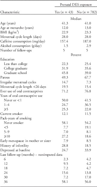
DES-exposed women were similar to unexposed women in terms of age, BMI, menstrual cycle length and the prevalence of early menopause in a mother or sister (Table 1). Consistent with the literature, DES-exposed women tended to have a slightly earlier age at menarche, greater menstrual cycle irregularity, reduced parity and a greater prevalence of infertility. The median number of completed follow-ups was identical in exposed and unexposed groups.
Baseline characteristics by DES exposure, Harvard Study of Moods and Cycles (1995–1999)

DES, diethylstilbestrol.
n=number of participants. Based on imputed data.
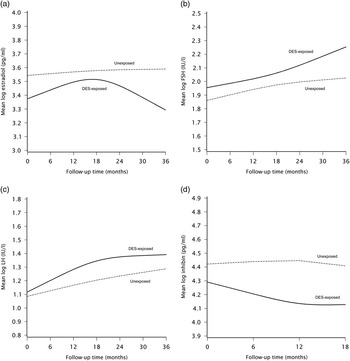
Unadjusted baseline, minimum, maximum and average FSH levels were consistently higher for DES-exposed women than unexposed women (Table 2). In contrast, estradiol and inhibin B levels were consistently lower for DES-exposed women relative to unexposed women. These findings agreed with spline graphs showing that exposed women had lower estradiol and inhibin B concentrations (Fig. 1a and 1d) and higher FSH concentrations (Fig. 1b) than unexposed women. The spline graphs showed little variation in DES-related hormone differences over time, with the exception of estradiol, where the differences in hormone levels appeared to diverge after 18 months of follow-up.
(a) Restricted cubic spline showing change in log-transformed mean estradiol concentrations over time among DES-exposed and unexposed women. Knots were placed at 6, 18 and 30 months. Plots are modeled at the median or modal value for age at baseline, age at menarche, parity, body mass index (BMI), current smoking, pack-years of smoking, alcohol, caffeine, years of oral contraceptive use, depression history and day of blood draw. (b) Restricted cubic spline showing change in log-transformed mean FH concentrations over time among DES-exposed and unexposed women. Knots were placed at 6, 18 and 30 months. Plots are modeled at the median or modal value for age at baseline, age at menarche, parity, BMI, current smoking, pack-years of smoking, alcohol, caffeine, years of oral contraceptive use, depression history and day of blood draw. (c) Restricted cubic spline showing change in log-transformed mean LH concentrations over time among DES-exposed and unexposed women. Knots were placed at 6, 18 and 30 months. Plots are modeled at the median or modal value for age at baseline, age at menarche, parity, BMI, current smoking, pack-years of smoking, alcohol, caffeine, years of oral contraceptive use, depression history and day of blood draw. (d) Restricted cubic spline showing change in log-transformed mean inhibin B concentrations over time among DES-exposed and unexposed women. Knots were placed at 6, 12 and 18 months. Plots are modeled at the median or modal value for age at baseline, age at menarche, parity, BMI, current smoking, pack-years of smoking, alcohol, caffeine, years of oral contraceptive use, depression history and day of blood draw.

Unadjusted geometric meansFootnote a of baseline, minimum, maximum and average hormone values within each woman by prenatal DES exposure

DES, diethylstilbestrol; FSH, follicle-stimulating hormone; LH, luteinizing hormone.
a Estimates are back-transformed means from log-transformed hormone values.
b Inhibin B data are based on 0–18 months of follow-up only.
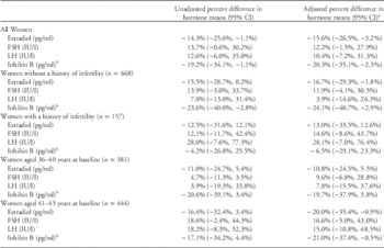
In multivariable-adjusted repeated measures analyses (Table 3), DES-exposed women had lower mean levels of estradiol (pg/ml) than unexposed women (% difference in means=−15.6%, 95% CI: −26.5%, −3.2%) and lower mean levels of inhibin B (pg/ml) than unexposed women (% difference in means=−20.3%, 95% CI: −35.1%, −2.3%). DES-exposed women had higher mean levels of FSH (IU/I) [% difference in means=12.2%, 95% confidence interval (CI): −1.5%, 27.9%] and LH (IU/I) (% difference in means=10.4%, 95% CI: −7.2%, 31.3%) than unexposed women; the LH results were the weakest of all four hormones assessed. Results did not vary appreciably by infertility history or age at baseline, with the exception of stronger results for FSH and LH among older reproductive-aged women, and stronger results for LH among women with a history of infertility.
Prenatal DES exposure in relation to reproductive hormones

DES, diethylstilbestrol; FSH, follicle-stimulating hormone; LH, luteinizing hormone; BMI, body mass index; CI, confidence interval.
Percent differences and 95% CI comparing DES-exposed with unexposed women.
a Adjusted for age at baseline, age at menarche, parity, BMI, current smoking, pack-years of smoking, alcohol, caffeine, years of oral contraceptive use, depression history and day of blood draw.
b Inhibin B data are based on 0–18 months of follow-up. All other missing data on hormones have been imputed.
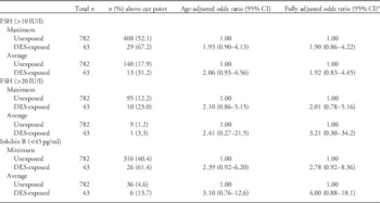
There was a positive association between DES exposure and measures of low ovarian reserve (Table 4). A higher proportion of DES-exposed women had FSH >20 IU/I, the traditional cut point used to define menopause, though numbers of exposed women were small. The adjusted ORs for maximum and average FSH >10 IU/I (less stringent cut point for low ovarian reserve) were 1.90 (95% CI: 0.86, 4.22) and 1.92 (95% CI: 0.83, 4.45), respectively, comparing exposed with unexposed women. The adjusted risk ratios for minimum and average inhibin B <45 pg/ml were 2.78 (95% CI: 0.92, 8.36) and 4.00 (95% CI: 0.88, 18.1), respectively.
Association between prenatal DES exposure and clinical indicators of low ovarian reserve

DES, diethylstilbestrol; FSH, follicle-stimulating hormone; CI, confidence interval.
Odds ratios and 95% CIs comparing DES-exposed with unexposed women.
a Adjusted for age at baseline, current smoking, pack-years of smoking, age at menarche, parity, BMI, depression history, year of oral contraceptive use, alcohol consumption and caffeine consumption.
Finally, we compared results from analyses that incorporated different ways of handling missing data (Supplementary Table S2). The results from the complete case and missing indicator approaches were generally weaker but in agreement with the analyses using multiple imputation. The multiple imputation analyses with and without imputation of DES exposure were nearly identical, indicating that differences in estimates were influenced mainly by the missing outcome data.
Discussion
The present prospective cohort study of premenopausal women aged 36–45 years from the Greater Boston area indicated that prenatal DES exposure was associated with increased FSH concentrations, and decreased estradiol and inhibin B concentrations, relative to unexposed women. The percent differences in mean hormone concentrations ranged from ∼10.4% to 20.3%, and were relatively consistent over follow-up. This range of effect was similar in magnitude to that observed for other established predictors of hormone values in this cohort (e.g. current smoking was associated with a 10.3% increase in FSH relative to never smoking). Prenatal DES exposure was also positively associated with clinically relevant changes in FSH (>10 and >20 IU/I) and inhibin B (<45 pg/ml), though these associations had limited precision. To the extent that high FSH and low inhibin B levels are indicative of lower ovarian reserve, these data suggest that DES-exposed women experience earlier ovarian decline than similarly aged women not exposed prenatally. These findings are consistent with a previous epidemiologic study in which women exposed prenatally to DES were more likely to experience early menopause;Reference Hatch, Troisi and Wise 6 sex steroid hormones were not measured in that study.
Our study findings generally agree with animal and human studies showing that females exposed prenatally to DES experience alterations in sex steroid hormones and gonadotropins that persist into adulthood, although the direction of associations is inconsistent. Animal data show that in vitro production of testosterone, total estrogen and progesterone by ovarian tissue was significantly greater in female mice exposed prenatally to DES, at all ages studied.Reference Haney, Newbold and McLachlan 10 Among female rhesus monkeys exposed prenatally to DES, abnormal gonadotropin patterns were observed during the first 500 days postnatally – specifically, LH and FSH concentrations were elevated after 4 and 5 months of age, respectively.Reference Fuller, Yates, Helton and Hobson 11 In humans, prenatal exposure to DES in female offspring was associated with elevated concentrations of serum testosterone,Reference Peress, Tsai, Mathur and Williamson 8 , Reference Wu, Mangan, Burtnett and Mikhail 9 but not appreciably associated with progesteroneReference Wu, Mangan, Burtnett and Mikhail 9 or estradiol.Reference Peress, Tsai, Mathur and Williamson 8 , Reference Wu, Mangan, Burtnett and Mikhail 9 FSH concentrations were materially elevated in DES-exposed women in two studies,Reference Peress, Tsai, Mathur and Williamson 8 , Reference Wu, Mangan, Burtnett and Mikhail 9 but no appreciable differences have been found in LHReference Peress, Tsai, Mathur and Williamson 8 , Reference Wu, Mangan, Burtnett and Mikhail 9 or the ratio of FSH to LH.Reference Peress, Tsai, Mathur and Williamson 8 Previous studies in humans comprised similar numbers of women compared with the current study, but participants tended to be younger. None of the studies timed blood collection with regard to the menstrual cycle.Reference Peress, Tsai, Mathur and Williamson 8 , Reference Wu, Mangan, Burtnett and Mikhail 9 Although one studyReference Wu, Mangan, Burtnett and Mikhail 9 stratified results according to phase of the menstrual cycle, such stratification yielded small numbers of women in each exposure window.
FSH has traditionally been used as a marker of ovarian reserve with values greater than 10 IU/I indicating perimenopause and values greater than 20 indicating postmenopause,Reference Lobo 24 but the validity of these thresholds has been criticized due to intra-woman variability across menstrual cycles.Reference Broekmans, Kwee, Hendriks, Mol and Lambalk 25 To avoid misclassification related to fluctuations in FSH across cycles within the same woman, we created a measure that accounted for any increase in FSH over follow-up. In recent years, inhibin B has been hailed as a more reliable measure of ovarian reserve. Circulating serum inhibin B in women is derived from granulosa cells and directly reflects ovarian reserve during the follicular phase. A decrease in serum inhibin B levels is one of the earliest indicators of depleted ovarian reserve associated with onset of menopause,Reference Welt, McNicholl, Taylor and Hall 26 , Reference Burger, Cahir and Robertson 27 with levels <45 indicating low ovarian reserve.Reference Groome, Illingworth and O'Brien 17 , Reference Diamandi, Krishna, Bodani, Mistry and Khosravi 18 , Reference Burger, Cahir and Robertson 27 , Reference Fawzy, Lambert and Harrison 28 Levels of inhibin B increase during the course of the follicular phase of the normal menstrual cycle,Reference Groome, Illingworth and O'Brien 17 alongside the proliferation of granulosa cells and increased antral follicle numbers.Reference Welt and Schneyer 29 Inhibin B levels during the regular menstrual cycleReference Seifer, Lambert-Messerlian and Hogan 30 , Reference Hall, Welt, Cramer and Inhibin 31 or during treatment with gonadotropinsReference Fawzy, Lambert and Harrison 28 , Reference Eldar-Geva, Robertson and Cahir 32 reflect follicle number and development, and are predictive of the number of oocytes retrieved after ovarian stimulation for in vitro fertilization. We did not measure potentially more valid biomarkers for assessing ovarian reserve (e.g. anti-mullerian hormone).
Unlike previous studies, the present study evaluated changes in hormone measurements longitudinally. Differences in hormones by DES status were relatively consistent over 3 years of follow-up. Results were similar across age strata (<41 v. ⩾41 years) for estradiol and inhibin B, but were more pronounced for the gonadotropins (FSH and LH) among older women. Consistent with this finding, estradiol levels decrease only slightly before the final menstrual period,Reference Longcope, Franz, Morello, Baker and Johnston 33 , Reference Longcope and Johnston 34 whereas gonadotropins – particularly FSH – increase appreciably as women enter perimenopause (typically in mid- to late-40s).Reference Lobo 24
A limitation of the present study is its reliance on a self-reported measure of prenatal DES exposure. Self-reports were not validated against prenatal records, and data were not available on dose or timing of exposure. Nevertheless, the characteristics of DES-exposed women in this cohort were consistent with those observed in previous studies of DES-exposed women.Reference Herbst, Ulfelder and Poskanzer 1 , Reference Kaufman, Adam and Hatch 4 , Reference Palmer, Hatch and Rao 5 For example, in the NCI-sponsored DES Follow-up Study,Reference Palmer, Hatch and Rao 5 where exposure was either confirmed by medical record review, DES-exposed women tended to have higher education, be less likely to smoke, and have lower parity than the unexposed. Moreover, the prevalence of DES exposure in this cohort (5%) agrees with what would be expected for this age group and geographic region.Reference Palmer, Wise and Hatch 35 These similarities indicate a reasonable accuracy of exposure classification. The extent to which underascertainment of DES exposure occurred or how it influenced our results is unclear. Women who experienced infertility may have been more likely to receive a clinical work-up (e.g. identifying a T-shaped uterus characteristic of DES exposure) or delve deeper into their prenatal histories regarding harmful exposures. If factors associated with increased detection of DES (e.g. infertility) were also related to alterations in reproductive hormones (e.g. low ovarian reserve), differential misclassification of exposure could have introduced bias. However, DES-related hormone differences persisted after restricting the cohort to women without a history of infertility. In the absence of any clinical outcome that may have influenced detection of DES exposure, nondifferential misclassification of exposure is more likely, which would generally attenuate effect estimates. The use of two independent instruments to collect data on exposure (questionnaire) and outcomes (hormone assays) reduced potential for dependent misclassification errors.Reference Kristensen 36 Thus, a woman’s knowledge of her DES exposure would not have biased her hormone measurements.
The present study controlled for a wide range of covariates in multivariable models. While many of these covariates are ‘downstream’ effects relative to the prenatal exposure (e.g. infertility, low parity, higher BMI), some are potential markers of variables that could have jointly caused prenatal DES exposure and late reproductive-age hormonal alterations (e.g. family history of early menopause or infertility could indicate a genetic predisposition to early ovarian decline, a true confounder). Thus, control for these factors may have reduced confounding. Nevertheless, we also presented simpler models adjusted only for age.
Multiple imputation is considered the most valid method for dealing with missing data when data are missing at random conditional on measured characteristics.Reference Zhou, Eckert and Tierney 22 , Reference Greenland and Finkle 37 Although the percentage of missingness for hormones measured late in follow-up was high, we imputed missing data using several covariates associated with loss to follow-up (e.g. smoking, depression, education), which may have partially controlled for bias arising from differential losses. In addition, use of the complete case analysis would have attenuated our results if missing hormone data was related to increasing menstrual irregularity in the later periods of follow-up. Such a phenomenon would systematically exclude those with abnormal hormone levels precipitated by low ovarian reserve. Nevertheless, analysis of the data using the complete case method produced similar results to our main results (Supplementary Table S2); thus, our choice of method for handling missingness made little difference in our final interpretation of the data. Furthermore, exposed and unexposed women had similar proportions of loss-to-follow-up, reducing potential for selection bias.
While there is biologic plausibility for long-term DES-related alterations in the hypothalamic–pituitary–ovarian axis and reproductive hormones,Reference Haney, Newbold and McLachlan 10 , Reference Fuller, Yates, Helton and Hobson 11 prenatal DES exposure may also be a marker of increased genetic susceptibility for premature depletion of ovarian reserve or early menopause, as the mothers of women given DES were often prescribed the drug to maintain pregnancyReference Kaufman, Adam and Hatch 4 and menopausal age is highly correlated between mothers and daughters.Reference Steiner, Baird and Kesner 38 , Reference van Asselt, Kok and Pearson 39 In the National Cancer Institute’s DES Collaborative Follow-up Study, the most common indications for DES use were spotting/bleeding (43.4%) and history of miscarriage, stillbirth or abortion (23.9%).Reference Hoover, Hyer and Pfeiffer 40 However, fewer than 50% of women in the DESAD cohort had an indication for use mentioned in their prenatal records. Routine use in normal pregnancies was common in subsequent years,Reference Smith and Smith 41 when the vast majority of women in our study would have been exposed. Thus, there is little support for the theory that mothers with genetic predisposition to early ovarian decline were more likely to take DES. In addition, our study found no evidence that DES-exposed women were more likely to have a family history of early menopause, and results did not vary appreciably by infertility history.
In conclusion, prenatal DES exposure was associated with variation in concentrations of FSH, estradiol and inhibin B among women of late reproductive age. The percent differences in mean hormone concentrations were relatively consistent over follow-up time. These data suggest that prenatal DES exposure is related to an altered hormonal profile that persists into adulthood. If these DES-related effects are causal, they may have implications for bone health and vasomotor symptoms as women enter and transition through the menopause.
Acknowledgments
The authors gratefully acknowledge the contributions of HSMC participants and Ms. Alison Vitonis, MPH.
Financial Support
This work was supported by NIH grants R01-MH50013 and R01-MH69732 (PI: Harlow). The content is solely the responsibility of the authors and does not necessarily represent the official view of the National Institutes of Health. The authors have no competing interests to report.
Conflicts of Interest
None.
Ethical Standards
The authors assert that all procedures contributing to this work comply with the ethical standards of the relevant national guidelines on human experimentation (United States) and with the Helsinki Declaration of 1975, as revised in 2008, and has been approved by the institutional committees of the Brigham and Women’s Hospital. No laboratory animals were used in the present study.
Supplementary material
To view supplementary material for this article, please visit http://dx.doi.org/10.1017/S2040174415000082