INTRODUCTION
Babesiosis is a zoonosis caused by Babesia species, which are tick-transmitted intraerythrocytic protozoan parasites. In immunocompetent human individuals, the disease is caused almost solely by Babesia microti which has long been regarded as a Babesia species that infects a wide range of field small mammals including rodents, insectivores and also rabbits (Spielman et al. Reference Spielman, Etkind, Piesman, Ruebush, Juranek and Jacobs1981; Goethert and Telford, Reference Goethert and Telford2003a), and has recently been recognized as a species complex that parasitizes a wider range of mammals (Goethert and Telford, Reference Goethert and Telford2003b). Most human babesiosis cases caused by B. microti have been reported from the northeast and the upper Midwest regions of the United States of America (US) (Gorenflot et al. Reference Gorenflot, Moubri, Precigout, Carcy and Schetters1998; Telford and Spielman, Reference Telford, 3rd and Spielman, Collier, Balows and Sussman1998; Homer et al. Reference Homer, Aguilar, Telford, Krause and Persing2000), although B. microti has been widely enzootic among rodents not only in other parts of the US, but also in Europe and Asia (van Peenen et al. Reference Van Peenen, Chang, Banknieder and Santana1977; Shiota et al. Reference Shiota, Kurimoto, Haguma and Yoshida1984; Goethert and Telford, Reference Goethert and Telford2003b; Gray, Reference Gray2006).
A few human cases caused by B. microti have recently been reported in Asia (Shih et al. Reference Shih, Liu, Chung, Ong and Wang1997; Saito-Ito et al. Reference Saito-Ito, Tsuji, Wei, He, Matsui, Kohsaki, Arai, Kamiyama, Hioki and Ishihara2000). We identified the first Japanese human babesiosis case in Kobe, Hyogo Prefecture in 1999 and showed that the occurrence attributed to blood transfusion from an asymptomatic donor whose residence was later disclosed to be Awaji Island, Hyogo Prefecture (Saito-Ito et al. Reference Saito-Ito, Rai, He, Kohsaki, Tsuji and Ishihara1999, Reference Saito-Ito, Tsuji, Wei, He, Matsui, Kohsaki, Arai, Kamiyama, Hioki and Ishihara2000; Wei et al. Reference Wei, Tsuji, Zamoto, Kohsaki, Matsui, Shiota, Telford and Ishihara2001). The Kobe-type small subunit rRNA gene (SSUrDNA) sequence of the aetiological parasite (the Kobe strain) was not identical but was remarkably homologous to the US-type SSUrDNA sequences of the strains originating from the human babesiosis-endemic areas of the US (Saito-Ito et al. Reference Saito-Ito, Tsuji, Wei, He, Matsui, Kohsaki, Arai, Kamiyama, Hioki and Ishihara2000). The Kobe strain was serologically differentiated clearly from the US-type strains (Saito-Ito et al. Reference Saito-Ito, Tsuji, Wei, He, Matsui, Kohsaki, Arai, Kamiyama, Hioki and Ishihara2000). The Kobe-type SSUrDNA also differed to a similar extent from the Otsu-type SSUrDNA of the Otsu strain, which was established from a B. microti-positive Apodemus speciosus mouse captured in 1999 at the same place in Otsu, Shiga Prefecture, where B. microti-like parasites were first discovered in Japanese rodents in the early 1980s (Shiota et al. Reference Shiota, Kurimoto, Haguma and Yoshida1984; Saito-Ito et al. Reference Saito-Ito, Yano, Dantrakool, Hashimoto and Takada2004). Very impressively, early studies showed that Kobe-type B. microti was identified in rodents only on Awaji Island, the true human babesiosis emergence place in Japan, while Otsu-type, which was reported as ‘Hobetsu-type’ by Tsuji et al. (Reference Tsuji, Wei, Zamoto, Morita, Arai, Shiota, Fujimagari, Itagaki, Fujita and Ishihara2001), was widely distributed in Japan, including Awaji Island and Kobe city (Tsuji et al. Reference Tsuji, Wei, Zamoto, Morita, Arai, Shiota, Fujimagari, Itagaki, Fujita and Ishihara2001; Saito-Ito et al. Reference Saito-Ito, Yano, Dantrakool, Hashimoto and Takada2004). Earlier than the Japanese case, the Taiwanese case was reported (Shih et al. Reference Shih, Liu, Chung, Ong and Wang1997) and has been recognized as the first well-documented human babesiosis case in Asia (Gorenflot et al. Reference Gorenflot, Moubri, Precigout, Carcy and Schetters1998). The case occurred in Minshiung, Chiayi County in west-central Taiwan in 1994. The aetiological Babesia species (designated as TW1) was suggested to be closely related to B. microti, which was found in Rattus coxinga rats in neighbouring areas, by morphological and immunological analyses (Shih et al. Reference Shih, Liu, Chung, Ong and Wang1997). In Taiwan, babesias had been identified in Bandicota indica and R. coxinga in the 1960s (Manwell and Kuntz, Reference Manwell and Kuntz1964). To our knowledge, however, the genetic features have not so far been reported for babesias identified in Taiwanese rodents including TW1. In mainland China, a definite human babesiosis case after kidney transplantation was reported in Hangzhou, Zhejiang Province in 2002, with documentation consisting of microscopic photos of the parasites, but no detailed descriptions of parasite characteristics (Su et al. Reference Su, Zhao and Ye2002).
Under the circumstances, we have attempted to identify or isolate rodent babesias in central Taiwan and southeastern mainland China, in the vicinities of definite Asian human babesiosis occurrences. Here, we report the distribution of Kobe-type B. microti in field rodents in both places and comparative data of Taiwanese, Chinese and Japanese Kobe-types on the internal transcribed spacer 1 to 2 (ITS1/2) sequences and serological characteristics.
MATERIALS AND METHODS
Field collections and sample preparation
Field rodents were collected at several sites in Chiayi, Kaohsiung, and Nantou Counties and also in Taipei city, Taiwan from 2002 to 2004 and in Hangzhou city and around Mt. Tiantai, Zhejiang Province and at the southern foot of the Wuyi Mountains, Fujian Province in mainland China in 2005 (Fig. 1). The rodents captured using Sherman live traps were identified referring to Chinese monographs (Ma et al. Reference Ma, Wang, Jin and Li1987; Zhang, Reference Zhang1997). DNA was extracted from an aliquot of blood (100 μl) taken by cardiac puncture or a piece of spleen (approx. 5 mg) using a nucleic acid purification kit (Mag Extractor™-Genome-; Toyobo, Osaka, Japan) according to the manufacturer's protocol. Another aliquot of blood (100 μl) was loaded with 20% glycerol in Alsever's solution (200 μl) and stocked as stabilates in liquid N2 for future establishment of parasite strains (see below). A thin blood smear was made and subjected to Giemsa staining. The remaining blood was used for sera preparation.
Map of field survey sites. Site numbers correspond to those shown in Table 1. The stars refer to the sites of human babesiosis occurrences. The closed circles indicate positive sites for the Kobe-type Babesia microti in rodents. The open circles indicate negative sites for B. microti.

Microscopical examination
Thin blood smears were examined for B. microti-like parasites under a light microscope at x1000 magnification. For the positive smears, percentage parasitaemia (%) was determined by counting the number of infected erythrocytes per 10 000 erythrocytes.
PCR and genotypic analysis by sequencing
B. microti was detected by 2 kinds of nested PCR, detection PCR and confirmation PCR, as described previously (Saito-Ito et al. Reference Saito-Ito, Yano, Dantrakool, Hashimoto and Takada2004). Briefly, in detection PCR, B. microti was detected using 2 B. microti-specific primer sets targeted on the SSUrDNA, Bab1-Bab4 and Bab2-Bab3 (Persing et al. Reference Persing, Mathiesen, Marshall, Telford, Spielman, Thomford and Conrad1992). Confirmation PCR was performed on detection PCR-positive samples using Babesia- or B. microti-specific primer sets of CF1-BabD and BabA-BabB, also targeted on the SSUrDNA (Saito-Ito et al. Reference Saito-Ito, Yano, Dantrakool, Hashimoto and Takada2004). To determine the SSUrDNA-type, we directly sequenced the first products of detection PCR and the second products of confirmation PCR, both of which contain the most divergent regions of SSUrDNA sequences between Kobe-, Otsu-, and US-types (Saito-Ito et al. Reference Saito-Ito, Yano, Dantrakool, Hashimoto and Takada2004). For several B. microti samples, overlapping fragments covering nearly the entire SSUrDNA sequence were amplified using 2 primer sets of (i) Anl-CR1 or Anl-CR2 and (ii) CF1-Bnl or CF2-Bnl or CF2-CR3 and directly sequenced as described elsewhere (Saito-Ito et al. Reference Saito-Ito, Kasahara, Kasai, Dantrakool, Kawai, Fujita, Yano, Kawabata and Takada2007). We also determined the more variable ITS1/2 sequences that were amplified with a primer set of RIB13-RIB3 based on the 3′ portion of the SSUrDNA of Babesia spp. and the 5′-portion of the large subunit ribosomal RNA gene conserved among a variety of eukaryotes, respectively (Zahler et al. Reference Zahler, Schein, Rinder and Gothe1998), as described elsewhere (Saito-Ito et al. Reference Saito-Ito, Kasahara, Kasai, Dantrakool, Kawai, Fujita, Yano, Kawabata and Takada2007). To avoid errors caused by the contamination of PCR products, original DNA was extracted, divided into several aliquots and used only in a safety cabinet placed in a room where PCR products had never been treated.
Indirect fluorescent antibody testing (IFAT)
IFAT was performed using a slightly modified version of the method described previously (Saito-Ito et al. Reference Saito-Ito, Tsuji, Wei, He, Matsui, Kohsaki, Arai, Kamiyama, Hioki and Ishihara2000). Briefly, a 50% suspension of erythrocytes from a B. microti-infected SCID mouse (C.B-17/Icr-scid Jcl; CLEA Japan, Tokyo, Japan) in phosphate buffered saline containing 50% fetal calf serum was smeared on a glass slide and fixed with acetone. Serial 2-fold dilutions (starting from x16 dilution) of antisera from laboratory ICR mice (Jcl: ICR; CLEA) were used as the first antibodies. Antisera were prepared more than 3 months after inoculation of the respective strains (after peak parasitaemia). Fluorescent isothiocyanate (FITC)-conjugated goat anti-mouse IgG (x64 dilution) (Sigma, St Louis, MO, USA) was used as the secondary antibody. For field rodents, sera of the first antibodies were diluted by 2- or 4-fold dilutions (starting from x128 dilution), and FITC-conjugated anti-mouse IgG (x64) plus anti-rat IgG (x32) (Sigma) were used as the secondary antibodies. The specificity difference (SPD), the index that is often used for serological typing of pathogens, was calculated by the formula described by Philip et al. (Reference Philip, Casper, Burgdorfer, Gerloff, Hughes and Bell1978).
B. microti strains
A Taiwanese Kobe-type B. microti strain (the Meishan strain) was established in a SCID mouse by inoculating a thawed frozen stabilate of Taiwanese R. coxinga blood infected with Kobe-type B. microti (RcMS202) (see Results section). The Kobe strain (Kobe-type) had been established in a Syrian hamster (Slc: Syrian; Japan SLC, Shizuoka, Japan) by intraperitoneal injection of the first Japanese patient blood (Saito-Ito et al. Reference Saito-Ito, Rai, He, Kohsaki, Tsuji and Ishihara1999). The Otsu strain (Otsu-type) was provided by Dr T. Shiota of Kyoto Prefectural University of Medicine. The Hachimaki strain (Otsu-type with the ITS1/2 sequence differing by only 1 nucleotide from that of the Otsu strain) had been established in a Syrian hamster by inoculating A. speciosus rodent blood infected with B. microti (AsRK/HM109) from the Rokko Mountains, Hyogo Prefecture (Saito-Ito et al. Reference Saito-Ito, Yano, Dantrakool, Hashimoto and Takada2004, Reference Saito-Ito, Kasahara, Kasai, Dantrakool, Kawai, Fujita, Yano, Kawabata and Takada2007). The GI strain (US-type) originating from a patient on Nantucket Island in the northeastern US (Piesman et al. Reference Piesman, Karakashian, Lewengrub, Rudzinska and Spielmank1986) was provided by Dr T. N. Mather of the Center for Vector-Borne Disease of the University of Rhode Island. The N/A strain (ATCC No. 30222)Footnote * was purchased from the American Type Culture Collection (ATCC; Manassas, VA, USA). The N/A strain is listed by ATCC as being established from a forest mouse (Thamnomys surdaster) in Kinshasa, Congo (Zaire), Africa, but our previous study showed that the strain possessed identical SSUrDNA (US-type) and ITS1/2 to those of the GI strain (Saito-Ito et al. Reference Saito-Ito, Kasahara, Kasai, Dantrakool, Kawai, Fujita, Yano, Kawabata and Takada2007). These B. microti strains were maintained in SCID mice, ICR mice and Syrian hamsters. All animal experiments were performed in accordance with the Laboratory Animal Experiment Guidelines of Kobe University.
Nucleotide sequence accession numbers
Sequences of SSUrDNA plus ITS1/2 determined in the present study were submitted to the DNA Databank of Japan (DDBJ). When identical sequences were obtained from multiple samples, the sequence from a single sample was submitted as a representative; 4 different sequences were submitted. The accession numbers are AB112050 for the Meishan strain (RcMS202) in Taiwan and AB241631, AB241632 and AB241633 for 3 isolates (NcHz509 NcTT512 and NcWY504) in mainland China.
RESULTS
Prevalence and genotyping of B. microti in field rodents
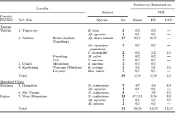
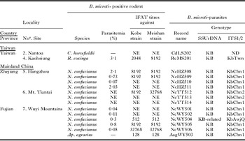
Table 1A and B provide a summary of the surveys in Taiwan and mainland China. B. microti was detected by PCR in 1 rodent and 1 insectivore in Taiwan and in 13 rodents in China (Table 1A). All PCR-positive B. microti samples except 1 were shown to be the Kobe-type (Table 1B). The SSUrDNA sequence of the exception (NcWY504) was a Kobe-related type, which differed from the Kobe-type by only 1 nucleotide (C) deletion at nucleotide-position 119 from the 5′-end after the primer Anl. B. microti-like parasites were observed microscopically with parasitaemia from 0·04 to 3·1% (Table 1B). In addition, intraerythrocytic protozoan parasites that were morphologically differentiated from B. microti were observed on the smear of a Niviventer confucianus rat from the Wuyi Mountains. For this rat, the detection PCR for B. microti was negative. The SSUrDNA sequence of this intraerythrocytic protozoon was more homologous to those of Cytauxzoon spp. or Theileria spp. than to Babesia spp.; thus, its details will be reported elsewhere. The ITS1/2 sequence (KbTwn-ITS1/2) of B. microti (RcMS201) identified in the Taiwanese R. coxinga rat was considerably different including an insertion in the ITS1 of 25 continuous nucleotides from the ITS1/2 sequence (KbAwj-ITS1/2) of the Kobe strain with 93·7% pairwise identity (Fig. 2). All Kobe-type B. microti samples in China were shown to possess either of 2 ITS1/2 sequences, KbChn1-ITS1/2 and KbChn2-ITS1/2, which differed by only 5 and 6 nucleotides from KbTwn-ITS1/2, respectively. On the other hand, the Kobe-related type sample (NcWY504) in China possessed an ITS1/2 sequence (KbAwjQ-ITS1/2) that differed by only 1 nucleotide (A) deletion from KbAwj-ITS1/2. Nucleotides and nucleotide-positions different between ITS1/2-genotypes are shown in Fig. 2.
Nucleotides and nucleotide-positions different between ITS1/2-genotypes. Nucleotide-positions are numbered from the 5′-end of ITS1/2-sequences that were aligned by Higgins' automatic sequence alignment program in DNASIS-Mac v3.5 (Hitachi Soft Engineering, Tokyo) and adjusted visually when necessary. Nucleotides different from those of KbAwj-ITS1/2 are shown in grey.

Summary of field rodent survey for Babesia microti-infection
(A) Positive ratio of B. microti-detection in field rodents by smear observation and PCR

(B) B. microti-positive rodents or insectivores and B. microti-parasites

a Site numbers correspond to those shown in Fig. 1. b The 6 rodents do not include a rodent in which intraerythrocytic protozoa different from B. microti were identified. c PCR could not be performed for one of the 6 B. microti-like parasite-positive rodents due to the lack of the DNA sample.
Abbreviations: R: Rattus; Ap: Apodemus; An: Anourosorex; C: Crocidura; M: Mus; S: Suncus; Ban: Bandicota; N: Niviventer; NE: not examined; ND: not determined; DT: detection PCR; CF: confirmation PCR; KB: Kobe.
Serological analysis
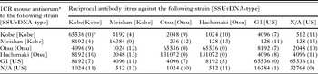
Table 2 shows IFAT titres for ICR mouse sera infected with each B. microti strain. The titres of antisera to the Kobe and Meishan strains against these homologous strains were 2–8 times higher than those against the heterologous strains. The SPD value between the Kobe and Taiwan strains was 4. With respect to rickettsial pathogens, the serotypes of 2 strains are assumed to be different when the index is 3 or more; when it is less than 3, the serotypes are assumed to be identical (Philip et al. Reference Philip, Casper, Burgdorfer, Gerloff, Hughes and Bell1978). Applying these criteria to B. microti infection, the serotype of the Meishan strain was different from that of the Kobe strain.
Indirect immunofluorescent test for Babesia microti strains

a Convalescent or chronic phase serum from a laboratory ICR mouse more than 3 months after inoculation.
b The numeral in the parentheses shows the specificity difference between two strains (Philip et al. Reference Philip, Casper, Burgdorfer, Gerloff, Hughes and Bell1978).
For sera of 8 B. microti-positive field rodents and 2 B. microti-negative field rodents, IFAT was performed against the Meishan, Kobe, GI, and Hachimaki strains (Table 1B). All sera of the B. microti-positive rodents were positive against the Meishan and Kobe strains and negative against the GI and Hachimaki strains at x128 dilution. The titres against the Meishan strain for the R. coxinga serum (x8192) and 1 Chinese N. confucianus serum (x32768) were 4 times higher than those against the Kobe strain. For the other 6 B. microti-positive sera, the titres against the Meishan and Kobe strains were the same. Considering that less than 4-fold-differences of titres could be masked, because sera of the first antibodies were often diluted by 4-fold dilutions for field rodents, the results were not considered to contradict the results for experimental ICR mice. Sera of both B. microti-negative rodents were negative against all strains at x128 dilution.
DISCUSSION
The present study demonstrated that Kobe-type B. microti, the aetiological SSUrDNA-type parasite of the first Japanese patient, was also distributed in central Taiwan and southeastern China, in the vicinities of definite Asian human babesiosis occurrences. The Taiwanese case in Minshiung, Chiayi County was reported as an asymptomatic case, although the case showed disordered symptoms and signs, which were attributed to concomitant gallstone disease (Shih et al. Reference Shih, Liu, Chung, Ong and Wang1997). However, it is possible that some of the symptoms were related to babesiosis. In the present survey, we caught only 3 Suncus murinus shrews free from B. microti at Minshiung. However, Kobe-type B. microti was identified in a R. coxinga rat from Meishan, Kaohsiung, approximately 50 km east from Minshiung. R. coxinga had been suggested to be the reservoir host of the aetiological parasite of the Taiwanese case. The Kobe-type was also identified in a Crocidura horsefieldii shrew in a separate location, Lushan, Nantou in central Taiwan. Thus, the Kobe-type is likely to have a fairly wide distribution in terms of both its host range and its geographical range. Meanwhile, in mainland China, a human case occurred 2 months after kidney transplantation in Hangzhou, Zhejiang. The patient was suggested to have contracted babesiosis through kidney transplantation or blood transfusion from donors (Su et al. Reference Su, Zhao and Ye2002). The present survey demonstrated that the Kobe-type is densely distributed not only in Hangzhou city but also around Mt. Tiantai, Zhejiang and at the southern foot of the Wuyi Mountains, Fujian. The distribution of the Kobe-type in the vicinities of occurrences in Taiwan and China as well as in Japan seems to be suggestive of involvement of the Kobe-type in Asian human babesiosis rather than to be extrinsic or accidental.
In the southern part of mainland China, 2 cases of human babesiosis had been reported from Daxing Community of Dima County, Yunnan Province in the early 1980s (Li et al. Reference Li, Meng, Wang, Dong and Wang1984). The aetiological babesia parasites were described precisely, in spite of the lack of photo documentation, although this paper, written in Chinese, has not been well publicized. Recently, a resident who had no history of travel abroad, splenectomy, or blood transfusion, was diagnosed with babesiosis in Gwalior in Mahya Pradesh in central India (Marathe et al. Reference Marathe, Tripathi, Handa and Date2005), again in southern Asia. In contrast, definite human babesiosis cases have rarely been reported in northern Asia, where US- or US-related types are distributed (Zamoto et al.Reference Zamoto, Tsuji, Kawabuchi, Wei, Asakawa and Ishihara2004a, Reference Zamoto, Tsuji, Wei, Cho, Shin, Kim, Leonova, Hagiwara, Asakawa, Kariwa, Takashima and Ishiharab). A case report in Inner Mongolia in 1996 (Shi et al. Reference Shi, Li, Gao and Ding1996) seems to lack convincing evidence. A recent Korean case was not a case caused by B. microti but by Babesia sp. classified in a group of large Babesia including Babesia divergens and B. divergens-related parasites, which cause human babesiosis almost exclusively in splenectomized individuals (Kim et al. Reference Kim, Cho, Joo, Tsuji, Cho, Park, Chung, Ju, Cheun, Lee, Lee and Kim2007). US-related types of B. microti are also distributed in Europe and have been suggested to be less pathogenic to humans than the US-type in the United States (Duh et al. Reference Duh, Petrovec, Trilar and Avsic-Zupanc2003; Goethert and Telford, Reference Goethert and Telford2003b; Gray, Reference Gray2006), because clinical cases caused by B. microti have been rarely reported in Europe, despite relatively high seropositive rates against B. microti (Foppa et al. Reference Foppa, Krause, Spielman, Goethert, Gern, Brand and Telford2002; Hunfeld et al. Reference Hunfeld, Lambert, Kampen, Albert, Epe, Brade and Tenter2002). Recently, however, a patient suffering from acute myeloid leukaemia in Germany was, for the first time, reported to have contracted human babesiosis caused by an autochthonous US-related type of B. microti in Europe (Hildebrandt et al. Reference Hildebrandt, Hunfeld, Baier, Krumbholz, Sachse, Lorenzen, Kiehntopf, Fricke and Straube2007). In any case, it is of great interest to reveal the radius of distribution of the Kobe-type in Asia or Eurasia.
In Japan, we have recently found 2 new foci of Kobe-type B. microti in Aomori, the northernmost prefecture on the Mainland of Japan (Honshu Island), significantly distant from Awaji Island (Saito-Ito et al. Reference Saito-Ito, Kasahara, Kasai, Dantrakool, Kawai, Fujita, Yano, Kawabata and Takada2007). Thus, the Kobe-type seems to be distributed dispersively in some restricted areas in Japan. In Asia, it is possible that Kobe-type B. microti is spread from southeastern China to Taiwan and Japan. Further surveys in Asia including the surrounding islands will clarify the spread mode of the Kobe-type in these areas.
The sequences of ITS1 and 2 are generally assumed to vary rapidly (Gerbi, Reference Gerbi1986; Hillis and Dixon, Reference Hillis and Dixon1991) and to be useful for differentiation or phylogenetic analysis at and below the species level (Hillis and Dixon, Reference Hillis and Dixon1991), although ITS sequences have so far been reported only for a few Babesia spp. (Zahler et al. Reference Zahler, Schein, Rinder and Gothe1998; Holman et al. Reference Holman, Bendele, Schoelkopf, Jones and Jones2003). Our recent study showed that all Japanese Kobe-type isolates possess identical ITS1/2 sequences to that of the Kobe strain (Saito-Ito et al. Reference Saito-Ito, Kasahara, Kasai, Dantrakool, Kawai, Fujita, Yano, Kawabata and Takada2007). The present study showed that the ITS1/2 sequences of the Taiwanese and Chinese Kobe-types were very similar but not identical to each other (99·2% pairwise identities) and considerably different from that of the Japanese Kobe-type (approx. 94% pairwise identities). The serological analysis showed that the serotype of the Taiwanese Meishan strain was somewhat different from that of the Japanese Kobe strain, although the difference was not so prominent. Recently, we have identified the Otsu-related type in Japan, which differs by only 2 nucleotides in the SSUrDNA sequence from the Otsu-type (Saito-Ito et al. Reference Saito-Ito, Kasahara, Kasai, Dantrakool, Kawai, Fujita, Yano, Kawabata and Takada2007). The ITS1/2 sequences of the Otsu-related type and Otsu-type differ with approx. 94% pairwise identity, about the same value as the identities between the Taiwanese or Chinese and Japanese Kobe-types. The Otsu-related type-strain (the Nagano strain) showed a different serotype and distinct host specificities from the Otsu strain (the Otsu-type) (Saito-Ito et al. Reference Saito-Ito, Kasahara, Kasai, Dantrakool, Kawai, Fujita, Yano, Kawabata and Takada2007). On the other hand, the Otsu strain and the Hachimaki strain with very similar ITS1/2 sequences (99·9% pairwise identity) showed the same serotype in the present analysis. The GI and the N/A strains with identical ITS1/2 sequences also showed the same serotype in this and the previous studies (Saito-Ito et al. Reference Saito-Ito, Kasahara, Kasai, Dantrakool, Kawai, Fujita, Yano, Kawabata and Takada2007). Therefore, it seems possible that the Taiwanese or Chinese and Japanese Kobe-types are different in some important biological characteristics such as host specificities, infectivity or pathogenicity. The ITS1/2 sequence seems to be a useful and sensitive genetic marker for discriminating, comparing and investigating parasites of the same SSUrDNA type.
Regarding vector tick species, we have just identified the tick species infesting field rodents in the present study. Nymphs of Ixodes granulatus were found infesting a B. microti-positive R. coxinga rat and 2 B. microti-positive and negative C. horsefieldii shrews in Taiwan. In mainland China, larvae and nymphs of Ixodes tanuki, Ixodes ovatus, Haemaphysalis bandicota, and Rhipicephalus haemaphysaloides were found infesting, in total, 6 B. microti-positive or negative N. confucianus rats in the Wuyi Mountains. Only tick species belonging to the genus Ixodes are known to transmit B. microti (Homer et al. Reference Homer, Aguilar, Telford, Krause and Persing2000). I. granulatus, Ixodes tanuki and Ixodes ovatus are thus likely to be candidates of vector species for Kobe-type B. microti. I. granulatus was reported to transmit Taiwanese B. microti-like parasites to laboratory rats (van Peenen et al. Reference Van Peenen, Chang, Banknieder and Santana1977). In the previous study, we detected Otsu-type but not Kobe-type B. microti in I. ovatus on Awaji Island in Japan (Saito-Ito et al. Reference Saito-Ito, Yano, Dantrakool, Hashimoto and Takada2004; Yano et al. Reference Yano, Saito-Ito, Dantrakool and Takada2005). I. tanuki was also sometimes found in rodents in B. microti-enzootic areas in Japan (unpublished data). More systematic studies for ticks in these areas will be required to determine vector tick species of Kobe-type B. microti in Asia.
In conclusion, we demonstrated for the first time the distribution of Kobe-type B. microti in rodents in the vicinities of occurrences in 2 human babesiosis emergence Asian countries, Taiwan and China. These findings suggest the involvement of the Kobe-type in Asian human babesiosis. We now plan to perform seroepidemiological surveys of residents for B. microti-infection using respective Kobe-types, with comparison of the biological characteristics of respective Kobe-types.
We thank Ms Atsuko Kawai, Ms Midori Kasahara, Mr Masatoshi Kasai and Dr Anchalee Dantrakool for helping with sequence analyses. We also thank Dr Hak Hotta, who gave helpful comments on the work. Dr Dar-Der Ji of the Institute of Preventive Medicine of the National Defense Medical Center in Taiwan helped collecting rodents in Taipei city. This work was supported in part by Grants-in-Aid for Scientific Research from the Ministry of Education, Science and Culture of Japan (13670244, 15590367, 16406008 and 17590369), a grant from the Ohyama Health Foundation, Inc., and a grant from the Takeda Science Foundation.