Introduction
Endobronchial tuberculosis is one of the serious complications of pulmonary tuberculosis. It was first described by the English physician Richard Morton in 1689.Reference Hudson and Heaf1
Endotracheal tuberculosis is a relatively uncommon (about 4 per cent), localised form of endobronchial tuberculosis which may cause acute respiratory failure.Reference Morrone and Abe2 Only 150 cases have been reported worldwide.Reference Belhassen, Boudaya, Ayadi, Ammar, Djilani and Mezni3 Early diagnosis and prompt treatment of endotracheal tuberculosis is important to prevent the development of fibrotic scarring and resultant tracheobronchial stenosis. In cases of endobronchial tuberculosis, the clinical and radiological presentation is not specific, and early diagnosis depends upon a high index of clinical suspicion.Reference Ip, So, Lam and Mok4
Case report
A 24-year-old man presented to us in September 2008 with breathing difficulty. He was a non-smoker and non-drinker with no history of diabetes, hypertension, bronchial asthma, pulmonary tuberculosis or any cardiac ailment.
He had previously been involved in a road traffic accident (3 May 2006), sustaining blunt chest injury, a fractured mandible and minor head injury. On 9 May, the mandibular fracture had been fixed at a local hospital, using arch bars, and the patient discharged on 11 May in a healthy state.
However, from 15 June 2006 the patient developed progressive breathing difficulty with stridor. On 22 June 2006, he was admitted to the same local hospital in cardiopulmonary arrest; an emergency tracheostomy was performed and the patient successfully revived. Afterwards, the patient was otherwise well but continued to suffer breathing difficulty, despite the tracheostomy in situ.
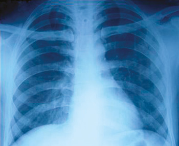
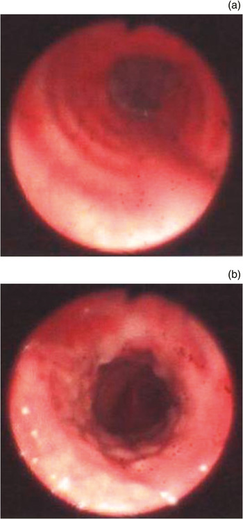
A chest X-ray taken at this stage appeared normal (Figure 1). Bronchoscopy revealed some granulation tissue around the tracheostomy stoma (Figure 2) but no tracheal stenosis.
Posterior-anterior chest X-ray showing normal lung markings.

Bronchoscopic views showing some granulation tissue around the tracheostomy stoma but no stenosis.

At this stage, the patient was referred to a tertiary hospital in Delhi, where the clogged tracheostomy tube was removed. Fibre-optic bronchoscopy showed no tracheal stenosis. Following tracheostomy removal, the patient was able to breathe normally, with no breathing difficulty. He was closely observed for 4 days and then discharged.
However, the patient was readmitted after 10 days with increasing breathing difficulty for the previous 5 days and stridor for the previous 3 days. On examination, he was in respiratory distress but was conscious, oriented and afebrile, with a respiratory rate of 32 breaths/minute, a pulse rate of 132 beats/minute and a blood pressure of 160/96 mmHg. He had paradoxical, laboured breathing with indrawing of the intercostal spaces and a tracheal tug. Systemic examination was unremarkable.
Chest X-ray and blood biochemistry results were both normal. Arterial blood gas analysis indicated respiratory acidosis.
The patient was placed on a mechanical ventilator with very high peak airway pressure with a small tidal volume. His arterial blood gases normalised.
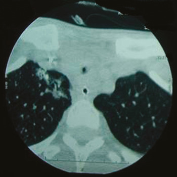
Fibre-optic bronchoscopy, performed via the endotracheal tube, revealed a tight tracheal stenosis about 3–4 mm diameter at midtracheal level, approximately 3 cm distal to the tracheostomy stoma (Figure 3).
Axial computed tomography scan showing the site of stenosis, approximately 3 cm distal to the tracheostomy stoma.

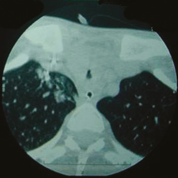
Computed tomography (CT) scanning confirmed the bronchoscopic findings and revealed the stenosis to be approximately 1.5 cm in length (Figure 4).
Axial computed tomography scan confirming the bronchoscopy findings, and showing the length of the stenosis to be approximately 1.5 cm.

Microbial cultures of bronchial lavage fluid were negative for acid-fast bacilli, pyogenic organisms and fungi.
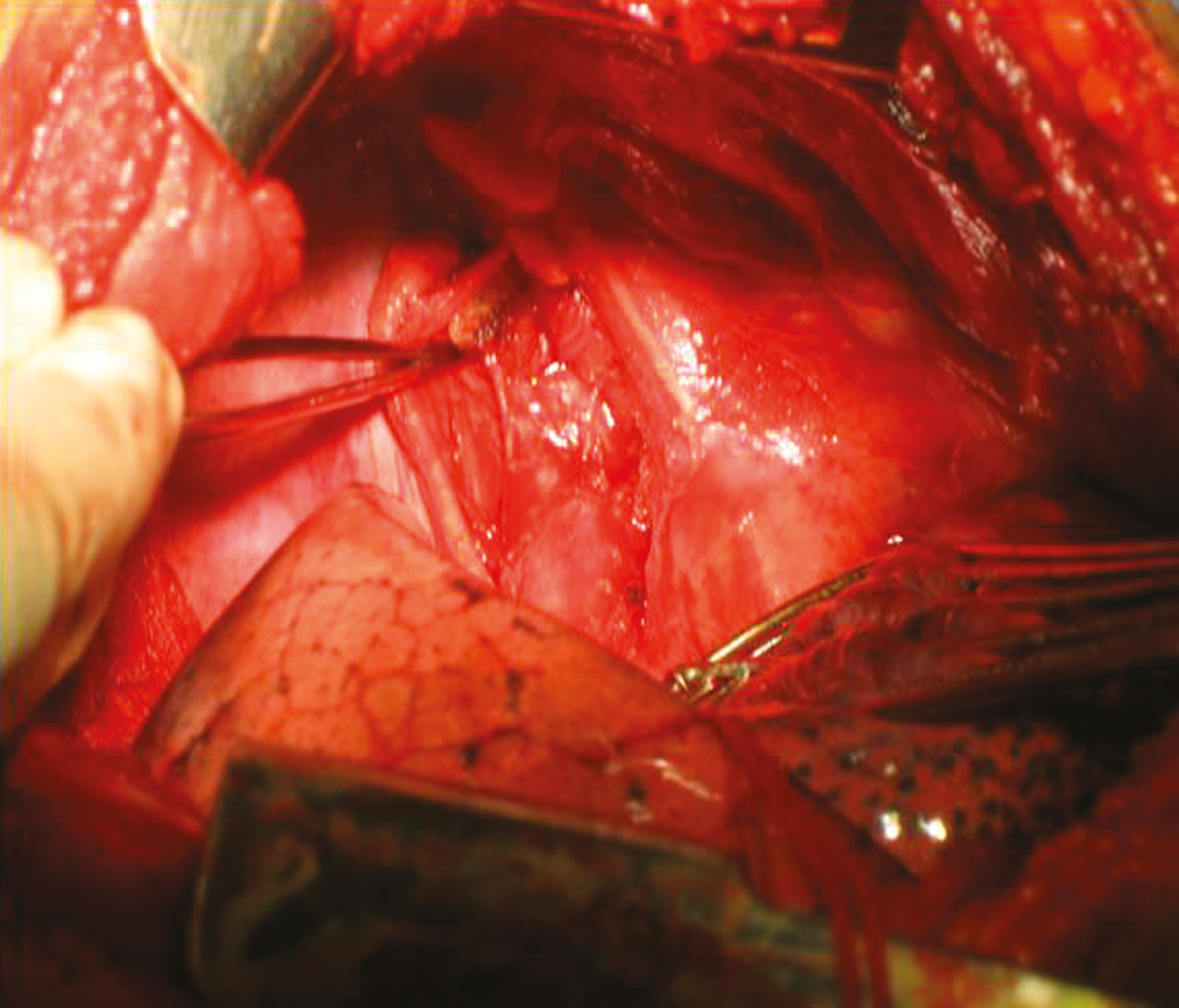
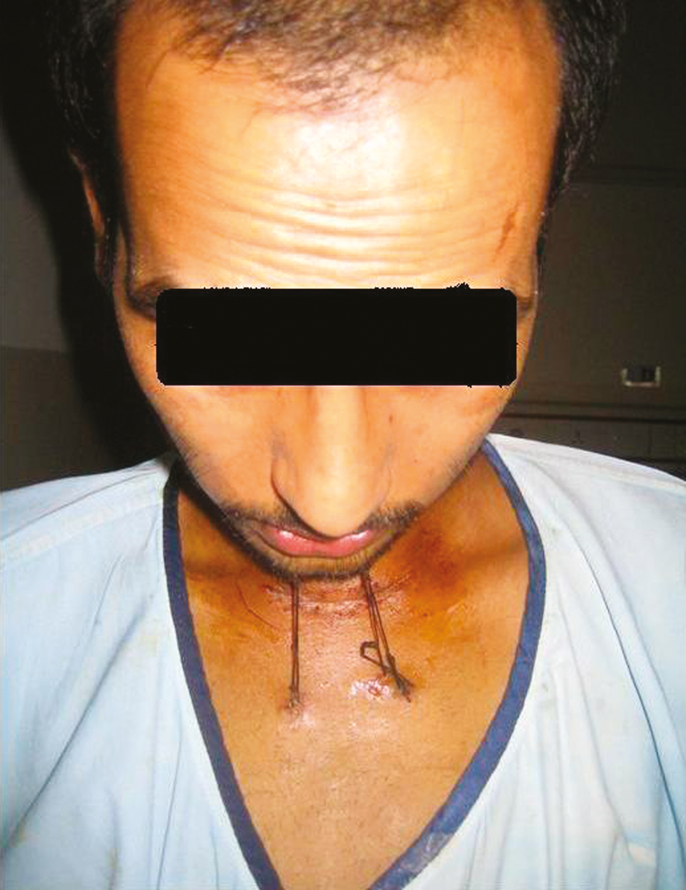
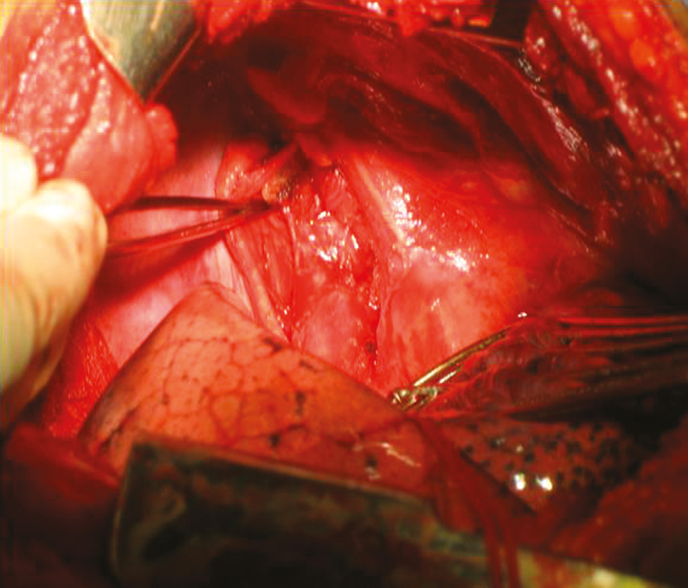
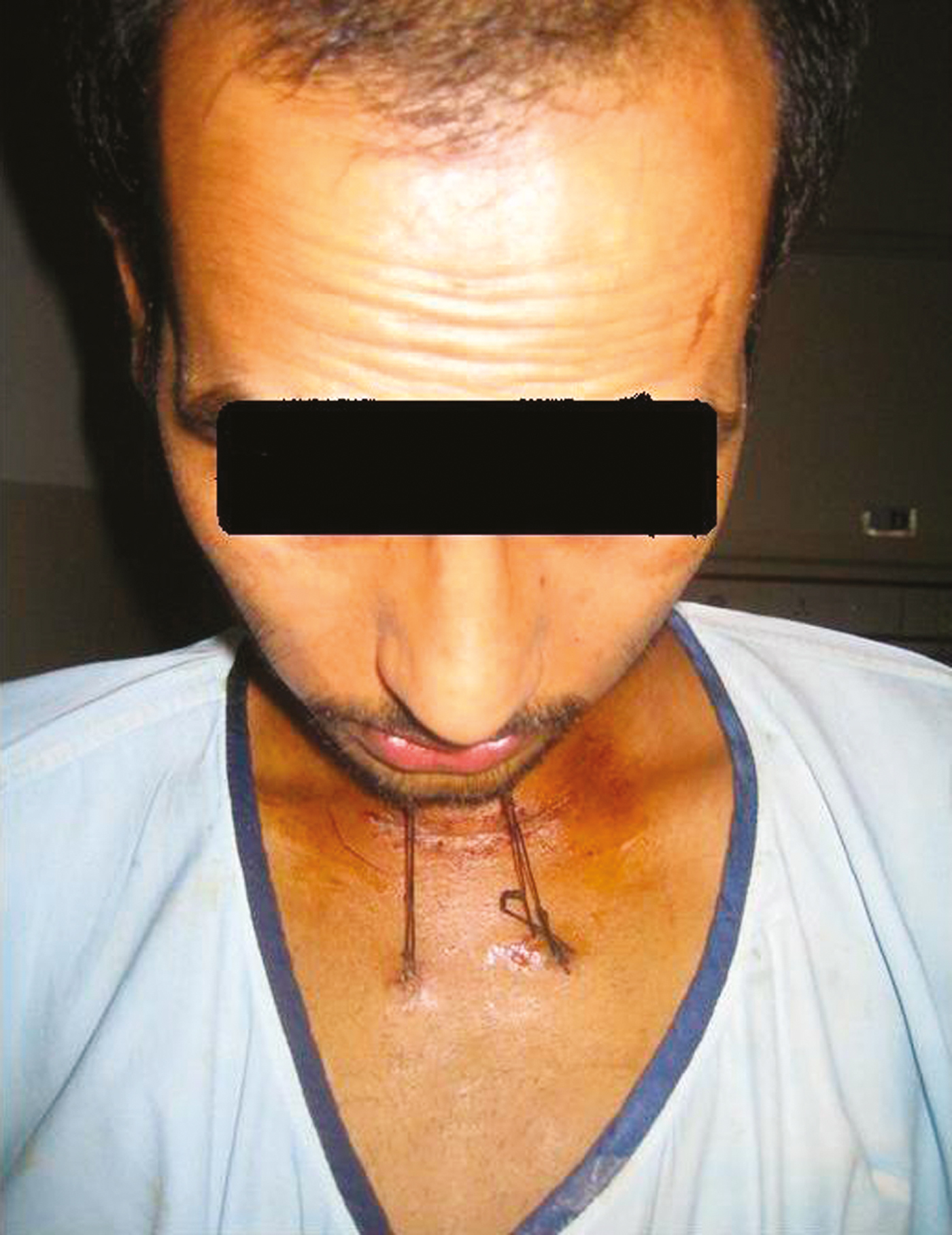
The patient underwent tracheal resection and anastomosis, while on the ventilator. Firstly, the tracheostomy stoma was excised and the cervical trachea mobilised via a cervical incision. As expected, peritracheal adhesions were noted in the neck. The patient was then repositioned for a right posterolateral thoracotomy, and the trachea mobilised. Unexpectedly, there were dense, tough, fibrous peritracheal adhesions throughout the length of intrathoracic trachea (Figure 5). Approximately 2.5 cm of stenotic trachea was excised, and an end-to-end anastomosis was created to restore airway continuity. A chin-to-manubrium heavy silk suture was applied to keep the neck in a flexed position (Figure 6).
Intra-operative photograph showing the dense, tough, fibrous peritracheal adhesions which were present throughout the length of the intrathoracic trachea.
Post-operatively, the patient's neck was held in flexion via a heavy silk suture fixed from the chin to the manubrium.
The patient was extubated immediately after the operation, and was observed in the intensive care unit for the next 4 days. Results for routine investigations and arterial blood gases were normal. The patient remained well, with no breathing difficulty whatsoever. He was discharged 10 days after surgery, and was eating, walking, talking and breathing normally, albeit with a forcibly flexed neck.
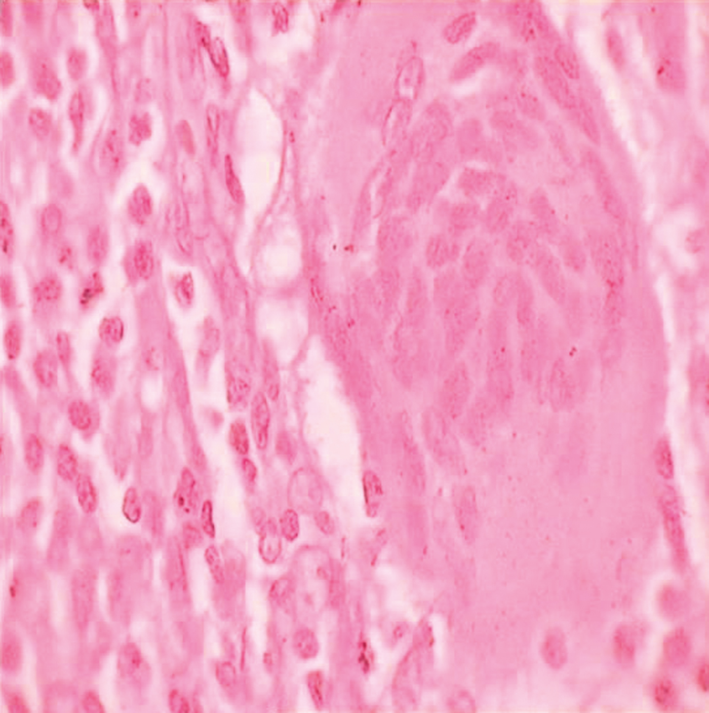
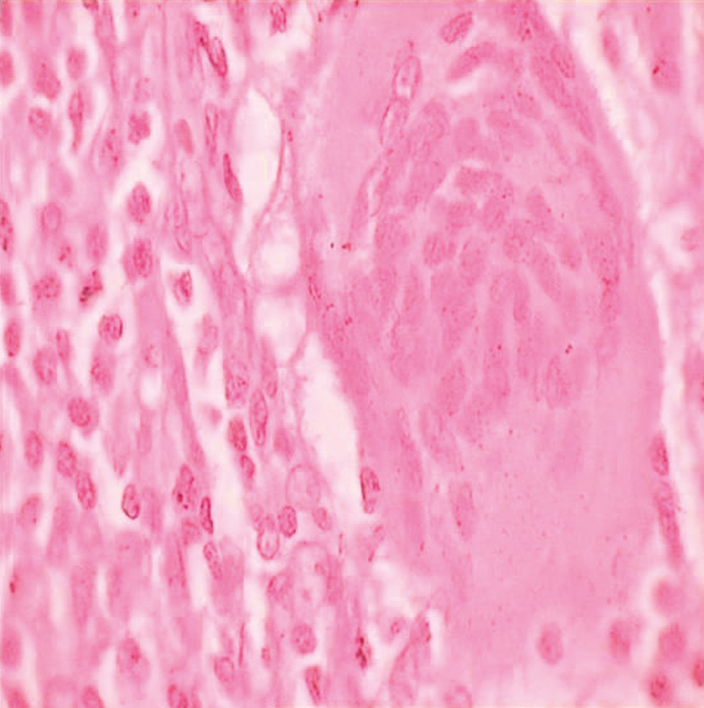
To our great surprise, histopathological examination of the resected tracheal segment revealed typical tubercular granulomas (Figure 7). In addition, acid-fast staining was positive for tubercle bacilli.
Photomicrograph of the resected tracheal segment, showing a typical tubercular granuloma. (Z and N Stain; ×400)
The patient was commenced on four-drug antitubercular treatment.
Neck flexion suture were removed after 2 weeks of discharge. More than three months after tracheal resection, he continued to do well.
Discussion
Bronchial strictures can develop despite effective treatment of endobronchial tuberculosis; this complication has been reported in 60–95 per cent of cases.Reference Ip, So, Lam and Mok4, Reference Park, Woo, Son, Lee, Kim and Mo5
The pathogenesis of endobronchial tuberculosis is not yet fully established. However, possible mechanisms include: direct implantation of tubercle bacilli into the bronchus from an adjacent pulmonary parenchymal lesion; direct airway infiltration from an adjacent tuberculous mediastinal lymph node; erosion and protrusion of an intrathoracic tuberculous lymph node into the bronchus; haematogenous spread; and extension to the peribronchial region via lymphatic drainage.Reference Chung and Lee6
Endobronchial tuberculosis is commonest in the third decade.
The clinical manifestations of endobronchial tuberculosis vary, and include chronic and productive cough, barking cough, chest pain, haemoptysis, generalised weakness, dyspnoea, and fever. Endobronchial tuberculosis can cause bronchogenic carcinoma, polypoidal mass, asthma, foreign body aspiration and pneumonia, and atelectasis.Reference Park, Woo, Son, Lee, Kim and Mo5
Tuberculous tracheobronchial stenosis is a serious clinical problem which can cause obstructive pneumonia and dyspnoea on exertion. Surgical resection and bronchoplastic reconstruction has long been the standard treatment.Reference Hsu, Hsu, Huang and Huang7
• Tracheal resection and end-to-end anastomosis remains the treatment of choice for benign tracheal strictures
• In cases of tuberculous tracheobronchial stenosis, early diagnosis and prompt treatment are important to prevent fibrotic scarring
• Biopsy has an important role in tuberculosis diagnosis, especially when clinical and radiological investigations are negative
Therapeutic interventions for endobronchial tuberculosis depend on the stage of the illness: whether active (imaging shows hyperplasic changes with oedema) or fibrotic (imaging shows smooth narrowing). This distinction is usually made on CT scanning.Reference Belhassen, Boudaya, Ayadi, Ammar, Djilani and Mezni3 Patients with active disease usually respond to antitubercular treatment and require no further intervention. Those in the fibrotic stage of the illness may require radiologically guided procedures or surgical intervention. Segmental tracheal resection with end-to-end anastomosis is the preferred treatment for benign stenosis, as seen in the presented case.Reference Grillo, Donahue, Mathisen, Wain and Wright8
Most tracheal stenosis cases occur after prolonged endotracheal intubation and tracheostomy. We believe our patient to be unique in that his cause of tracheal stenosis was very unusual. Although he had a history of trauma, our patient never received endotracheal intubation. Even with a tracheostomy, he had breathing difficulty, indicating the presence of a pre-existing tracheal lesion which worsened with retained secretions and emergency tracheostomy. The presence of extensive, tough, fibrous adhesions throughout the length of the trachea also points to pre-existing tracheal tuberculosis and resulting stenosis.
Tracheal resection and end-to-end anastomosis remains the treatment of choice for benign tracheal strictures. In expert hands, post-operative mortality and morbidity rates are extremely low. Tracheal stents should not be considered in such cases until the patient has been thoroughly evaluated by a thoracic surgeon experienced in tracheal surgery.
Conclusion
In patients with endotracheal tuberculosis, early diagnosis and prompt treatment are necessary to prevent fibrotic scarring and resultant tracheobronchial stenosis. However, the clinical and radiological presentation of this condition is not specific, and early diagnosis thus depends on a high index of clinical suspicion.