Introduction
Perceived loneliness is increasingly recognized as an important societal issue that affects mental health (Cacioppo et al., Reference Cacioppo, Grippo, London, Goossens and Cacioppo2015a). Defined as persistent feeling of unsatisfactory social relationships that are insufficient for meeting one's social needs (Hawkley and Cacioppo, Reference Hawkley and Cacioppo2010), perceived loneliness affects up to 40% of middle- to older-aged individuals, and is associated with high prevalence of various physical and psychological issues, including major depressive disorder (MDD) (Cacioppo et al., Reference Cacioppo, Grippo, London, Goossens and Cacioppo2015a). Thus, identifying the key psychobiological processes of loneliness has great implications for promoting mental health for the broad society.
A large body of research associates MDD with impaired affective processing and regulation, characterized by bias toward negative emotions (Gotlib and Joormann, Reference Gotlib and Joormann2010; Admon and Pizzagalli, Reference Admon and Pizzagalli2015). General reduction in both positive and negative affective processing was also suggested, with the reduction being greater for positive- than for negative-valenced processing (Bylsma et al., Reference Bylsma, Morris and Rottenberg2008). The affective symptoms are accompanied by disconnected functioning of the lateral prefrontal cortex (LPFC)-anterior cingulate cortex (ACC)-parietal-cerebellar pathway implicated in both cognitive control and affect regulation (Fitzgerald et al., Reference Fitzgerald, Laird, Maller and Daskalakis2008; Frodl et al., Reference Frodl, Bokde, Scheuerecker, Lisiecka, Schoepf, Hampel, Möller, Brückmann, Wiesmann and Meisenzahl2010; Guo et al., Reference Guo, Liu, Xue, Gao, Liu, Xiao, Chen and Zhao2013). MDD patients also show altered connectivity within the default mode network (DMN) at rest. Certain connectivity within the DMN encompassing the dorsomedial prefrontal cortex, the precuneus and the hippocampus was found to be more positive in MDD patients compared to controls, which possibly reflects greater bias toward self-oriented and autobiographic processing (Kaiser et al., Reference Kaiser, Andrews-Hanna, Wager and Pizzagalli2015; Mulders et al., Reference Mulders, van Eijndhoven, Schene, Beckmann and Tendolkar2015). On the other hand, functional connectivity (FC) between the anterior and posterior DMN, and between the DMN and the networks involved in cognitive control and affect regulation (e.g. the dorsal and rostral ACC), was often found to be decreased in MDD (Dutta et al., Reference Dutta, McKie and Deakin2014; Kaiser et al., Reference Kaiser, Andrews-Hanna, Wager and Pizzagalli2015; Mulders et al., Reference Mulders, van Eijndhoven, Schene, Beckmann and Tendolkar2015), possibly indicating reduced top-down regulation of internal affective processing. However, none of these studies controlled for the loneliness levels of MDD patients and healthy controls (HCs). Abnormal functioning of the cognitive control network underlies both cognitive (Vasic et al., Reference Vasic, Walter, Sambataro and Wolf2009; Brzezicka, Reference Brzezicka2013) and affective impairments (Fitzgerald et al., Reference Fitzgerald, Laird, Maller and Daskalakis2008; Frodl et al., Reference Frodl, Bokde, Scheuerecker, Lisiecka, Schoepf, Hampel, Möller, Brückmann, Wiesmann and Meisenzahl2010) in MDD, and could itself be a consequence of dysregulated affective reactivity (Brzezicka, Reference Brzezicka2013). In particular, the prefrontal-parietal connectivity is strongly implicated in superordinate cognitive control functions such as inhibition, attention orientation and conflict processing (Roberts and Hall, Reference Roberts and Hall2008; Niendam et al., Reference Niendam, Laird, Ray, Dean, Glahn and Carter2012; Harding et al., Reference Harding, Yücel, Harrison, Pantelis and Breakspear2015), and in emotion regulatory functions (Ochsner et al., Reference Ochsner, Bunge, Gross and Gabrieli2002; Müller et al., Reference Müller, Cieslik, Laird, Fox and Eickhoff2013; Kohn et al., Reference Kohn, Eickhoff, Scheller, Laird, Fox and Habel2014), and was found to be decreased in MDD (Schutter et al., Reference Schutter, d'Alfonso, Van Honk and Schweitzer2003; Vasic et al., Reference Vasic, Walter, Sambataro and Wolf2009). In accordance, MDD is considered to be principally characterized by an inability to disengage from negative stimuli, highlighting reduced goal-directed, top-down executive control (Gotlib and Joormann, Reference Gotlib and Joormann2010; Disner et al., Reference Disner, Beevers, Haigh and Beck2011).
Loneliness often co-occurs with and predicts MDD onset (Hawkley and Cacioppo, Reference Hawkley and Cacioppo2010; Cacioppo et al., Reference Cacioppo, Grippo, London, Goossens and Cacioppo2015a). Existing evidence tends to suggest that loneliness is associated with reduced affective sensitivity and arousal response to both positive and negative stimuli (Cacioppo et al., Reference Cacioppo, Ernst, Burleson, McClintock, Malarkey, Hawkley, Kowalewski, Paulsen, Hobson, Hugdahl and Spiegel2000, Reference Cacioppo, Norris, Decety, Monteleone and Nusbaum2009; Wong et al., Reference Wong, Liu, Lin, Huang, Wai, Lee and Lee2016), suggesting possible dissociated affective profiles for loneliness and MDD (Cacioppo et al., Reference Cacioppo, Grippo, London, Goossens and Cacioppo2015a). Such findings seem, at first glance, at odds with the existing prevalent model of loneliness. Specifically, it is proposed that lonely individuals are eager to connect with others, yet exhibit implicit hypervigilance to social threats, leading to bias toward negative processing and memories about social interactions (Hawkley and Cacioppo, Reference Hawkley and Cacioppo2010). According to this view, loneliness may be characterized by enhanced automatic, bottom-up attention bias toward negative social stimuli, consistent with evidence indicating heightened visual activities in lonely individuals when processing negative social stimuli (Cacioppo et al., Reference Cacioppo, Norris, Decety, Monteleone and Nusbaum2009, Reference Cacioppo, Balogh and Cacioppo2015b), and increased resting-state connectivity in the neural networks implicated in salience and tonic alertness (Layden et al., Reference Layden, Cacioppo, Cacioppo, Cappa, Dodich, Falini and Canessa2017). However, the increased bottom-up negative affective bias may be counteracted by simultaneous increase of top-down affect regulatory functions, as discussed below.
To reconcile the existing evidence on reduced general affective reactivity on one hand, but enhanced automatic negative attention bias on the other hand in loneliness, we hypothesized the following model. Specifically, it could be that among lonely individuals, enhanced negative-biased bottom-up attentional processes performed by the inferior parietal-cerebellum circuitry (D'Angelo and Casali, Reference D'Angelo and Casali2013; Humphreys and Lambon Ralph, Reference Humphreys and Lambon Ralph2015) are (over)compensated by upregulated top-down inhibition and conflict-resolution functions, which are performed by the LPFC-ACC-superior parietal circuitries (Roberts and Hall, Reference Roberts and Hall2008; Niendam et al., Reference Niendam, Laird, Ray, Dean, Glahn and Carter2012; Humphreys and Lambon Ralph, Reference Humphreys and Lambon Ralph2015). Consistent with this, a recent study reported that loneliness positively predicted functional integrity of the ACC-parietal-operculum circuitry at rest after controlling for depression scores (Layden et al., Reference Layden, Cacioppo, Cacioppo, Cappa, Dodich, Falini and Canessa2017). However, existing research examining cognitive control and neural circuitry functions in lonely individuals is lacking and inconclusive, particularly for non-elderly populations (Boss et al., Reference Boss, Kang and Branson2015). Such knowledge is critical for fully understanding the potentially altered affective processing and regulation in lonely individuals (Gotlib and Joormann, Reference Gotlib and Joormann2010).
Thus, a critical gap exists in our current knowledge on the precise nature and possible mechanisms of the association between MDD and loneliness. Importantly, we need to know not only the independent processes of MDD and loneliness, but also how they might exert interactive influence when co-occurring (or not). Such knowledge is essential for understanding why a considerable proportion of lonely individuals might be subsequently prone to MDD development, and the implication of high loneliness in patients already diagnosed with MDD, yet only one study to our knowledge explicitly investigated the joint influence of MDD and loneliness on neural mechanisms during affective processing in elderly individuals (Wong et al., Reference Wong, Liu, Lin, Huang, Wai, Lee and Lee2016). Therefore, we investigated the unique and interactive effects of MDD and loneliness across cognitive and resting states to characterize their respective neural profiles, toward the ultimate goal of delineating the psychobiological basis for their close association. Cognitive control was assessed using the Stroop color-word task that taps into selective attention, inhibition and conflict-resolution functions (Roberts and Hall, Reference Roberts and Hall2008; Niendam et al., Reference Niendam, Laird, Ray, Dean, Glahn and Carter2012). Although we did not include any tasks that overly measure affective processes, our study could still tap into the affective system given (1) the fronto-parietal-cerebellar circuitries are implicated in both cognitive control and affect regulatory functions (D'Angelo and Casali, Reference D'Angelo and Casali2013; Kohn et al., Reference Kohn, Eickhoff, Scheller, Laird, Fox and Habel2014; Kong et al., Reference Kong, Wei, Li, Cun, Xue, Zhang and Qiu2015); (2) the DMN is often implicated in affective processing and regulatory processes, likely related to its dysfunction in MDD patients (Sheline et al., Reference Sheline, Barch, Price, Rundle, Vaishnavi, Snyder, Mintun, Wang, Coalson and Raichle2009; Ho et al., Reference Ho, Connolly, Blom, LeWinn, Strigo, Paulus, Frank, Max, Wu, Chan and Tapert2015); (3) recent evidence highlights common neural substrates for mental processes across cognitive and emotional domains (Depue et al., Reference Depue, Orr, Smolker, Naaz and Banich2016), and meta-analysis suggests highly overlapping neural networks for cognitive and emotional regulatory processes (Kohn et al., Reference Kohn, Eickhoff, Scheller, Laird, Fox and Habel2014) and (4) DMN function at rest could reflect primarily self-oriented affective processing in the DMN, and regulation of such activities by the cognitive control networks (Northoff et al., Reference Northoff, Wiebking, Feinberg and Panksepp2011). Based on these arguments, our pre-selected regions-of-interest (ROIs) covered the LPFC, ACC, parietal and cerebellar regions, all of which are strongly implicated in both cognitive control and affect regulation. Further, the rostral ACC, inferior parietal cortex and the cerebellum are all part of and/or closely associated with the DMN (Long et al., Reference Long, Zuo, Kiviniemi, Yang, Zou, Zhu, Jiang, Yang, Gong, Wang and Li2008; Habas et al., Reference Habas, Kamdar, Nguyen, Prater, Beckmann, Menon and Greicius2009; Sripada et al., Reference Sripada, King, Welsh, Garfinkel, Wang, Sripada and Liberzon2012), allowing us to assess the functional patterns of the DMN, cognitive control and affect regulation networks in both cognitive and resting states.
We hypothesized impaired Stroop performance along with reduced functional integrity (i.e. reduced FC) of the prefrontal-ACC-parietal circuitries for MDD patients, and the opposite pattern for lonely individuals. A recent study showed that during negative affective processing, loneliness negatively predicted LPFC activities in MDD patients but exerted the opposite effect in HCs (Wong et al., Reference Wong, Liu, Lin, Huang, Wai, Lee and Lee2016). Thus, we predicted a similar interactive influence of loneliness and MDD on task-related cognitive control activities. Furthermore, we hypothesized that the MDD patients would show altered FC patterns within the DMN, and reduced connectivity between the DMN and the networks involved in cognitive control and affect regulation at rest, while tentatively predicting that lonely individuals would again exhibit the opposite patterns.
Materials and methods
Participant and measures
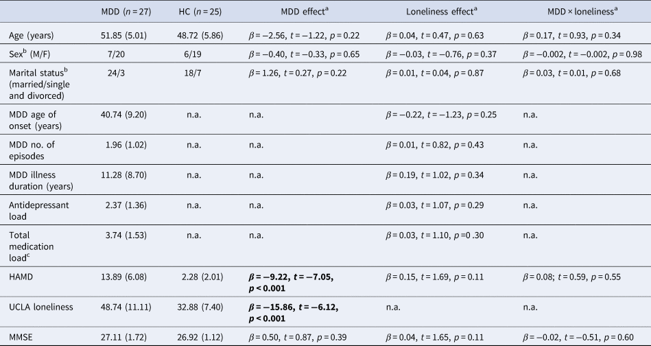
This study was approved by the institutional review board of Chang Gung Memorial Hospital and complied with the declaration of Helsinki. Twenty-seven right-handed, middle-aged individuals (age = 39–59 years) diagnosed with MDD, and 25 age- and gender-matched (p > 0.22) HCs were recruited (Table 1). The patients previously had on average two depressive episodes but no comorbidities, and were on antidepressants and/or hypnotic medications during the study. All HCs were free of current or previous major physical, neurological or psychological illnesses. All participants provided written informed consent for participation. See online Supplementary Materials for further details of participants.
Demographic, illness and psychometric characteristics of MDD patients and HCs

MDD, major depressive disorder; HC, healthy controls; M/F, males/females; HAMD, Hamilton Rating Scale for Depression; MMSE, Mini-Mental State Examination.
Both mean and standard deviation (in brackets) are shown. Significant effects at p < 0.05 are marked in bold.
a The main effects of MDD and loneliness, and the MDD × loneliness effect were all evaluated using linear or logistic regression analyses (MDD coded as 0, HC coded as 1). Unstandardized coefficient (β) and t statistics (β/s.e.) are reported. Bootstrapping procedure (5000 times) was applied to alleviate potential data non-normality and/or heteroscedasticity (Erceg-Hurn and Mirosevich, Reference Erceg-Hurn and Mirosevich2008).
b The effects of dichotomous variables were evaluated using binary logistic regression.
c Total medication includes both antidepressants and hypnotics.
All participants scored >23 on the Mini-Mental State Examination (MMSE) (Chiu et al., Reference Chiu, Lee, Chung and Kwong1994), indicating the absence of general cognitive impairment. The MDD patients and HCs showed overall comparable MMSE scores (p = 0.39). Perceived trait loneliness was measured using the 20-item UCLA loneliness scale (Wu et al., Reference Wu, Sun, Sun, Zhang, Tao and Cui2010) (current-sample Cronbach's α = 0.95). Depressive symptoms were measured using the 17-item Hamilton rating scale for depression (HAMD) (Zheng et al., Reference Zheng, Zhao, Phillips, Liu, Cai, Sun and Huang1988). All HCs scored <7, indicating the absence of depressive symptomology.
The Stroop color-word task
Participants performed a Stroop color-word task during functional magnetic resonance imaging (fMRI) scanning (Fig. 1a). The task consisted of two blocks of each of the neutral and incongruent conditions, with 12 trials in each block, intermixed with 12-s fixation periods. During incongruent trials, participants needed to respond as accurately and quickly as possible on whether the ink color of the upper word matched the color meaning of the lower word. The meaning and ink color of the upper word were always conflictive. In the neutral trials, the upper words were affectively neutral and color-unrelated (online Supplementary Materials). The incongruent and neutral blocks were delivered in pseudo-random orders, provided that blocks of the same type could not occur as the first or last two blocks (i.e. no AABB or BBAA). See online Supplementary Materials for further details of task.
(a) The Stroop color-word task. The task consisted of two blocks of each of the neutral and incongruent conditions, interspersed with 12-s fixation periods. In the incongruent trials, participants were presented with a pair of colored words above and below a central fixation point (in figure: ‘blue’ and ‘red’ above and below the center, respectively) and needed to indicate as accurately and quickly as possible whether the ink color of the upper word matched the color meaning of the lower word. Critically, the upper word was always printed in a different color from the color it read as, creating a cognitive incongruence, whereas the lower word was always printed in white. In the neutral trials, the upper color word was substituted by an affectively neutral, color-unrelated word (in figure: ‘division’). All button-press responses needed to be made within 2 s following stimuli onset. Each trial lasted for 3 s in total. (b) The loneliness score modulated the color Stroop interference effect on RT. Higher UCLA loneliness scores were associated with significantly reduced RTs for the incongruent trials (in red, p = 0.04), but insignificantly increased RTs for the neutral trials (in blue, p = 0.32), leading to a significant incongruence × loneliness effect (p = 0.02). The effect remained significant, even after controlling for the (insignificant) incongruence × MDD effect (p = 0.02). The model additionally controlled for the intercept, block number (first or second), within-block trial order (1–12) and block-specific mean accuracy. *p < 0.05.

Behavioral analysis
MDD, loneliness and MDD × loneliness effects on demographic and psychometric scores were evaluated using linear or binary logistic regression in SPSS v.21. A bootstrapping procedure (5000 times) was applied to correct for any potential data non-normality and/or heteroscedasticity (Erceg-Hurn and Mirosevich, Reference Erceg-Hurn and Mirosevich2008).
For the Stroop task, three participants were excluded due to low task performance (<60% accuracy) or task incompletion, leaving 49 participants in task-related analyses (25 MDD, 24 HCs). In all analyses on Stroop task reaction time (RT), the block-specific mean task accuracies were entered as nuisance variables. Note that a multi-level regression model (as described below) was not employed to analyze accuracy data as the mean accuracy was computed as a compound measure for a given task condition, so no accuracy variable existed at the individual trial level.
Participants' RTs on correct trials were analyzed with a random-intercept, multi-level mixed-effect linear regression model implemented in MLwIN v2.28 (Rasbash et al., Reference Rasbash, Charlton, Browne, Healy and Cameron2009). The first level of the model captured the data variances across individual task trials within each participant, and the second level incorporated the between-participant variances. The main effects of incongruence, MDD and loneliness, as well as their interactions were assessed while controlling for within-block trial order (1–12), block number (1 or 2) and trial type- and block-specific accuracy. The statistical threshold was set at p < 0.05, two-tailed.
Imaging acquisition and analysis
Task and resting-state fMRI and T1-weighted structural images were acquired. See online Supplementary Materials for detailed imaging acquisition parameters and preprocessing steps. Imaging preprocessing and analyses were conducted using DPARSFA v. 4.3 (Yan et al., Reference Yan, Wang, Zuo and Zang2016) and Statistical Parametric Mapping (SPM12) software (Wellcome Department of Cognitive Neurology, London, UK).
In the first-level analysis of Stroop-related imaging data, the entire durations of the incongruent and neutral blocks were modeled, together with the six motion parameters and a mean regressor. Two contrasts of interest were generated (incongruence > neutral, neutral > incongruence), which were then forwarded to two group-level independent-samples t test models. The first model assessed the main effects of MDD and loneliness, while controlling for mean accuracy difference of the incongruent and neutral trials across participants. Such procedure minimized the possibility that any effect due to MDD or loneliness on task activations was confounded by their effects on performance accuracy, and was employed for all subsequent imaging analyses. The second model assessed the interactive effect of MDD and loneliness. These effects were of a priori interest for the current study. Statistical thresholds were determined using the threshold-free-cluster-enhancement (TFCE) method (Smith and Nichols, Reference Smith and Nichols2009), and were whole-brain familywise-error (FWE)-corrected at p < 0.05 (online Supplementary Materials).
The imaging results were evaluated primarily based on a priori anatomical, unbiased ROIs, although whole-brain analyses are also reported for completeness sake. Such a procedure ensured that a consistent set of theory driven ROIs are evaluated for each imaging analyses [task fMRI, generalized psycho-physiological interaction (gPPI), resting-state connectivity], allowing comparability across findings. Four ROIs were constructed, namely LPFC, ACC, parietal cortices and cerebellum (online Supplementary Fig. S1). As discussed above, these regions constitute the key networks for cognitive and affective control. The ROI templates were generated using WFU-Pickatlas software. The LPFC mask included the superior, middle and inferior frontal gyri (BA8, 9, 44, 45, 46, 47). The ACC mask included both rostral and dorsal ACC (BA 24, 32). The parietal cortices included both superior and inferior parietal lobule (BA7, 39, 40). The cerebellum mask encompassed the lateral hemispheres, the intermediate zone and the vermis. All ROIs were bilateral. A false-discovery rate (FDR) procedure was additionally employed on the FWE-corrected p values to further correct for the number of ROIs. Exploratory regression and correlation analyses with FDR correction were conducted to further characterize the significant signals and to assess their associations with behavioral RT. The corrected statistical threshold was set at p < 0.05, two-tailed.
In order to assess the network-level function (i.e. inter-region communications) of key regions where activations on the Stroop task were significantly influenced by MDD, loneliness or their interaction, we subsequently conducted gPPI analyses (McLaren et al., Reference McLaren, Ries, Xu and Johnson2012), using 6-mm spherical seeds centered at the locus of maxima of significant clusters to the effects of interest within predefined ROIs. The group-level analysis models, thresholding, ROIs, multiple-testing corrections and further analyses on extracted parameters were identical to those for the regional activation analyses.
Finally, we characterized the network-level function of the key regions implicated in cognitive control on the Stroop task where activations were significantly affected by MDD, loneliness or their interaction, when the participants were at rest. Fifty participants were included in the resting-state data analysis (26 MDD, 24 HCs, two participants were excluded due to technical failure). Details about image acquisition and preprocessing are included in online Supplementary Materials. We focused on examining the resting-state FC (rsFC) patterns of the same seed regions as those used for the gPPI analyses. The resulted rsFC maps were then analyzed using the same group-level models, thresholding, ROIs and multiple-testing corrections, and significant values were extracted and underwent the same further analyses, as in the task-based analyses.
Supplementary analyses
Exploratory analyses were conducted to assess the effects of the HAMD score and illness-related characteristics (age of onset, number of episodes, illness duration, antidepressant load, total medication load) on task performance and neural signals for the MDD patients, utilizing the linear regression approach with bootstrapping (5000 times).
Results
Demographic and behavioral analyses
MDD patients reported significantly greater HAMD scores than HCs (t 49 = 7.05, p < 0.001), but no loneliness or MDD × loneliness effect was detected (ps > 0.1). Patients also reported higher perceived loneliness (t 49 = 6.12, p < 0.001), but collinearity diagnosis indicated that MDD and loneliness were still statistically separable (tolerance = 0.57). No MDD, loneliness or MDD × loneliness effect was observed for the demographic variables or MMSE score (ps > 0.1). Loneliness had no significant effect on illness-related variables among MDD patients (ps > 0.25).
We proceeded to test our hypotheses that the MDD and loneliness would respectively show negative and positive effects on Stroop performance measures. On the Stroop task, linear regression analyses with bootstrapping revealed no significant effect of MDD on average accuracy in neutral trials (MDD: 90.8% ± 11.5%; HC: 94.3% ± 6.0%; t 46 = 1.14, p = 0.27), but patients showed significantly lower accuracy for incongruent trials (MDD: 74.2% ± 13.2%; HC: 87.7% ± 11.8%; t 46 = 3.26, p = 0.002). No effect of loneliness, or MDD × loneliness, was observed for either the neutral or incongruent trial accuracy (ps > 0.64).
The Stroop task RT showed normal distribution within each block (Kolmogorov–Smirnov test, ps > 0.39). A positive incongruence effect was observed (z = 8.02, p < 0.001), indicating a classic interference effect. MDD patients showed greater overall RTs than HCs (z = 3.52, p < 0.001), while loneliness had no significant effect on overall RTs (p > 0.54). Importantly, a significant incongruence × loneliness effect was observed (z = −2.42, p = 0.016), even after controlling for the incongruence × MDD effect (z = −2.19, p = 0.029). Similar results were obtained when additionally controlling for the HAMD scores (z = −2.55, p = 0.011), while the HAMD and loneliness × HAMD effects were non-significant (p > 0.95). Simple-effect analyses revealed that loneliness negatively predicted RT on incongruent trials (z = −2.04, p = 0.041), while the association reversed to insignificantly positive on neutral trials (z = 0.99, p = 0.322) (Fig. 1b). In contrast, MDD did not moderate the incongruence effect (p > 0.39). The three-way incongruence × MDD × loneliness effect was insignificant (p > 0.63).
Stroop fMRI regional analysis
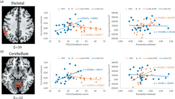
In this section, we tested the MDD, loneliness and the MDD × loneliness effects on the Stroop task-related activities in the frontoparietal and cerebellar circuitries. ROI analyses on the incongruence > congruence contrast revealed significant activations in the rostral and dorsal ACC (maxima = 6, 12, 24, voxels = 91, TFCE = 128.4), LPFC (maxima = −36, 24, 24, voxels = 2988, TFCE = 1167.16), parietal cortices (maxima = 54, −45, 51, voxels = 879, TFCE = 451.31) and cerebellum (maxima = 6, −78, −33, voxels = 206, TFCE = 292.77) (all p corrected < 0.05). The reverse contrast elicited no significant activation. No main effect of MDD or loneliness on the incongruence > neutral contrast was discovered. However, ROI analyses revealed significant MDD × loneliness effect on activations in the left inferior and superior parietal cortices (maxima = −57, −42, 39; voxels = 117, TFCE = 164.65), as well as in the bilateral cerebellum encompassing anterior and posterior vermis and lateral hemispheres (maxima = −12, −75, −27, voxels = 502, TFCE = 226.99) (both p corrected < 0.05). The whole-brain results on task activations are included in online Supplementary Materials.
To further characterize the significant interactive effects, parameter estimates for the incongruence > neutral contrast were extracted for the significant parietal and cerebellar clusters and subjected to linear regression analyses. Note that these follow-up analyses should not be viewed as being independent from the brain analyses presented above. After controlling for performance accuracy, loneliness showed significant (parietal: t 22 = −2.32, p corrected = 0.04; cerebellum: t 22 = −2.39, p corrected = 0.04) negative relations with neural signals in MDD patients, but significant (parietal: t 21 = 2.93, p corrected = 0.02; cerebellum: t 21 = 3.06, p corrected = 0.02) positive relations with neural signals in HCs (Fig. 2). The same results were obtained in the MDD group after controlling for total medication load and HAMD scores (p corrected < 0.05 for both parietal and cerebellum signals). No significant HAMD or HAMD × loneliness effect on the parietal or cerebellar signals was observed (p > 0.42). No main effect of MDD or loneliness survived FDR correction (p corrected > 0.08).
Two ROIs showed a significant MDD × loneliness effect on signals to the incongruent > neutral contrast, among 25 MDD patients and 24 HCs. (a) A region of the left inferior parietal lobule (BA40) and (b) bilateral cerebellar regions exhibited signals that were significantly and negatively correlated with loneliness in MDD patients, but significantly and positively associated with loneliness in HCs (middle panel). The same signals were positively correlated with participants' RT differences in incongruent v. neutral trials in MDD patients, but not in HCs (negative insignificant association) (right panel). *p corrected < 0.05.

Exploratory brain-behavior analyses revealed that signals in both clusters correlated significantly and positively with performance RT difference in incongruent v. neutral trials (parietal: t 22 = 2.61; cerebellum: t 22 = 3.12; both p corrected < 0.05) in the MDD patients, while the associations became insignificantly negative in HCs (t 21 = −0.15 and −0.39) (Fig. 2). The same positive correlations were obtained in the MDD group after controlling for total medication load and HAMD scores (p corrected < 0.05 for both parietal and cerebellum signals). No significant HAMD effect was observed (p > 0.21). The moderating effect of MDD on brain–RT relationship was marginal for cerebellum (t 44 = 2.02, p corrected = 0.075).
Stroop fMRI gPPI analysis
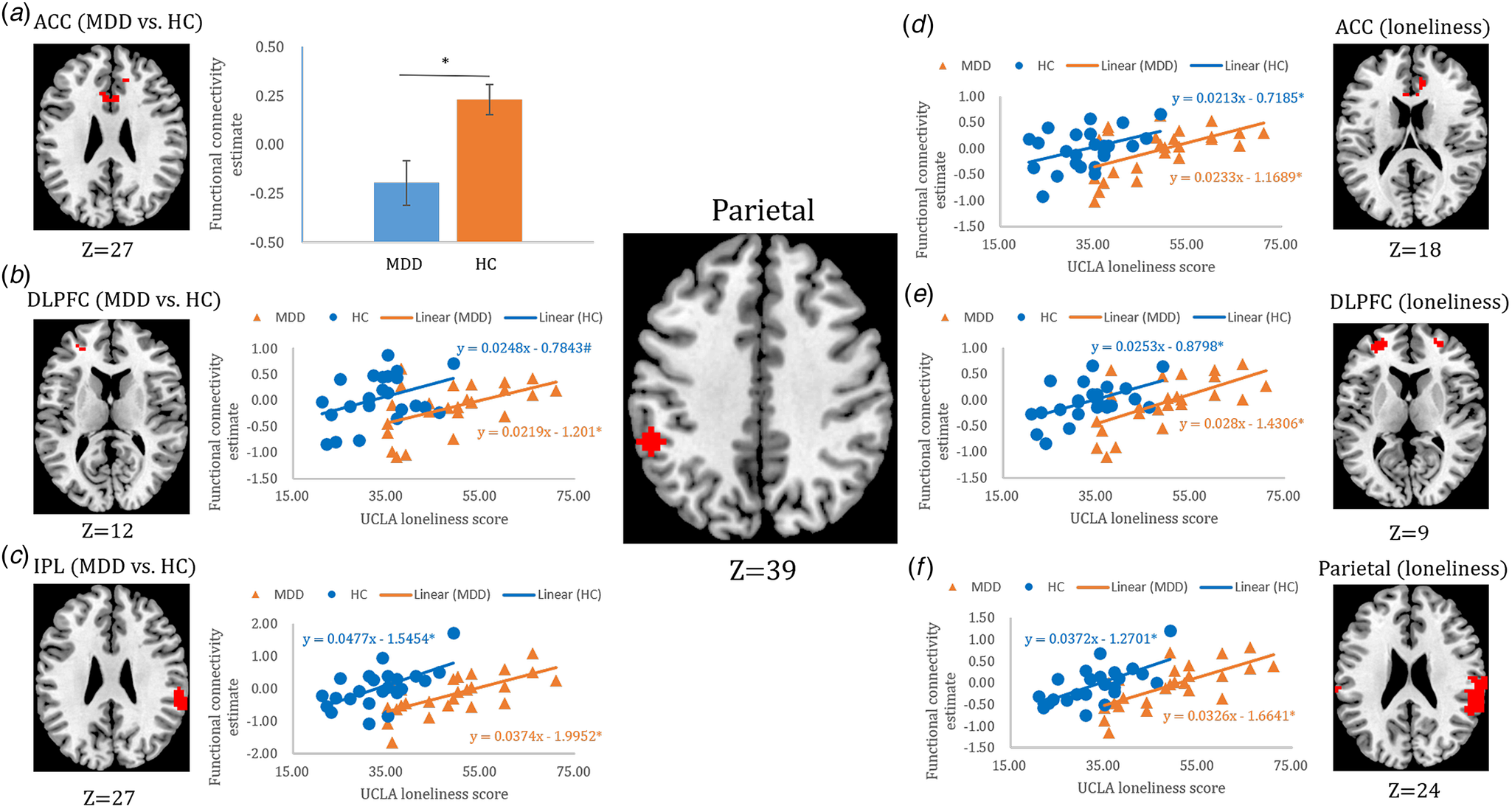
We then tested our hypotheses that the MDD patients would show reduced functional integrity of the frontoparietal and cerebellar networks during performing the Stroop task, while loneliness would have the opposite effect. To this end, the gPPI analysis was conducted based on 6-mm seeds centered at the locus of maxima of the parietal (inferior) and cerebellar (Crus I) clusters significant to the MDD × loneliness effect. ROI analyses revealed that compared to MDD patients, HCs exhibited significant or marginal increases of parietal connectivity with the dorsal ACC (maxima = −6, 21, 24, voxels = 22, TFCE = 88.61, p corrected < 0.05), right inferior parietal cortex (maxima = 63, −39, 27, voxels = 100, TFCE = 182.47, p corrected < 0.05) and left DLPFC (maxima = −30, 48, 9, voxels = 5, TFCE = 147.21, p corrected = 0.056) in incongruent v. neutral trials (Fig. 3). Similarly, across all participants, significant positive loneliness effects were observed for parietal connectivity with the rostral and dorsal ACC (maxima = 9, 36, 18, voxels = 33, TFCE = 87.15), left DLPFC (maxima = −30, 51, 9, voxels = 117, TFCE = 222.79) and the right (and to lesser extent, left) inferior and superior parietal cortices (maxima = 63, −27, 30, voxels = 494, TFCE = 451.63) to the same contrast (all p corrected < 0.05) (Fig. 3). No significant MDD × loneliness effect on parietal connectivity, and no significant MDD, loneliness or MDD × loneliness effect on cerebellar connectivity, were observed. Whole-brain analysis results are included in online Supplementary Materials.
MDD and loneliness main effects on parietal task-related connectivity in incongruent v. neutral trials, among 25 MDD patients and 24 HCs. (a) An ACC region showed more positive connectivity with the parietal seed in HCs than in MDD patients; (b–f) the left DLPFC, right IPL, ACC, bilateral DLPFC and bilateral parietal regions in which connectivity with the parietal seed was more positive in the HCs than in MDD patients, and showed a positive relationship with loneliness. Please note that the separation of the blue (representing HCs) and orange (representing MDD patients) trend lines in each scatterplot denotes the MDD main effect. ACC: anterior cingulate cortex. DLPFC: dorsolateral prefrontal cortex. IPL: inferior parietal lobule. *p < 0.05. #p = 0.058. Error bar represents 1 standard error of the mean.

Further exploratory analyses revealed that, in the right inferior parietal and left DLPFC regions, where FC with the parietal seed in incongruent v. neural trials was more positive in HCs than in MDD patients, the connectivity strength also showed a significant positive association with loneliness across all participants (both t 45 > 3.52, p corrected < 0.05) (Fig. 3). Similarly, in the ACC, left DLPFC and bilateral parietal regions, where connectivity with the parietal seed showed positive relationships with loneliness, the connectivity strengths were also significantly more positive in HCs than in MDD patients (all t 45 > 2.55, p corrected < 0.05) (Fig. 3). All the above results remained significant after additionally controlling for the HAMD scores (p corrected < 0.05). No significant HAMD or HAMD × loneliness effect on the FC measures was observed (p > 0.05). No MDD × loneliness effect was found (all ps > 0.56). No significant correlation was found between the connectivity estimate and RT difference in incongruent v. neutral trials in MDD patients or in HCs (pscorrected > 0.36).
Resting-state functional connectivity analysis
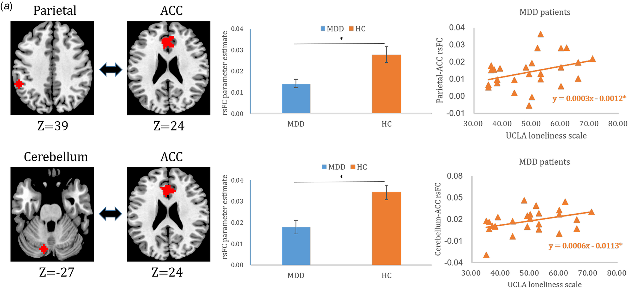
Finally, we tested the hypotheses that MDD and loneliness would exert opposite effects on the functional integrity of the DMN, and on the connectivity between the DMN and the networks involved in cognitive control and affect regulation, during resting state. The same parietal and cerebellar seeds used in the gPPI analysis were applied in the rsFC analyses. ROI analyses revealed more positive rsFC in HCs than in MDD patients between the parietal seed and rostral and dorsal ACC (maxima = −3, 45, 12, voxels = 195, TFCE = 153.54, p corrected < 0.05), and between the cerebellar seed and rostral and dorsal ACC (maxima = −3, 30, 24, voxels = 178, TFCE = 158.65, p corrected < 0.05) (Fig. 4). No loneliness or MDD × loneliness effect survived the TFCE correction. No other significant cluster was observed at the whole-brain level.
(a) Parietal- and (b) cerebellum-seeded rsFC analysis, among 26 MDD patients and 24 HCs. HCs showed more positive parietal-ACC and cerebellum-ACC rsFC than MDD patients (middle panel). Among MDD patients, loneliness positively predicted parietal-ACC and cerebellum-ACC rsFC (right panel). *p < 0.05. Error bar represents 1 standard error of the mean.

Further exploratory analyses on the rsFC of the significant ACC clusters revealed no significant loneliness (t 47 = 1.28 and 1.56) or MDD × loneliness effect across all participants (ps > 0.1), but loneliness showed positive effects on both parietal-ACC and cerebellum-ACC rsFC in the MDD patients (pscorrected < 0.05) (Fig. 4). After additionally controlling for medication load, the positive effects of loneliness in MDD patients remained marginal (pscorrected = 0.06 and 0.08). The positive loneliness effect in MDD patients also remained marginally significant after controlling for the HAMD scores (pscorrected = 0.065 and 0.065). No significant HAMD or HAMD × loneliness effect was observed (ps > 0.05). The parietal-ACC and cerebellum-ACC rsFC that showed significant MDD effect positively correlated with each other across all participants (r = 0.645, bootstrapping CI 0.44–0.79). Moreover, the parietal-ACC rsFC and the parietal-ACC Stroop-related connectivity that were both more positive in HCs than in MDD patients showed marginal positive correlation across all participants (r = 0.239, bootstrapping CI 0.01–0.51) (online Supplementary Fig. S2).
Supplementary analyses
In the MDD patients, no significant effect of MMSE, HAMD scores or illness-related variables was detected on the RT difference between incongruent and neutral trials (ps > 0.14). No significant effect of HAMD scores or illness-related variables was observed for any regional activity or FC measures (pscorrected > 0.12).
Discussion
Loneliness and MDD exerted distinct and opposing effects on Stroop task behaviors, Stroop-related FC of the prefrontal-ACC-parietal circuitry, as well as interactive effects on parietal and cerebellar activities during Stroop performance. Moreover, MDD patients showed decreased resting-state connectivity between the ACC and both the inferior parietal cortex and medial cerebellum, while loneliness positively predicted the same connectivities among patients. Collectively, our findings reveal distinct neural mechanisms implicated in cognitive control and affect regulation for loneliness and MDD.
The greater incongruence-specific accuracy reduction among MDD patients suggested compromised executive control functions (Vasic et al., Reference Vasic, Walter, Sambataro and Wolf2009; Hammar et al., Reference Hammar, Sørensen, Årdal, Oedegaard, Kroken, Roness and Lund2010; Chantiluke et al., Reference Chantiluke, Halari, Simic, Pariante, Papadopoulos, Giampietro and Rubia2012). However, no effect of MDD on Stroop RT interference was observed, concurring with past evidence on unmedicated, acute-phase MDD patients (Wagner et al., Reference Wagner, Sinsel, Sobanski, Köhler, Marinou, Mentzel, Sauer and Schlösser2006; Holmes and Pizzagalli, Reference Holmes and Pizzagalli2008). The specific impairment on accuracy may reflect selective reduction of conflict-resolution and performance-monitoring functions (Holmes and Pizzagalli, Reference Holmes and Pizzagalli2008; Hammar et al., Reference Hammar, Sørensen, Årdal, Oedegaard, Kroken, Roness and Lund2010; Chantiluke et al., Reference Chantiluke, Halari, Simic, Pariante, Papadopoulos, Giampietro and Rubia2012). In contrast, perceived loneliness specifically reduced the RT interference effect on incongruent trials. Based on our speculative model, lonely individuals might show elevated automatic, bottom-up attention (Cacioppo et al., Reference Cacioppo, Ernst, Burleson, McClintock, Malarkey, Hawkley, Kowalewski, Paulsen, Hobson, Hugdahl and Spiegel2000) but upregulated compensatory top-down cognitive control and inhibitory functions, allowing for quick disengagement from the irrelevant stimulus dimension toward the relevant dimension and the initiation of appropriate responses (Ravizza and Carter, Reference Ravizza and Carter2008).
Across all participants, Stroop incongruence elicited pronounced activations in the prefrontal-ACC-parietal-cerebellum circuitry (Mohanty et al., Reference Mohanty, Engels, Herrington, Heller, Ringo Ho, Banich, Webb, Warren and Miller2007; Roberts and Hall, Reference Roberts and Hall2008; O'Halloran et al., Reference O'Halloran, Kinsella and Storey2012). Both the rostral and dorsal ACC perform error detection and attention control functions (Menon et al., Reference Menon, Adleman, White, Glover and Reiss2001), and communicate with the DLPFC and the superior parietal cortex to enable appropriate and flexible response selection (Milham et al., Reference Milham, Banich and Barad2003; Humphreys and Lambon Ralph, Reference Humphreys and Lambon Ralph2015). No significant main effect of MDD or loneliness emerged, consistent with previous research (Videbech et al., Reference Videbech, Ravnkilde, Gammelgaard, Egander, Clemmensen, Rasmussen, Gjedde and Rosenberg2004; Wong et al., Reference Wong, Liu, Lin, Huang, Wai, Lee and Lee2016). However, MDD significantly modulated the loneliness effect in inferior and superior parietal cortices (primarily left-lateralized but was also present to lesser extent on the right), and in anterior and posterior cerebellar vermis and lateral hemispheres. Furthermore, the parietal and cerebellar signals positively predicted RT interference in MDD patients but not in controls. Among older adults during negative affective processing, loneliness negatively and positively predicted LPFC activities in MDD patients and HCs, respectively (Wong et al., Reference Wong, Liu, Lin, Huang, Wai, Lee and Lee2016), a pattern that was replicated on parietal and cerebellar signals in our sample. The quantitatively greater effect on the left parietal cortex might be due to the Stroop task that used lexical stimuli, since both phonological and semantic processing may engage the left parietal cortex to greater extent (Humphrey and Lambon Ralph, Reference Humphreys and Lambon Ralph2015). The posterior cerebellum, particularly Crus I of lobule VII, is implicated in cognitive control functions including attention, inhibition and error detection (Buckner, Reference Buckner2013; D'Angelo and Casali, Reference D'Angelo and Casali2013), while the anterior and medial cerebellum (vermis) are respectively associated with sensorimotor and affect regulation functions (Stoodley and Schmahmann, Reference Stoodley and Schmahmann2010; D'Angelo and Casali, Reference D'Angelo and Casali2013). The cerebellum works closely with the inferior and superior parietal cortices, which are respectively implicated in automatic bottom-up and goal-directed top-down attention processes (Cabeza et al., Reference Cabeza, Ciaramelli, Olson and Moscovitch2008; Ciaramelli et al., Reference Ciaramelli, Grady and Moscovitch2008; Roberts and Hall, Reference Roberts and Hall2008; Ghajar and Ivry, Reference Ghajar and Ivry2009). Thus, among MDD patients, greater parietal and cerebellar activities associated with increased RT interference might indicate reduced cognitive control efficiency and/or increased affective interference (Brzezicka, Reference Brzezicka2013), while their negative relationships with loneliness might indicate enhanced cognitive and affective control processes in lonelier depressed individuals. The positive association between loneliness and parietal-cerebellar activities in HCs might suggest increase in both automatic bottom-up attention and top-down attention control functions during incongruence processing. The parietal-cerebellar signals showed no association with Stroop performance in controls, consistent with previous findings (Wagner et al., Reference Wagner, Sinsel, Sobanski, Köhler, Marinou, Mentzel, Sauer and Schlösser2006), suggesting possible stable neural features that are relatively independent of performance fluctuations.
FC in the prefrontal-ACC-parietal network showed distinct and opposite effects of MDD and loneliness, with the former being associated with decreased and the latter with increased connectivity when resolving incongruence-elicited cognitive conflict. The pivotal role of the prefrontal-ACC-parietal circuitry in conflict processing is well established (Roberts and Hall, Reference Roberts and Hall2008; Niendam et al., Reference Niendam, Laird, Ray, Dean, Glahn and Carter2012; Müller et al., Reference Müller, Cieslik, Laird, Fox and Eickhoff2013; Harding et al., Reference Harding, Yücel, Harrison, Pantelis and Breakspear2015), with the inferior parietal cortex relaying information about stimulus salience to the PFC and communicating with the dorsal executive network for high-order control of bottom-up attention (Miller and Cohen, Reference Miller and Cohen2001; Humphreys and Lambon Ralph, Reference Humphreys and Lambon Ralph2015). Decreased PFC-inferior parietal connectivity during cognitive control was previously also found in schizophrenic patients (Yoon et al., Reference Yoon, Minzenberg, Ursu, Walters, Wendelken, Ragland and Carter2008; Fornito et al., Reference Fornito, Yoon, Zalesky, Bullmore and Carter2011). Thus, while lonely individuals showed closer communications in the cognitive control network, MDD patients exhibited disconnected PFC-parietal network, possibly underlying attenuated top-down attention control (Vasic et al., Reference Vasic, Walter, Sambataro and Wolf2009; Gotlib and Joormann, Reference Gotlib and Joormann2010; Disner et al., Reference Disner, Beevers, Haigh and Beck2011).
MDD is associated with dysregulated self-focused affective processes such as rumination (Gotlib and Joormann, Reference Gotlib and Joormann2010). We found that at rest, MDD patients exhibited attenuated inferior parietal and cerebellar connectivity with the ACC, both of which were positively associated with loneliness. Moreover, the parietal-ACC connectivities across the cognitive and resting states were positively correlated, implicating overlapping cognitive control and affect regulation networks. The ACC, the inferior parietal cortex, and the Crus I of the cerebellum are consistently implicated in cognitive control of negative affective processing (Ochsner et al., Reference Ochsner, Bunge, Gross and Gabrieli2002; Mohanty et al., Reference Mohanty, Engels, Herrington, Heller, Ringo Ho, Banich, Webb, Warren and Miller2007; Schutter and van Honk, Reference Schutter and van Honk2009; Pizzagalli, Reference Pizzagalli2011; Keren-Happuch et al., Reference Keren-Happuch, Chen, Ho and Desmond2014; Kohn et al., Reference Kohn, Eickhoff, Scheller, Laird, Fox and Habel2014). All these regions are also part of or closely associated with the DMN (Long et al., Reference Long, Zuo, Kiviniemi, Yang, Zou, Zhu, Jiang, Yang, Gong, Wang and Li2008; Habas et al., Reference Habas, Kamdar, Nguyen, Prater, Beckmann, Menon and Greicius2009; Sripada et al., Reference Sripada, King, Welsh, Garfinkel, Wang, Sripada and Liberzon2012). Altered cerebellum-DMN connectivity has been observed in MDD (Liu et al., Reference Liu, Zeng, Li, Ma, Li, Shen and Hu2012). The cerebellum-rostral ACC circuitry was selectively activated during the anticipation of negative stimuli (Ploghaus et al., Reference Ploghaus, Becerra, Borras and Borsook2003), with the cerebellum conveying emotion-predictive signals to the ACC, which then performs attention control and reappraisal functions (Wiech et al., Reference Wiech, Ploner and Tracey2008). Collectively, MDD is characterized by altered DMN integrity and reduced connectivity between the DMN and the cognitive and emotion control networks at rest (Guo et al., Reference Guo, Liu, Xue, Gao, Liu, Xiao, Chen and Zhao2013; Kaiser et al., Reference Kaiser, Andrews-Hanna, Wager and Pizzagalli2015; Mulders et al., Reference Mulders, van Eijndhoven, Schene, Beckmann and Tendolkar2015), which was independent of depressive symptomology, possibly representing stable, trait-like affect regulatory deficits (Hammar et al., Reference Hammar, Sørensen, Årdal, Oedegaard, Kroken, Roness and Lund2010; Mulders et al., Reference Mulders, van Eijndhoven, Schene, Beckmann and Tendolkar2015). Conversely, loneliness positively predicted parietal- and cerebellum-ACC resting-state connectivities, which might mean that lonely depressed individuals display greater recruitment of cognitive control circuitries over automatic attention and hypervigilance, toward reducing general affect sensitivity (Cacioppo et al., Reference Cacioppo, Norris, Decety, Monteleone and Nusbaum2009; Wong et al., Reference Wong, Liu, Lin, Huang, Wai, Lee and Lee2016).
Our findings are generally consistent with our proposed model that lonely individuals recruit greater high-level cognitive control processes to regulate the also enhanced bottom-up automatic attention processes. This is reflected by (1) lonely individuals exhibited generally superior Stroop performance as reflected by their shorter RTs, indicating enhanced cognitive control functioning; (2) activities of the parietal-cerebellum circuitry positively predicted Stroop RT interference in MDD patients, and the same activities were negatively associated with loneliness among patients, suggesting that loneliness might be related to better cognitive control in MDD; (3) loneliness in HCs was associated with increased activities in both superior and inferior parietal cortices, networks that were respectively involved in top-down and bottom-up attention processes (Roberts and Hall, Reference Roberts and Hall2008; Ghajar and Ivry, Reference Ghajar and Ivry2009; Humphreys and Lambon Ralph, Reference Humphreys and Lambon Ralph2015) and (4) loneliness positively predicted FC strength of the inferior parietal cortex with the dorsal frontoparietal network and the cerebellum during Stroop performance and at rest, suggesting upregulated control of automatic attention during both external- and internal-oriented processing. Yet, the alignment of the current findings to the proposed model is admittedly preliminary and limited, suffering from the potential issue of reverse inference (inferring on functions based on neural patterns). Therefore, our model needs further testing using paradigms that directly assess bottom-up and top-down cognitive control processes, and affect regulation functions.
Our current study does not offer direct evidence on the neurocognitive mechanism for the association between loneliness and MDD. However, we speculate that while lonely individuals may recruit greater cognitive resource to regulate the enhanced bottom-up attention process as a compensatory strategy in the short term, as loneliness-related stressors accumulate over time, the cognitive control resource may gradually become exhausted, resulting in dysregulated affective processes and MDD-like characteristics. Such hypothetical transitional model remains to be tested by future longitudinal studies that follow up lonely individuals across unaffected and disease phases.
Several limitations need to be noted. First, the current cross-sectional study cannot ascertain the causal effect of either MDD or loneliness on cognitive or neural processes, which needs to be addressed by future longitudinal research. Second, our MDD patients were on medications and showed heterogeneous symptomology levels, although none of the key behavioral or neural measures showed significant relationships with medication- or illness-related variables. Third, our sample sizes were modest, although they were comparable or superior to those of a number of recent relevant studies (Wagner et al., Reference Wagner, Sinsel, Sobanski, Köhler, Marinou, Mentzel, Sauer and Schlösser2006; Mitterschiffthaler et al., Reference Mitterschiffthaler, Williams, Walsh, Cleare, Donaldson, Scott and Fu2008; Cacioppo et al., Reference Cacioppo, Norris, Decety, Monteleone and Nusbaum2009; Vasic et al., Reference Vasic, Walter, Sambataro and Wolf2009; Chantiluke et al., Reference Chantiluke, Halari, Simic, Pariante, Papadopoulos, Giampietro and Rubia2012; Müller et al., Reference Müller, Cieslik, Laird, Fox and Eickhoff2013; Wong et al., Reference Wong, Liu, Lin, Huang, Wai, Lee and Lee2016). Finally, our participants were all middle-aged, which may limit the generalization of the current findings to other-aged samples.
In summary, we provide novel evidence on distinct neural profiles in MDD and loneliness, implicating potential dissociations in key cognitive and affective processes. Our findings lay the path for future research on the association of loneliness and MDD, toward promoting mental wellbeing among the substantial populations with unfulfilled social needs.
Supplementary material
The supplementary material for this article can be found at https://doi.org/10.1017/S0033291719002782.
Data
The data that support the findings of this study are available from the corresponding author upon reasonable request.
Financial support
This work was supported by (1) the Medical Research Grants CMRPG3C0041 and CMRPG3C0042 from the Chang Gung Memorial Hospital to Chemin Lin and (2) The University of Hong Hong May Endowed Professorship in Neuropsychology and the Science and Technology Program of Guangdong (Ref: 2018B030334001) to Tatia Lee.
Author contributions
R.S. contributed to data analysis and manuscript drafting; H.-L.L. contributed to study design and manuscript drafting; C.-M.H. contributed to study conceptualization and design; Y.-L.C. contributed to data collection; M.G. contributed to data analysis and manuscript drafting; S.-H.L. contributed to study conceptualization, design and data collection; C.L. contributed to study conceptualization, design and data collection; T.M.C.L. contributed to study conceptualization, design and manuscript drafting. All authors have read and approved the final version of the manuscript.
Conflict of interest
None.