Pulmonary arterial hypertension is a rare disease with an incidence of 2–3 per million and a prevalence of 25–50 per million.Reference Berger, Beghetti and Humpl 1 Although recent developments of pulmonary hypertension-targeted therapies result in an improvement of prognosis, mortality is still high.Reference Barst, McGoon, Elliott, Foreman, Miller and Ivy 2 Pulmonary arterial hypertension is characterised by pathologic remodelling of the pulmonary arterioles with intima thickening and media hypertrophy, resulting in increased pulmonary vascular resistance and pressure, and eventually right ventricular failure.Reference Berger, Beghetti and Humpl 1 , Reference Galiè, Manes and Branzi 3 The aetiology of pulmonary arterial hypertension is multifactorial; however, endothelin-1, a potent and long-lasting vasoconstrictive peptide, is thought to play a central role in the remodelling process.Reference Giaid, Yanagisawa and Langleben 4
Endothelin-1 is produced and secreted into circulation by the endothelial cells of the pulmonary vessels after transformation from its precursor form big endothelin-1 by the endothelin-1-converting enzyme. Although the exact mode of endothelin-1 inactivation has not been fully clarified, it is assumed that lysosomal enzymes are involved in its degradation.
Elevated plasma endothelin-1 levels have been detected in diverse forms of pulmonary arterial hypertension and also in experimental disease models.Reference Galiè, Manes and Branzi 3 Different stimuli, such as hypoxia, ischaemia, shear stress, catecholamines, and insulin were shown to increase the synthesis of endothelin-1 in pulmonary arterial hypertension. In addition, augmented endothelin-1-converting enzyme levels and upregulation of endothelin-1 receptors have also been described,Reference Galiè, Manes and Branzi 3 and in patients with severe pulmonary arterial hypertension, usually all components of the endothelin system are upregulated secondary to increased endothelin-1 production within the vascular endothelial cells.Reference Galiè, Manes and Branzi 3
On the basis of theoretical considerations, hampered degradation should also cause pulmonary arterial hypertension by augmenting endothelin-1 plasma levels, but this has not yet been demonstrated. Therefore, we report on a patient with severe pulmonary arterial hypertension and mucolipidosis type II, a disorder characterised by impaired enzyme transport into the lysosome, whose endothelin-1 and big-endothelin-1 values suggest that reduced degradation instead of increased production contributed to pulmonary arterial hypertension.
The patient is the second child of healthy consanguineous Turkish parents. At the age of 25 months, a lysosomal storage disorder was suspected because of developmental delay, short stature, Hurler-like facial appearance, contractures, hip dislocation, and dysostosis multiplex. His older sister was similarly affected, but had a milder phenotype. In the boy, the diagnosis of mucolipidosis type II was made after measuring significantly increased activities of various lysosomal hydrolases in plasma, and after determining reduced activities of several lysosomal enzymes in lymphocytes and fibroblasts.
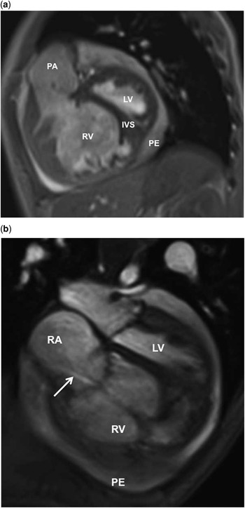
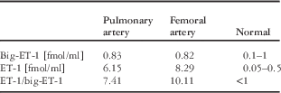
Serial echocardiograms during regular follow-up demonstrated normal cardiac anatomy. However, at the age of 9 years, a chest X-ray, performed because of repeated cough, revealed cardiomegaly. A subsequent echocardiogram showed right ventricular dilation with reduced systolic right ventricular function, left ventricular compression (systolic left ventricular eccentricity index of 1.5), dilated pulmonary arteries, and slight non-circular pericardial effusion. Computed tomography of the chest demonstrated no signs of pulmonary thromboembolism and displayed only mild fibrotic alterations in both lungs. Lung function testing showed a vital capacity of 75% predicted. Catheterisation confirmed marked precapillary pulmonary arterial hypertension – mean pulmonary arterial pressure of 63 mmHg; ratio of mean pulmonary and systemic arterial pressure of 0.85; pulmonary capillary wedge pressure 8 mmHg – without significant response to vasodilator testing – minimal ratio of mean pulmonary and systemic arterial pressure of 0.68. Remarkably, endothelin-1 levels were significantly increased in both the pulmonary artery and in the femoral artery, whereas big endothelin-1 levels lay within normal range, as shown in Table 1. This resulted in markedly elevated endothelin-1/big endothelin-1 ratios in both vascular beds. The boy was treated orally with a dual ET receptor antagonist in combination with a phosphodiesterase-5 inhibitor. This resulted in a gradual and sustained improvement of his clinical condition during the following months. A further catheterisation had to be performed at the age of 11 years, as the patient developed signs of right ventricular failure, accompanied by pericardial effusion as shown in Figure 1 and markedly increased B-type natriuretic peptide values – maximum 3100 pg/ml. Interventional reopening and dilatation of the foramen ovale up to a diameter of 10 mm in conjunction with an add-on therapy consisting of inhalative iloprost, digoxin, furosemide, and spironolactone stabilised his cardiac function. Currently, at the age of 13 years, the boy is in a stable condition (World Health Organization Functional class 3).
Cardiac magnetic resonance imaging. Short axis (a) and four-chamber view (b) showing the dilated and hypertrophied right ventricle (RV) with enlarged right atrium (RA) and the interventricular septum (IVS) bowing to the left ventricle (LV). Please also note the circular pericardial effusion (PE) and the moderate tricuspid regurgitation (arrow). PA=pulmonary arterial trunk.

ET-1 and big ET-1 levels assessed in the pulmonary and femoral artery (fmol/ml).

ET-1=endothelin-1
Mucolipidosis type II is a rare autosomal recessive Hurler-like disorder caused by deficiency of the enzyme Uridine diphosphate-N-acetylglucosamine-1-phospho-transferase. This leads to hampered phosphorylation of lysosomal enzymes inside the golgi apparatus, secondary to insufficient synthesis of the mannose 6-phosphate recognition marker. Lack of this marker affects targeting and trafficking of lysosomal hydrolases from the cytosol and endoplasmatic reticulum to the lysosome, resulting in their leakage into the extracellular compartment and accumulation of lysosomal substrates in various tissues of the body. The disorder shows a wide interindividual variability and is characterised clinically by short stature, skeletal abnormalities, cardiomegaly, and developmental delay. Pulmonary complications reported in mucolipidosis type II include airway obstruction due to abnormal storage of lysosomal substrates from the tongue to the trachea, congestions and focal indurations secondary to bronchopneumonia, lipid granulomata, pulmonary bleeding, chronic respiratory infections, and restrictive lung disease due to small thoracic cage and lung fibrosis.Reference Ishak, Zambrano, Bazzy-Asaad and Esquibies 5 Pulmonary arterial hypertension, which is not infrequent in other lysosomal storage disorders such as Gaucher disease or mucopolysaccharidosis, has only rarely been observed in mucolipidosis type II.Reference Ishak, Zambrano, Bazzy-Asaad and Esquibies 5 , Reference Kovacevic, Schranz, Meissner, Pillekamp and Schmidt 6
In contrast with its synthesis, degradation of endothelin-1 has only incompletely been clarified. It is known that endothelin-1 can be internalised by the endothelin-B receptor and transported into the lysosome, where the peptide is further degraded.Reference Deng and Jeng 7 As endothelin-B receptor does not traffic back to the cell surface, it is assumed that endothelin-B works as a pure clearance receptor. Alternatively, endothelin-1 can also be internalised by the endothelin-A receptor, which is known to recycle to the cell surface. To date, it is unclear whether endothelin-1 internalised by endothelin-A receptor is recycled, or also transported into the lysosome for further degradation.Reference Wang, Chiou, Gagne and Wu-Wong 8 , Reference Paasche, Attramadal, Sandberg, Johansen and Attramadal 9 Finally, plasma membrane-bound neutral peptidases degrading endothelin-1 have also been identified. Although there are obviously different modes of endothelin-1 inactivation, there is clear evidence that lysosomal enzymes are involved in endothelin-1 degradation in vascular smooth muscle cells.Reference Bermek, Peng, Angelova, Ergul and Puett 10 , Reference Itoh, Kase, Shimmoto, Satake, Sakuraba and Suzuki 11
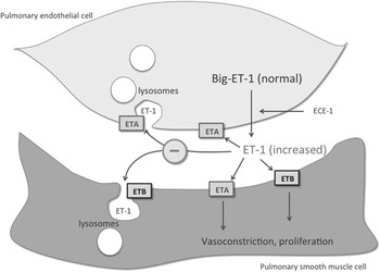
In our patient, lung function testing and chest computer tomography excluded embolism and significant pulmonary fibrosis as potential causes of his severe pulmonary arterial hypertension. In addition, the constellation of normal big-endothelin-1 and increased endothelin-1 levels in conjunction with lacking upregulation of other endothelin system components argues against increased synthesis, but is consistent with impaired lysosomal endothelin-1 degradation, as shown in Figure 2.
Cartoon depicting ET-1 synthesis and degradation. ET-1 levels were markedly increased whereas big ET-1 levels, the precursor form of ET-1, were within the normal range. This suggests that impaired degradation of ET-1 instead of increased synthesis resulted in the observed elevated ET-1 levels. Since ET-1 degradation takes place in the lysosome, it appears likely that lysosomal dysfunction caused by the underlying disease mucolipidosis II contributed to elevated ET-1 levels in this patient and led to the development of pulmonary arterial hypertension due to ET-1-mediated vasoconstriction of the pulmonary smooth muscle cells. ET-1=endothelin-1; ETA=endothelin-1 receptor A; ETB=endothelin-1 receptor B; ECE-1=endothelin-1 converting enzyme.

In conclusion, these findings provide for the first time evidence that impaired endothelin-1 degradation represents a potential mechanism for the development of pulmonary arterial hypertension. Our findings also underscore the necessity for regular cardiac follow-up examinations on mucolipidosis type II demonstrate that lysosomal dysfunction can contribute to pulmonary arterial hypertension, and add pulmonary arterial hypertension to the list of pulmonary complications that have to be taken into account in this disorder.
Acknowledgements
The authors thank Professor Dr Joachim Kreuder, Dr Heiner Latus, and Professor Dr Dietmar Schranz for their inspirations, critical comments, and discussion of the manuscript.
Financial Support
This research received no specific grant from any funding agency, commercial, or not-for-profit sectors.
Conflicts of Interest
None.