Case report
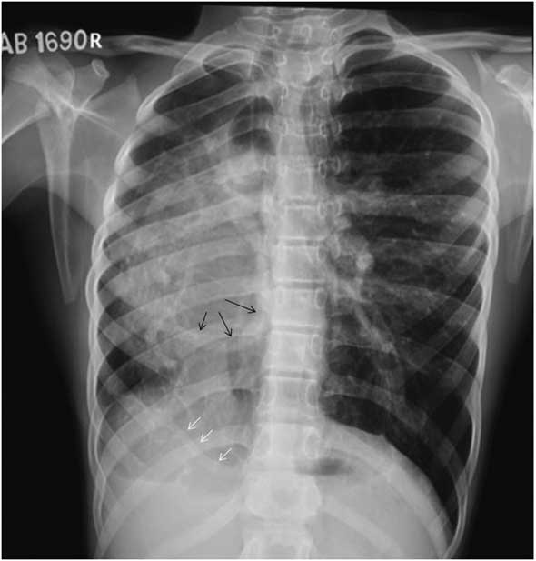
A 15-year-old boy, diagnosed to have a large ventricular septal defect at 6 years of age, presented with complaints of NYHA Class II dyspnoea on exertion. There was no history of cyanosis. On examination he had a saturation of 96% in all four limbs. The right precordium was hyperdynamic. On auscultation, he had a loud pulmonary component of second heart sound and a grade 3/6-ejection systolic murmur in the right parasternal border. Other system examinations were normal. Chest X-ray (Fig 1) showed dextrocardia/dextroposition with situs solitus, trachea shifted to the right, hypoplasia of the right lung, abnormal vascularity on the right side, increased vascularity on the left side, and relatively high position of the right dome of the diaphragm. There were at least two abnormal vascular structures seen on X-ray: one tortuous vessel coming from the right upper lobe and descending, and another vessel coming from below the right diaphragm and ascending towards the lower lateral part of the right lung.
Chest Radiograph showing loss of volume in the right lung with shift of mediastinum to the right. Two abnormal vessels are seen one coming from right upper lobe and descending down (black arrows) and another vessel coming from below right diaphragm and ascending towards lower lateral part of the right lung (white arrows).

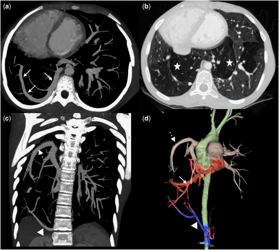
Echo showed situs solitus, mesocardia, large subaortic ventricular septal defect with bidirectional shunt, and severe pulmonary artery hypertension. The pulmonary venous anatomy and interatrial septum was not convincingly visualised owing to the abnormal orientation of the heart and lung anomaly. Hence, a CT scan (Fig 2) was performed, which showed atrial situs solitus with dextroposition of the heart with ventricular and atrial septal defects. The right bronchus showed trifurcation morphology. There was loss of volume in the right upper lobe. The right lower lobe was located in midline with medial herniation, separated from the left lung by pleura, which is a cross-over lung segment with pseudo horseshoe lung, and was supplied by a systemic arterial branch from the coeliac axis – dual arterial supply. The left lung was drained by two left pulmonary veins. The right apical lobe had a separate small pulmonary vein draining to the left atrium. The remaining right lung along with the pseudo horseshoe component drained to the left atrium through a long right lower pulmonary vein, coursing from the upper part of the right lung to the right diaphragm and then turning upwards to join the left atrium. The main and left pulmonary arteries were dilated and the right pulmonary artery was smaller than the left. The right pulmonary artery was seen to descend from the right upper lateral position towards the diaphragm to supply the medially placed right lower lobe.
Images from CT chest with contrast. Axial images (a and b) showing the horseshoe morphology right lower lobe (star) and unusual course of the right inferior pulmonary vein draining into the left atrium (arrow). Coronal maximum intensity projection image (c) and 3D rendered image (d) showing the right pulmonary artery (broken arrow) and a branch from the celiac artery (arrow head).

In view of the large ventricular septal defect with bidirectional shunt, cardiac cath studies were conducted to assess pulmonary vascular reactivity, which showed reactive pulmonary vascular bed. He was subsequently operated for atrial and ventricular septal defect closure, and the postoperative course was uneventful.
Discussion
Scimitar syndrome or pulmonary venolobar syndrome is a rare pulmonary anomaly that consists of anomalous pulmonary venous drainage of the right lung to the inferior caval vein – giving rise to the scimitar sign – anomalous systemic arterial supply of the right lower lobe from either the thoracic or abdominal aorta, hypoplasia of the right lung, with resultant cardiac dextroposition, and right pulmonary artery hypoplasia.Reference Sanger, Taylor and Robicsek 1 On a frontal chest X-ray film, a large pulmonary vein running caudad and medially along the right heart border to the diaphragm is referred to as a scimitar sign, commonly indicating partial anomalous pulmonary venous return to the inferior caval vein or low right atrium. However, there have been cases reportedReference Morgan and Forker 2 in which the scimitar sign and features of scimitar syndrome were present, but the aberrant pulmonary vein ultimately drained normally into the left atrium. The scimitar sign is often seen with scimitar syndrome, and it can also be rarely seen in anomalous intrapulmonary venous connection to the superior caval vein, obstruction of a major pulmonary vein with development of a distended intrapulmonary collateral, and an anomalous inferior caval vein with normal pulmonary venous drainage.Reference Herer, Jaubert, Delaisements, Huchon and Chretien 3 The X-ray in our patient had two vessels, which put together mimicked a scimitar vein, wherein the right pulmonary artery descended from the right lateral position towards the diaphragm to supply the medially placed right lower lobe. The second vessel traversed cephalad from the abdominal aorta to supply the right lower lobe. There was further meandering course of the right pulmonary vein, which turned cephalad near the right diaphragm, to ascend up and join the left atrium (Fig 1).
Rodrigues et alReference Rodrigues, Ritchie and Murchison 4 have recently reviewed and proposed a nomenclature review of scimitar syndrome and conditions with radiological findings similar to scimitar syndrome but with variation in the draining pattern of the anomalous pulmonary veins to the right atrium, inferior caval vein, or left atrium. According to that nomenclature, the case we describe is of meandering pulmonary veins. A total of 32 cases of similar anomalies, under various names, have been described and reviewed by Odenthal et al,Reference Odenthal and Sarikwal 5 with only a few cases reported in the paediatric population. Association with CHD is rare and that provoked early recognition of the anomaly in our patient, who subsequently underwent successful surgical repair. Hypoplasia of the right lung was definitively contributory to the pulmonary artery hypertension, but whether the abnormal course of the pulmonary venous drainage normally to left atrium was contributory is speculative.
It has been suggested that meandering pulmonary veins and related anomalies occur as a consequence of atresia or hypoplasia of one of the pulmonary veins before pulmonary segmentation, with drainage of the entire lung subsequently occurring via the remaining vein. The frequent association of the anomaly with a hypoplastic hemithorax supports this mechanism.Reference Hanson, Wood, Seymour and Petheram 6
The importance of knowing this entity is manifold. The fact that scimitar syndrome and meandering pulmonary veins both represent variations within the same spectrum of anomalies is evident by the presence of some additional anatomical variants shared between both anomalies. Both are frequently associated with a hypoplastic right lung, cardiac dextroposition, and scimitar sign on X-ray. Anomalous systemic arterial supply to the lower right lung, classically associated with scimitar syndrome, has also been described in meandering pulmonary vein, including our case, and in anomalous unilateral single pulmonary vein. Meandering pulmonary vein is not only rare but also potentially benign as compared with scimitar syndrome and may not require treatment for its presence per se, as there is no intra- or extra-cardiac shunt. Tomographic imaging with either CT angiography or MRI is the single most important investigation for diagnosing such pathologies, and the practicing clinician should spare unnecessary investigations for those patients presenting with the scimitar sign on chest radiograph.
Acknowledgement
None.
Financial Support
This research received no specific grant from any funding agency, commercial or not-for-profit sectors.
Conflicts of Interest
None.