Introduction
To initiate a study on the occurrence of predatory dorylaimid nematodes in Iran, several soil samples were collected from natural regions of the country in August 2008. One sample of wet soil from the rhizosphere of grasses from Sahand Mountains, Eastern Azarbaijan (north-western Iran) yielded a population of an undescribed species of Paractinolaimus which is described herein. In addition to the morphological description, two ribosomal genes (partial 18S and 28S D2/D3) were sequenced to characterize the species. According to Andrássy (Reference Andrássy2006), 27 species can presently be ascribed to this genus of the family Actinolaimidae. To our knowledge, no extensive molecular phylogenetic study on members of the family Actinolaimidae has been carried out so far, since the only sequences deposited refer to Paractinolaimus macrolaimus (De Man, Reference De Man1880). However, the molecular approach, besides facilitating the future identification of the species, will contribute to providing a set of molecular data concerning the species of Paractinolaimus, and possibly of all Actinolaimidae, which could help in the future to reconstruct the phylogeny of this interesting taxon of nematodes. As far as we know, this is the first extensive study and report of Actinolaimidae in Iran.
Materials and methods
Collection and examination of nematodes
Soil samples were collected from the rhizosphere of grasses and mosses and naturally grown wild plants to a depth of 15–20 cm. Nematodes were extracted by suspending the soil samples in water and using sieves with 60 (250 μm aperture), 100 (150 μm aperture) and 200 (74 μm aperture) meshes to screen the material. The nematodes collected on the last sieve were hand picked under a stereomicroscope and fixed by adding hot 4% formaldehyde solution. The fixed nematodes were processed to anhydrous glycerin according to the method of De Grisse (Reference De Grisse1969). Permanent slides were made and measurements taken using an Olympus BX-41 light microscope. Digital images were captured using a DP50 digital camera attached to the microscope, with sharpness and brightness managed using Adobe® Photoshop® CS2 software. Drawings were done using Corel® Draw 12 software and Microsoft® Office® Picture Manager software.
Molecular analysis
For molecular analysis, a single female nematode was put in 50 μl of AE buffer (10 mm Tris-Cl, 0.5 mm EDTA; pH 9.0) and crushed with a pipette tip. The DNA sample was stored at − 20°C until use as a polymerase chain reaction (PCR) template. Different sets of primers were used in the PCR reactions. Primers for 18S amplification were: forward primer 18S-G18S4 (5′-GCTTGTCTCAAAGATTAAGCC-3′) and reverse primer 18S-18P (5′-TGATCCWKCYGCAGGTTCAC-3′) (De Ley et al., Reference De Ley, De Ley, Vierstraete, Karssen, Moens and Vanfleteren2002; Dorris et al., Reference Dorris, Viney and Blaxter2002); forward primer SSUF07 (5′-AAAGATTAAGCCATGCATG-3′) and reverse primer SSUR26 (5′-CATTCTTGGCAAATGCTTTCG-3′) (Floyd et al., Reference Floyd, Abeb, Papert and Blaxter2002); forward primer 18s965 (5′-GGCGATCAGATACCGCCCTAGTT-3′) and reverse primer 18s1573R (5′-TACAAAGGGCAGGGACGTAAT-3′) (Mullin et al., Reference Mullin, Harris and Powers2005). Primers for 28S D2/D3 amplification were forward primer D2a (5′-ACAAGTACCGTGAGGGAAAGT-3′) (Vrain et al., Reference Vrain, Wakarchuk, Levesque and Hamilton1992) and reverse primer D3b (5′-TGCGAAGGAACCAGCTACTA-3′) (Nunn, Reference Nunn1992). The 25 μl PCR contained 12.5 μl 2 × GoTaq DNA polymerase mix (Promega Corporation, Madison, Wisconsin, USA), 1 μl each of 0.4 μm forward and reverse primers, and 1 μl of DNA template. The thermal cycling programme was as follows: denaturation at 95°C for 6 min, followed by 35 cycles of denaturation at 94°C for 30 s, annealing at 55°C for 30 s and extension at 72°C for 1 min. A final extension was performed at 72°C for 10 min. PCR products were cleaned by Montage™ PCR Centrifugal Filter Devices (Billerica, Massachusetts, USA). PCR primers were used for direct sequencing by dideoxynucleotide chain termination using an ABI PRISM BigDye terminator cycle sequencing ready reaction kit (Applied Biosystems, Foster City, California, USA) in an Applied Biosystems 377 automated sequencer (Applied Biosystems) by Eurofins MWG Operon (Huntsville, Alabama, USA). DNA sequence was deposited in GenBank and was compared with other nematode species stored at the GenBank using the BLAST homology search program.
Results and discussion
Paractinolaimus sahandi n. sp.
Type locality and habitat. Sahand Mountains, Eastern Azarbaijan, north-western Iran. Wet soils about the rhizosphere of grasses.
Type material. Holotype female and several paratype males and females deposited at the Nematode Collection of University of Tabriz, and four paratypes (two females and two males) deposited in the collection of the Dipartimento di Biologia Animale, Università di Catania (Italy), the other voucher specimens were deposited in the following collections: USDA Nematode Collection, Beltsville, MD, USA; Nematode Collection of University of California, Riverside Collection; Nematode Collection of University of California, Davis Collection; Harold W. Manter Laboratory of Parasitology, University of Nebraska State Museum and Canadian National Collection of Nematodes, Ottawa, Ontario, Canada.
Etymology. The species has been named after Sahand Mountains, the geographic region where it has been found.
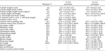
Morphometrics. See table 1.
Morphometric data (mean±SD) of Paractinolaimus sahandi n. sp. All measurements in μm except for L.

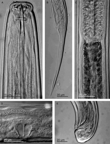
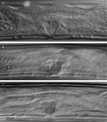
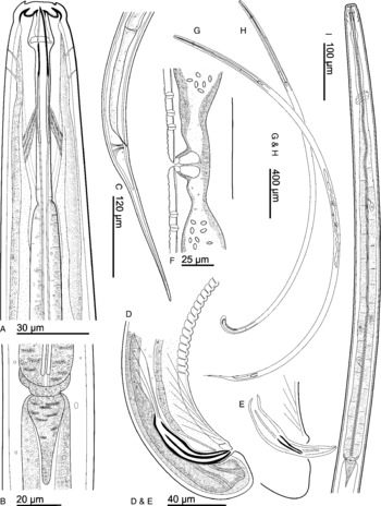
Female. Body long and unusually slender, in the shape of a more or less open C ventrally curved after heat relaxation, tapering very slightly towards both ends. Cuticle appearing smooth at light microscopy, 2.5–3.5 μm thick in the odontostyle region (about as thick as odontostyle), 3.0–4.5 μm at mid-body and 4.5–5.5 μm in the ventral side of the body posterior end, anterior to anus. Two body pores are present both dorsally and ventrally at odontostyle level. Lip region truncate, 2.8–3.6 times as wide as high, separated from the rest of body by a weak depression. Cheilostome well sclerotized with the usual four onchia and several pointed denticles. Amphids stirrup-shaped with wide openings (51–57% of lip region width). Odontostyle dorylaimoid, 3.0–3.7 μm wide, with aperture occupying 48–53% of odontostyle length; odontophore simple and rod shaped. Retractor muscles distinct. Pharynx muscular, very gradually increasing in diameter; attaining its maximum width at about 40% of pharynx length. The outlets of five pharyngeal glands usually distinct but the first pair of nuclei not visible. The locations of pharyngeal gland nuclei, following Andrássy's formula (Reference Andrássy1998) are as follows: D: 45.5–48.0%; AS1: 42.5–47.0%; AS2: 46.0–51.5%; PS: 62.5–68.0. Nerve ring at 155.5–179.5 μm from anterior end. Cardia conoid. Female reproductive system amphidelphic, with two branches practically equal in length (anterior branch 420–511 μm and posterior one 428–495 μm long), each composed of a thin-walled uterus 127–215 μm long, in the shape of a simple tube, usually filled with ellipsoid sperms, a sphincter, an oviduct 280–357 μm long, and a reflexed ovary variable in size (85.5–101.5 × 22.0–25.5 μm for anterior branch and 82.5–98.5 × 22–28 μm for posterior one). Vagina perpendicular to body axis, extending for 48.0–52.5% of corresponding body width and composed of three parts: pars distalis vaginae 4.0–5.0 μm long, pars refringens vaginae with two irregularly rounded pieces in lateral view as high as wide; 5.5–6.5 μm long and pars proximalis vaginae 15–19 μm long and 17–20 μm wide. Vulva longitudinal. Advulval papillae generally present in front of vulva and behind it, their number varying from 1 to 4 anteriorly and 1 to 3 posteriorly. Besides advulval papillae, numerous tiny papillae without innervation observed in pre- and post-vulval position, affecting only the outer layer of cuticle, in all the observed specimens. Prerectum 4–6 anal body widths long and rectum about as long as anal body width. Tail straight to ventrally curved and elongate–conoid (figs 1–3).
Paractinolaimus sahandi n. sp.: (A) anterior end, (B) female tail, (C) cardia, (D) vagina, (E) male tail.

Paractinolaimus sahandi n. sp.: (A–C) vulval area with papillae and tiny papillae.

Paractinolaimus sahandi n. sp.: (A) anterior end; (B) pharynx – intestine junction and cardia; (C) female tail; (D) and (E) male tails; (F) vulval region, vagina and uterus with sperms; (G) and (H) female and male total body; (I) pharynx region.

Male. As abundant as females and functional, with general morphology similar to that of females except for reproductive system and tail. The reproductive system composed of paired, symmetrical testes, 290–345 μm long; sperms ellipsoid, 5.5–6.5 μm long; relatively slender spicules 1.6–1.8 anal body width long, with well-developed lateral accessory pieces; supplements composed of a precloacal pair at 3.5–6.5 μm from cloacal opening and a series of 15–17 ventromedian contiguous supplements; the most posterior at 58.0–78.5 μm from cloacal pair. Tail rounded, shorter than anal body diameter (figs 1–3).
Differential diagnosis
Paractinolaimus sahandi n. sp. is a large-sized species of the genus characterized by having a 3.5–4.7 mm long body; odontostyle length of 27.0–32.5 μm; a index of 74.5–88.5; females with tail 4.5–7.5 times as long as anal body width; advulval papillae, both anterior and posterior, variable in number; and numerous, superficial, tiny papillae in front and behind vulva; males numerous with 15–17 ventromedian supplements.
By having a relatively long body and advulval papillae, the new species comes close to four species, namely P. macrolaimus (De Man, Reference De Man1880) Andrássy, Reference Andrássy1964, P. longidrilus Eveleigh, Reference Eveleigh1982, P. spanithelus Eveleigh, Reference Eveleigh1982 and P. rafiqi Khan & Jairajpuri, Reference Khan and Jairajpuri1998 (the last one lacking advulval papillae). By having a very slender body (a 74.5–88.5) and numerous tiny papillae in front and behind the vulva, the new species can be differentiated from all the above species. Moreover, the new species can be morphologically differentiated from P. longidrilus by having a longer body (3.5–4.7 versus 2.97–3.71 mm), a shorter female tail (body length/tail length c. 16.2–22.0 versus 10.4–13.8), smaller spicules (62.5–81.3 versus 83 μm in length), and a lower number of male supplements (15–17 versus 19). From P. spanithelus, the new species can be distinguished by having males with a rounded tail, 25.0–32.5 μm long, versus a convex–conoid tail, 44 μm long, and 15–17 male supplements versus 12. Paractinolaimus rafiqi can be differentiated from the new species by its longer body (4.4–5.3 versus 3.5–4.7 mm), posteriorly located guiding ring (24–26 versus 14.5–18.8 μm), longer odontostyle (33–35 versus 27.0–32.5 μm), longer pharynx (1012–1066 versus 781.5–894 μm), longer rectum and prerectum (48–54 versus 25.4–41.7 and 222–329 versus 143.0–209.5 μm, respectively), absence of advulval papillae and 21 male supplements versus 15–17 in P. sahandi n. sp. From P. macrolaimus, the new species, besides a more slender body and the advulval cuticular conformation, can be distinguished on a morphological basis only by having shorter female tail (body length/tail length c. 16.2–22.0 versus 8–16). However, the diagnostic characters of P. macrolaimus are not well defined because of its great morphological and morphometric variability and due to some discrepancies in the descriptions concerning it. The morphometrics of this species, considered cosmopolitan, have a range so wide that they could include many of the described species. It is reasonable to suspect that some attributions were not proper, as stated also by Thorne (Reference Thorne1939). In such cases the morphological characters can be of some help, but not as much in this specific case: neither de Man nor Loof (Reference Loof1961), who redescribed the species from type material belonging to the collection of de Man, reported the presence of advulval papillae, which are, however, reported by Andrássy (Reference Andrássy1964) for an African population and by Altherr for the species P. intermedius Altherr, Reference Alther1968, which the former author considers a synonym of P. macrolaimus. Still, P. sahandi shows some characters which are not included in this variability, therefore we consider it a good species; moreover we consider it highly recommendable not to add further confusion to the identity of P. macrolaimus until a good and fine characterization of this taxon is available.
Molecular characterization
For molecular analysis, 1246 bp of the partial 18S rDNA and 844 bp of the D2/D3 expansion segment of 28S rDNA were sequenced. The DNA sequences of P. sahandi n. sp. are available in the GenBank database under the accession numbers GU178030 (18S) and GU178031 (28S). A BLASTN search of the 18S of P. sahandi n. sp. revealed 99% identity with 6–10 bp differences compared with the following species: Paractinolaimus macrolaimus (GBINV: 34106022, 7 bp), P. macrolaimus (62910992, 8 bp differences), Paractinolaimus sp. (45550261, 10 bp), Aporcelaimellus cf. paraobtusicaudatus (34106008, 6 bp), Aporcelaimellus obtusicaudatus (72537657, 6 bp), Aporcelaimellus sp. (57833997, 6 bp), Aporcelaimellus sp. (57833996, 6 bp), A. obtusicaudatus (34106007, 7 bp), Oxydirus nethus (146220382, 10 bp), Oxydirus oxycephalus (34106021, 8 bp), Dorylaimus stagnalis (34105973, 7 bp), Allodorylaimus andrassyi (34105997, 8 bp), Ecumenicus monohystera (34105979, 8 bp), Mesodorylaimus sp. (34105976, 8 bp), Dorylaimoides limnophilus (51094035, 8 bp), Mesodorylaimus centrocercus (146220379, 9 bp), Ecumenicus sp. (34105977, 9 bp), Dorylaimoides sp. (51094036, 9 bp), Ecumenicus sp. (34105978, 10 bp) and Prodorylaimus sp. (146220377, 10 bp). The ten species with nearest identity from a BLASTN search of the 28S D2/D3 of P. sahandi n. sp. include: P. macrolaimus (GBINV: 51093890; identities = 811/849 (95%); gaps = 7/849 (0%)), P. macrolaimus (51093891; 771/809 (95%); 5/809 (0%)), P. macrolaimus (51093892; 743/783 (94%); 5/783 (0%)), Labronema vulvapapillatum (51093889; 746/811 (91%); 9/811 (1%)), Dorylaimus stagnalis (51093886; 749/813 (92%); 14/813 (1%)), Opisthodorylaimus sylphoides (51093902; 739/811 (91%); 12/811 (1%)), D. stagnalis (51093887; 723/787 (91%); 13/787 (1%)), O. sylphoides (51093901; 738/811 (90%); 12/811 (1%)), O. sylphoides (51093900; 738/811 (90%); 12/811 (1%)) and L. vulvapapillatum (51093888; 718/785 (91%); 9/785 (1%)). These results revealed that 28S D2/D3 is a more variable site than 18S. Sequences of the 18S and 28S D2/D3 of P. sahandi n. sp. have distinct differences from those of P. macrolaimus, the only available species from GenBank in this genus. Furthermore, no species of other genera of Actinolaimidae were sequenced, thus further phylogenetic analysis using these sequences was not pursued.
Acknowledgements
We thank our colleagues, Dr Toyoshi Yoshiga, Professor John Chitambar, Professor Wilfrida Decraemer and Dr Antoinette Swart for kindly providing some papers, Professor David J. Chitwood, Mrs Carol Masler from USDA and Mrs Safia Siddiqi for sending useful old papers, Mr En. Farhad Shojayi Tabrizi for his help in sampling and Mr En. Ali Norouzi for technical help.