Implications
This review presents a survey of pre-analytical factors of variation affecting results of laboratory blood analysis with special respect to farm animal veterinary diagnostics in routine procedures or scientific studies. It highlights critical animal-related and technique-related factors during the pre-analytical period and demonstrates consequences for the diagnostic outcome. Moreover, this review gives support to minimize the impact of pre-analytical factors of variation.
Introduction
Advances in instrument technology and automation have greatly improved many aspects in laboratory diagnostics. However, important sources of variation in the laboratory testing process are errors within the pre-analytical phase before specimen analysis in the laboratory. The number of pre-analytical factors of variation is very high as published and itemized by Young (Reference Young2007). According to estimations in human laboratory diagnostics, the lack of standardized procedures in specimen acquisition, handling and storage account for the majority of all errors currently encountered within the entire diagnostic process (Lippi et al., Reference Lippi, Guidi, Mattiuzzi and Plebani2006a; Wallin et al., Reference Wallin, Söderberg, Van Guelpen, Stenlund, Grankvist and Brulin2008; Szecsi and Odum, Reference Szecsi and Odum2009; Goswami et al., Reference Goswami, Singh, Chawla and Mallika2010).
Concurrently, the number of blood samples analyzed in veterinary practice and research projects has increased rapidly. However, veterinarians and scientists very often disregard the important impact of the pre-analytical procedure. The pre-analytical period covers preparation of blood sampling, the sampling procedure itself, as well as all specimen handling until the beginning of the specific laboratory analysis.
Working close to the guidelines from human medicine published, for example, by the World Health Organization (WHO, 2002), the International Council for Standardization in Haematology (ICSH, 1993) or the Clinical Laboratory Standard Institute (CLSI, 2007) seem to be suitable for veterinarians in general, but particularly in small animal practice because of comparable sampling condition such as indoor, room temperature, optimal lighting or skilled staff for animal restraint. Although some guidelines for veterinary clinical pathology were established, for example, by the American Society for Veterinary Clinical Pathology (ASVCP, 2009), special conditions in farm animal veterinary diagnostics were not considered in particular.
The aim of this review was to highlight the importance and source of pre-analytical factors with special respect to farm animal diagnostics. The factors of variation during the pre-analytical period are complex and difficult to categorize. However, the knowledge of these factors is a basic requirement to control their impact.
Animal-related changes
A multitude of physiological or pathophysiological factors affect diagnostic outcomes. Findings obtained from history, general inspection and clinical examination may provide laboratory diagnosticians with information that will help determine whether there are animal-related pre-analytical factors that should be considered when reporting results. Unfortunately, very often, these data are not transmitted to the analyzing laboratory and cannot be considered in the comments on the results.
Effects of time
Consideration of the time of sampling is important. Periodic changes are observed for most plasma hormones and reproductive cycle hormones in particular. For example, a study in 2-year-old mares showed that serum osteocalcin and IGF-I concentrations exhibited a significant circadian rhythm (Jackson et al., Reference Jackson, Blumsohn, Goodship, Wilson and Price2003). Others documented a significant diurnal variation of the mean total serum globulin concentration in a group of six dairy cows (Chorfi et al., Reference Chorfi, Lanevschi-Pietersma, Girard and Tremblay2004). Seasonal changes in the bone mineral density and bone tumor markers were described in sheep (Arens et al., Reference Arens, Sigrist, Alini, Schawalder, Schneider and Egermann2007). Seasonal variation associated to state of pregnancy should be considered separately. Besides, there is an ongoing discussion concerning time-dependent reference limits in veterinary (Geffré et al., Reference Geffré, Friedrichs, Harr, Concordet, Trumel and Braun2009), as well as in human medicine (Henny, Reference Henny2009). For example, during late pregnancy and early lactation in cows (Doornenbal et al., Reference Doornenbal, Tong and Murray1988; Dubreuil and Lapierre, Reference Dubreuil and Lapierre1997; Ingvartsen et al., Reference Ingvartsen, Dewhurst and Friggens2003; Quiroz-Rocha et al., Reference Quiroz-Rocha, LeBlanc, Duffield, Wood, Leslie and Jacobs2009; LeBlanc, Reference LeBlanc2010) and sheep (Obidike et al., Reference Obidike, Aka and Okafor2009), where is the diagnostic cut-point between physiological regulation and pathological disorder? This is important, for example, for β-hydroxybutyrate, fatty acids and calcium, because determined reference limits for the weeks before and after calving are markedly different from those for peak- or mid-lactation (Quiroz-Rocha et al., Reference Quiroz-Rocha, LeBlanc, Duffield, Wood, Leslie and Jacobs2009).
Effects of meal/fasting
External time-dependent events like feeding or recent changes in food composition have to be taken into account for several analytes. For example, serum concentrations of glucose and insulin increase about 10% and 25% in cows at 3 h after grain feeding (Jenny and Polan, Reference Jenny and Polan1975). Non-esterified fatty acids in serum of cows 1 week before calving were shown to decrease from 0.20 mmol/l at 1 h before feeding to 0.14 mmol/l at 4 h after feeding (Quiroz-Rocha et al., Reference Quiroz-Rocha, LeBlanc, Duffield, Jefferson, Wood, Leslie and Jacobs2010). In pigs, a decrease of free fatty acids in serum was seen within 1 h after feeding (Houpt et al., Reference Houpt, Reimers and Boyd1986), and insulin in plasma was found to increase postprandial (Koopmans et al., Reference Koopmans, van der Meulen, Dekker, Corbijn and Mroz2005). Moreover, plasma urea varies throughout the day in relation to the time of feeding (Gustafsson and Palmquist, Reference Gustafsson and Palmquist1993; Rodriguez et al., Reference Rodriguez, Stallings, Herbein and McGilliard1997; Piccione et al., Reference Piccione, Foa, Bertolucci and Caola2006). In sheep, Caldeira et al. (Reference Caldeira, Almeida, Santos, Vasques and Vaz Portugal1999) published variation of most routine analytes depending on feeding time and level of feed intake; however, in this study, ewes had access to food just once a day, which may not be comparable with ordinary conditions, for example, at pasture.
In all animals, but extremely obvious in horses, a special form of icterus is believed to result from fasting/anorexia, producing hyperbilirubinemia because of unconjugated bilirubin. The precise mechanism is unknown. Increased concentration of plasma free fatty acids may cause a competition between free fatty acids and bilirubin for ligandin binding and hepatic uptake (Naylor et al., Reference Naylor, Kronfeld and Johnson1980). Competition with free fatty acids for this carrier-mediated uptake seems likely, particularly because horses and ponies experience high degrees of hyperlipidemia during fasting. However, other studies do not fully support this being the sole mechanism responsible for this phenomenon, as shown by Engelking (Reference Engelking1993). Turbidity caused by lipaemic samples may alter laboratory results because of inhomogenity, water displacement and interference with photometric procedures (Randall et al., Reference Randall, Garcia-Webb and Beilby1990).
Gamma-glutamyl-transferase (GGT) activity transiently increases after first colostrum intake by factor 10 to 100 within 1 day, reflecting the absorption of colostral GGT (Thompson and Pauli, Reference Thompson and Pauli1981; Braun et al., Reference Braun, Tainturier, Laugier, Benard, Thouvenot and Rico1982; Zanker et al., Reference Zanker, Hammon and Blum2001).
Effects of age
An example of an age-related analyte is plasma alkaline phosphatase: high values during the growth phase are associated with osteoblastic mineralization (Hadlich and Kolb, Reference Hadlich and Kolb1975; Sugawara et al., Reference Sugawara, Suzuki, Koshikawa, Ando and Iida2002). Generally, several analytes require special reference intervals for different age groups as shown in cattle (Lumsden et al., Reference Lumsden, Mullen and Rowe1980; Kurz and Willett, Reference Kurz and Willett1991; Dubreuil and Lapierre, Reference Dubreuil and Lapierre1997; Mohri et al., Reference Mohri, Sharifi and Eidi2007), sheep (Bickhardt et al., Reference Bickhardt, Dudziak, Ganter and Henze1999) and pigs (Dubreuil and Lapierre, Reference Dubreuil and Lapierre1997).
Effects of gender
Apart from the obvious differences observed for the reproductive analytes, gender differences seem to have little impact. Nevertheless, gender-dependent differences were described, for example, in plasma cholinesterase activities (Evans, Reference Evans2009), as well as in selenoproteins (Riese et al., Reference Riese, Michaelis, Mentrup, Gotz, Kohrle, Schweizer and Schomburg2006) in rats.
Effects of altitude
Altitude may play a role for animals raised at high elevations, which may cause variation in some blood analytes, for example, higher results (about 8%) in packed cell volume and hemoglobin (Hb) at 1400 m above sea level as reported in humans (Narayanan, Reference Narayanan2000).
Effects of anesthetics/drug treatment
Anesthetics are known to produce alterations in blood pressure, respiration, posture and excitement causing tissue hypoxia, cell damage and enzyme efflux into extracellular compartments. For example, isoflurane, which is used routinely in general anesthesia in small ruminant and pig research studies, may cause a slight postoperative increase of aspartate-aminotransferase as shown in goats (McEwen et al., Reference McEwen, Gleed, Ludders, Stokol, Del Piero and Erb2000), as well as elevated GGT activities in serum, probably due to temporary hepatic dysfunction as shown in dogs (Topal et al., Reference Topal, Gul, Ilcol and Gorgul2003). Intravenous fluid commonly contains high concentrations of electrolytes, glucose or drugs and may cause spurious increase in analyte concentration in blood samples (Bellamy and Olexson, Reference Bellamy and Olexson2000).
Effects of physical exercise including stress
There are several possible causes of exertion, which may alter blood analyte concentrations. Muscle activity is known to elevate, for example, blood pressure, heart rate and plasma lactate concentration, and thus the time lag of sampling and rigorous exercise from training or animal trapping has to be taken into account to avoid misinterpretation. Volume shift from the intravascular to the interstitial compartment or volume depletion due to sweating during exercise can increase serum albumin concentrations (Narayanan, Reference Narayanan2000).
Stress during sampling procedure is known as well to alter blood values, for example, a significant increase of packed cell volume (0.28 to 0.33 l/l), lactate (from 0.40 up to 2.69 mmol/l) and decrease of β-hydroxybutyrate (0.247 to 0.139 mmol/l) in sheep (Gohary and Bickhardt, Reference Gohary and Bickhardt1979). An increase of packed cell volume (0.31 to 0.41 l/l), lactate (0.84 to 8.13 mmol/l) and glucose (4.48 to 4.97 mmol/l) within minutes due to restraint has been described in pigs (Bickhardt and Wirtz, Reference Bickhardt and Wirtz1978). Generally, restraint is unavoidable in farm animals, but good practice in animal care minimizes stress.
Effects of coinfection with parasites
Classification of parasitic infections as pre-analytical factors of variation is debatable, because any disease varies, more or less, the concentration of analytes. Nevertheless, parasitic infections are omnipresent in farm animals. Clinical relevance of endoparasites varies; however, they may have concealed side effects on biochemical values analyzed to detect other diseases. Internal parasitic coinfections may be due to decreased plasma protein concentrations and protein-bound components, as well as, for example, decreased packed cell volume (0.35 to 0.27 l/l) in sheep after infection with Hemonchus contortus (Barger and Dash, Reference Barger and Dash1987). Infections with blood parasites or hemophilic bacteria (e.g. Mycoplasma ovis, Mycoplasma suis) are known to cause in vivo as well as in vitro glucose consumption and should be taken into account when interpreting low glucose concentrations in serum or plasma (Sutton, Reference Sutton1977; Smith et al., Reference Smith, Cipriano and Hall1990; Burkhard and Garry, Reference Burkhard and Garry2004).
Technique-related changes
Effects of serum v. plasma
The use of serum or plasma for common clinical chemistry measurements is an old (Ladenson et al., Reference Ladenson, Tsai, Michael, Kessler and Joist1974; Lum and Gambino, Reference Lum and Gambino1974), but ongoing debate in human and veterinary medicine, and there seems to be a tendency to prefer serum for most of the analytes (Miles et al., Reference Miles, Roberts, Putnam and Roberts2004). If prolonged contact (>1 h) of plasma or serum with cells is unavoidable, as in many cases for large animal veterinary practitioners, the use of serum was recommended because of the higher instability of plasma analytes (Boyanton and Blick, Reference Boyanton and Blick2002). Nevertheless, serum samples have one inherent problem: incomplete clot formation may cause the serum to gel because of latent fibrin formation (Aielle, Reference Aielle1998; Magee, Reference Magee2005), causing the risk of fibrin clot interference on automated analyzers, especially those with no clot detection system (Roche Diagnostics, Mannheim, Germany and Medizintechnik Frank Guder GmbH, Bad Oeynhausen, Germany, personal communication).
The differences in results for certain analytes between serum and heparinized plasma are related to the consumption of fibrinogen and the lysis of cellular elements during the process of clotting. Thus, potassium is generally higher in serum, whereas in contrast, total protein is higher in plasma. In cows, serum inorganic phosphate (Pi) concentrations were slightly higher than plasma Pi, but both were still highly correlated (Montiel et al., Reference Montiel, Tremblay, Girard and Chorfi2007). Copper was reported to be lower (about 3 μmol/l) in serum with high inter-individual variation (Laven et al., Reference Laven, Lawrence and Livesey2007).
Effects of anticoagulants and gel separators
A general challenge is the frequent mismatch of the sort of sample sent to the laboratory and the desired analysis. If questionable, it is advisable to consult the laboratory in advance.
Various salts of heparin, such as lithium, sodium and ammonium, are available for routine clinical chemistry determinations. There is the perception that sodium heparin may overestimate sodium concentrations analyzed by flame photometry (Shek and Swaminathan, Reference Shek and Swaminathan1985). Heparin should not be used as an anticoagulant in molecular biological analysis of blood because of interference with PCR technique (Yokota et al., Reference Yokota, Tatsumi, Nathalang, Yamada and Tsuda1999), whereas sodium heparin is the favored anticoagulant for studying granulocytic surface markers (Elghetany and Davis, Reference Elghetany and Davis2005).
Ethylenediaminetetraacetic acid (EDTA) functions as an anticoagulant by chelating bivalent calcium ions and is commonly used for hematological determinations. EDTA is unsuitable for analysis of bivalent ions (e.g. Ca2+, Mg2+, Cu2+, Zn2+, Fe2+) by common routine procedures. Moreover, results of enzymatic determinations that use bivalent cations, for example, p-nitrophenol method to analyze alkaline phosphatase uses Mg2+, will be falsely low. K2EDTA and Na2EDTA salts are commonly used in dry form. K3EDTA is used in the liquid form and causes red blood cell shrinkage and plasma dilution, especially with increasing concentrations (see anticoagulant/blood ratio). K2EDTA spray-dried on the wall of the tube was recommended as the anticoagulant of choice in specimen collection for blood cell counting and sizing (ICSH, 1993; Patel, Reference Patel2009).
Trisodium citrate is used as an anticoagulant for blood specimens intended for global coagulation tests. Citrate has virtually replaced sodium oxalate in this field, as factor V is more stable in citrate than in oxalate (Narayanan, Reference Narayanan2000). The addition of sodium fluoride to the anticoagulant oxalate will halt the glycolysis by complexing enzymatic cofactors for about 72 h (Chan et al., Reference Chan, Swaminathan and Cockram1989).
The use of separator gels prevents metabolism effects by forming a barrier between the cellular and the serum component when the sample is centrifuged. This is an advantage if the serum cannot be separated immediately. However, if trace metal analysis is anticipated, it may be preferable to avoids gels that could contain trace metal contaminations. Moreover, centrifuging with horizontal rotors is preferable to fixed angle rotors in cases of separator gels, because with the latter gaps can occasionally occur, allowing some contact between serum and cells (Bellamy and Olexson, Reference Bellamy and Olexson2000).
Effects of the anticoagulant/blood ratio
The anticoagulant/blood ratio has critical effects in some laboratory tests. If too little blood is obtained, osmotic effects can result in cell shrinkage. Effective alteration of cell size is mainly evident for high concentrations of K3EDTA, if a tube is not filled to its full volume of draw. As a consequence, the packed cell volume is lowered and there may be artifacts on erythrocyte morphology (Narayanan, Reference Narayanan2000).
Underfilling citrated blood collection tubes can have significant effects on hemostatic laboratory tests: prothrombin time, activated partial thromboplastin time, as well as thrombin clotting time were significantly prolonged by decreasing the citrate-to-blood ratio from 1 : 9 to 1 : 7 or 1 : 5. Moreover, plasma factor concentrations (Factor VIII and Willebrand factor antigen) were significantly underestimated. None of the hemostatic tests was significantly affected by increasing the citrate-to-blood ratio from 1 : 9 to 1 : 11 as shown in dogs (Johnstone, Reference Johnstone1993).
Effects of the blood collection procedure
Blood collection procedures have a significant impact on the results of laboratory blood analysis. Obtaining adequate volume is usually not an issue in ruminants and pigs, a general guideline being to take less than 7.5% of total blood volume for weekly repetitive sampling (Evans, Reference Evans2009).
The effect of venipuncture site may be important for some analytes: a study of inorganic phosphate in cows showed that jugular concentrations were 19% lower than coccygeal ones (Montiel et al., Reference Montiel, Tremblay, Girard and Chorfi2007). Moreover, mean serum globulin concentration in samples obtained from the jugular vein was 2.35 g/l higher than that in samples obtained by coccygeal venipuncture (Chorfi et al., Reference Chorfi, Lanevschi-Pietersma, Girard and Tremblay2004). Inserting the needle beside the vein may result in injury of the adjacent muscles, inducing artificial elevation in creatine kinase activity (about 125% of result with correct technique) as shown in dogs and horses (Fayolle et al., Reference Fayolle, Lefebvre and Braun1992).
Ideally, blood should be taken from the unmodified blood flow; however, in most procedures, veins are occluded to facilitate a proper blood draw. Blood stasis longer than 1 min during blood collection results in water and electrolytes moving from the intravascular to the extravascular fluid space, causing an increase in the concentration of cells, proteins and plasma components bound to proteins (Statland et al., Reference Statland, Bokelund and Winkel1974; Bellamy and Olexson, Reference Bellamy and Olexson2000; Narayanan, Reference Narayanan2000).
To minimize blood sampling and handling effects, standardized procedures should be developed, especially for consecutive sampling in scientific studies. If several individuals are responsible for collecting samples, the collection order should be randomized.
If several different tubes have to be filled consecutively, it is important to be aware about possible consequences because of the order of draw. Generally, blood for coagulation testing (anticoagulant citrate) should not be the first tube collected because tissue thromboplastin can contaminate the initial venipuncture (Calam and Cooper, Reference Calam and Cooper1982). The CLSI (2007) and the WHO (2002) recommend collecting non-additive tubes first and EDTA tube finally to avoid potassium and EDTA contamination from liquid K3-EDTA into, for example, serum tubes causing decreased calcium and increased potassium concentrations, as published by Calam and Cooper (Reference Calam and Cooper1982). Subsequently, others could not approve statistically significant effects of the order of draw during venipuncture for calcium, potassium, magnesium, zinc, iron, alkaline phosphatase and EDTA concentrations (Majid et al., Reference Majid, Heaney, Padmanabhan and Spooner1996; Sulaiman et al., Reference Sulaiman, Cornes, Whitehead, Othonos, Ford and Gama2011). Therefore, they suggested that the high potassium and the low calcium measurements reported in other studies were due to the local factors (e.g. local tissue damage, experience of the phlebotomist) rather than the order of draw. However, the discussion is still ongoing (Stevens et al., Reference Stevens, Laina, Wong and Coulson2008; Sharratt et al., Reference Sharratt, Gilbert, Cornes, Ford and Gama2009; Cornes et al., Reference Cornes, Davidson, Darwin, Gay, Redpath, Waldron, Ford and Gama2010; Ijaz et al., Reference Ijaz, Maqsood ul, Khan, Saeed and Tariq2010).
However, the established order of draw in human medicine is not automatically preferable for farm animals. Generally, the sampling procedure is more difficult in farm animals because of, for example, movement of the animal, bad light conditions in barn, difficult access to the vein, for example, in sheep with wool or anatomical characteristics of deep vein position, in sows, requiring a 75 to 100 mm needle or vena cava puncture in sheep (Ganter et al., Reference Ganter, Herrmanns and Waibl2005). Clipping of hair/wool is a routine procedure in hospitalized animals, but not at farm conditions (see e.g. manual of the Canadian Food Inspection Agency; CFIA, 2011). The ideal outcome, penetrating the vessel on the first attempt to minimize the activation of the clotting process, depends on the individual experience of the veterinarian as well. Therefore, the more variable and time-consuming procedures in farm animals promote artificial clotting of the anticoagulated samples or, to a minor extent, formation of micro-clots. Major clotting is a cause for rejection, micro-clots may cause determination problems using hematological analyzers, including the risk of erroneous results or plugging of the analyzer aspiration needle. From our own experience, the micro-clots seem to be most evident in EDTA-anticoagulated blood from pigs. This is in accordance with Mason and Read (Reference Mason and Read1971), who described porcine anticoagulated blood as much more difficult to obtain devoid of incipient clotting than human anticoagulated blood.
To summarize, it might be advantageous to start with the most sensitive specimen (generally cell analysis/hematology; anticoagulant EDTA), followed by a second anticoagulant, finally taking serum in farm animal blood sampling, especially in pigs, to avoid artificial clotting of anticoagulated blood. However, if there is a special diagnostic focus on potassium or divalent ions, the veterinarian should be aware of the possible contamination effect of potassium-EDTA and may adapt the sampling order.
Effects of specimen handling
To avoid red cell fragmentation (hemolysis), blood aspiration and transferring to the sample tube has to proceed without excessive force. Sufficient mixing (inverting 5 to 8 times) immediately after filling anticoagulated tubes is critical to anticoagulation. Moreover, plastic tubes for serum with kaolin-coated plastic granulate coagulation accelerator should be inverted as well. Shaking of specimens should be strictly avoided. If serum tubes are used, 30 to 60 min for complete clot formation at room temperature in an upright position should be allowed before centrifugation.
The concentration of free Hb in plasma can markedly affect the outcome of laboratory tests (Randall et al., Reference Randall, Garcia-Webb and Beilby1990) by interfering with the spectrophotometric absorbance of biochemical tests read at wavelengths within the absorbance range for Hb (400 to 440 nm). Moreover, hemolysis releases intracellular components into the serum/plasma, causing an artificial increase of these analytes in serum, for example, potassium, phosphate, lactate dehydrogenase, acid phosphatase, prostatic phosphatase, magnesium or glutathione peroxidase (Yucel and Dalva, Reference Yucel and Dalva1992, Lippi et al., Reference Lippi, Salvagno, Montagnana, Brocco and Guidi2006b; Livesey et al., Reference Livesey, Ellis and Evans2008). Creatine kinase activity can be elevated artificially because of the release of erythrocyte adenylate kinase (Horder et al., Reference Horder, Elser, Gerhardt, Mathieu and Sampson1991).
Hemolysis is visible from >0.2 g Hb/l upwards. Although the amount of regular free Hb was estimated to be 0.02 and 0.05 g Hb/l for plasma and serum, respectively (Lippi et al., Reference Lippi, Blanckaert, Bonini, Green, Kitchen, Palicka, Vassault and Plebani2008), routine samples from in-patients, for example, of our clinic, generally show higher concentrations (0.1 to 0.2 g Hb/l). Regarding specimens sent to our laboratory, only 4% of the submitted non-centrifuged serum showed free Hb concentrations lower than 0.2 g Hb/l (own unpublished data).
Although quantified amounts of free Hb can be used to correct test results affected by hemolysis, this approach was characterized to be unsuitable (Yucel and Dalva, Reference Yucel and Dalva1992). Alternatively, a qualitative comment should be added to the results in the laboratory report. An exceptional case may be analytes (e.g. glutathione peroxidase activity), which are measured in a whole blood hemolysate, as well as in serum/plasma. Considering the Hb values in whole blood hemolysate and serum/plasma, a calculation to correct test results is possible.
Although hemolysis predominantly occurs in vitro, in vivo hemolysis has to be taken into account additionally. For example, chronic copper poisoning in sheep is known to cause a severe intravascular hemolysis at the final stage of the disease (Gopinath and Howell, Reference Gopinath and Howell1975; Bundza et al., Reference Bundza, Sugden, Nielsen and Hartin1982; Suttle, Reference Suttle2010).
Pooling of samples from several animals may help assess metabolic status of a group of animals (Van Saun, Reference Van Saun2004), but pooling hides inter-individual variation, and thus these data are missing for evaluation.
Effects of contamination
Generally, one-way tubes with caps avoid contamination, as well as evaporation. Closing open tubes with tape, as occasionally found in transmitted serum or plasma, is unacceptable. In cases of sampling from an injection line (catheter), contamination of the initial samples can appear. Subsequent sampling and discarding reduces the error to essentially zero by the third sample (Ponto et al., Reference Ponto, Graham, Richmond, Ward, Clermont, Schmitt, Clark, Conklin, Weldon, Watkins, Madsen and Hichwa2002; May et al., Reference May, Nolen-Walston, Utter and Boston2010). The infusion should be stopped 1 to 2 min before sampling. For some analytes, the tube material was found to contaminate the sample, for example zinc (increase of about 250%) from the rubber stoppers in serum vacuum tubes (Keyzer et al., Reference Keyzer, Oosting, Wolthers, Muskiet, Hindriks and van der Slik1983). Moreover, some disposable plastic syringes of blood collection systems were found to release distinct amounts of cadmium and zinc. Even some stainless steel needles were reported to be able to contaminate serum (e.g. with Co, Zn, Cd, Mo, Mn), the use of teflon needles was recommended for these elements (Minoia et al., Reference Minoia, Pietra, Sabbioni, Ronchi, Gatti, Cavalleri and Manzo1992).
Effects of specimen labeling
Proper labeling of the collected blood specimen seems to be the simplest procedure, but is a very common cause of pre-analytical errors. Correct identification of specimens is indispensable. Bar code systems generally reduce clerical errors and can save considerable time in the laboratory (Bellamy and Olexson, Reference Bellamy and Olexson2000). If the laboratory offers no bar code system, it might be wise to label tubes for one flock before sampling with permanent markers simply in a sequence (e.g. 1 to 10) and, additionally, to fill out a pre-printed collection list during sampling to note the identification of the individual animal (e.g. no. 1 = number of the ear tag). This system is very time saving for the laboratory staff and avoids errors, because handwritten ear tag identification on the specimen during sample collection on a farm is sometimes illegible and tends to result in inaccurate assignment. Moreover, many tubes are contaminated with blood and direct contact of the laboratory technicians with the original tube, even wearing disposable gloves, should be minimized as far as possible.
Effects of storage and serum/plasma separation
Generally, specimens should be stored in dark and refrigerated (4°C); however, in the case of samples for hematology, storage for a maximum of 2 days at room temperature is preferable. Heating of the collected blood, for example, in the car during summer time, should be strictly avoided. In contrast, in case of analysis of blood platelet disorders, cooling is unfavorable because of platelet aggregation (Olsen et al., Reference Olsen, Bladbjerg, Jensen and Hansen2001). A spurious increase of Pi and potassium was found if whole blood samples without anticoagulant were allowed to clot at refrigerator temperature (Riley, Reference Riley1992), possibly due to elevated cell lysis. Bilirubin will be degraded by light, causing false-negative results.
However, during farm visit tours, the best procedure may be (i) to store anticoagulated blood in the truck refrigerator and (ii) allow serum clotting for 30 min in an upright position at room temperature at farm/in the truck and thereafter store serum tubes in the truck refrigerator as well. In addition, (iii) to save appropriate material for hematological cell morphology to detect abnormalities or hemoparasites, it is advisable to prepare 3 to 5 air-dried blood smears immediately after sample collection and send them to the diagnostic laboratory along with the refrigerated aliquots as suggested by Polizopoulou (Reference Polizopoulou2010).
Serum and plasma should be separated by centrifugation for at least 10 min at 1500 × g and 15 min at 2000 to 3000 × g, respectively. The temperature should not drop below 15°C or exceed 24°C (WHO, 2002). Serum/plasma should be transferred to another tube within one hour of collection because of the occurrence of erythrocytic glycolysis and the leakage of potassium and phosphate from erythrocytes, as well as several enzymes mainly from platelets (Friedel and Mattenheimer, Reference Friedel and Mattenheimer1970). Centrifugation at 7700 × g for 10 min was sufficient to precipitate platelets completely. Thus, enzyme release (e.g. creatine kinase) from platelets can be prevented as shown in rats (Suzuki et al., Reference Suzuki, Kondo, Orita and Suhara1983). Contamination by the buffy coat must be avoided, for example, for catecholamine analysis as platelets contain high concentrations (Livesey et al., Reference Livesey, Ellis and Evans2008). Separated serum/plasma is stable for 48 to 72 h for most routine analytes if stored at 4°C. If storage/transportation is unavoidable, it is necessary to estimate the individual conditions: for hematology, Ihedioha and Onwubuche (Reference Ihedioha and Onwubuche2007) demonstrated that blood samples from cattle and goats for packed cell volume analysis can be stored for up to 12 h at both 30°C or 5°C, whereas porcine blood can be stored up to 8 h at both temperatures without any significant changes. Blood samples from all three species can be stored for more than 24 h without significant changes in Hb concentration, red blood cell count and total white blood cell count at room temperature.
Cyclical freezing and thawing should be avoided because this denatures proteins. Freezing of aliquots is suitable for repeated work with the same sample (Livesey et al., Reference Livesey, Ellis and Evans2008). Cryopreservation in liquid nitrogen stops any biological activity and is preferable for enzyme analysis. Standard long-term storage for as long as 90 days is acceptable for common biochemical analytes at −20°C, prolonged sample storage should occur at less than −70°C (Cray et al., Reference Cray, Rodriguez, Zaias and Altman2009). Ordinary −20°C freezer, but notably ‘frost-free/nonfrost’ freezer may go through temperature cycles, which lead to analyte degradation. That should be considered, although one study stated that biochemical analytes in serum samples from rats stored in frost-free and nonfrost-free −20°C freezer did not differ significantly through day 90 (Cray et al., Reference Cray, Rodriguez, Zaias and Altman2009), but this study offers no information about the effective temperature in the freezer. However, some laboratories recommend strictly avoiding ‘frost-free’ freezer for serum/plasma storage, for example, the Animal Health Diagnostic Centre, Cornell University, USA (AHDC, 2010). In summary, the stability of the analytes varies because of storage conditions. Each veterinary clinic, as well as each laboratory should evaluate the stability under the current operating conditions, for example, by supervising freezer temperature by an extra inside thermometer.
Effects of transportation
Transportation to the laboratory is a problem, especially in farm animal practice and field studies because of long driving distances. Excessive turbulence during transport can lead to hemolysis; this risk can be partly reduced by completely filling the specimen tube up to levels specified by the manufacturer. Generally, mailing of non-centrifuged serum and plasma samples should be avoided. Sampling and immediate posting at the beginning of the week avoids a break in transportation at weekends. Consistently, inadequately closed and carelessly cased specimens reach the laboratory; in many cases, analysis of the samples is not feasible because of loss of material. Cooling of specimens with cold packs or, in some cases, postage on dry ice is advisable. Some analytes demand very short transportation time, for example, ammonia in plasma is stable for 15 min/2 h at 22°C/2°C to 4°C, respectively (WHO, 2002).
If arterial blood for blood gas analysis is taken, time to analysis, syringe type and temperature during transport are important pre-analytical factors: On-ice storage of arterial blood samples in plastic tubes for delayed analysis is inappropriate because oxygen partial pressure (pO2) significantly increases in plastic tubes stored for 30 min at 4°C (Knowles et al., Reference Knowles, Mullin, Hunter and Douce2006). Blood collected for blood gas analysis in plastic tubes should be analyzed within 10 min, because O2 seems to have a greater chance to diffuse across plastic than across glass (Wiwanitkit, Reference Wiwanitkit2006). Accordingly, glass syringes placed on ice are preferable for blood gas analysis, if there is any need of transportation (Picandet et al., Reference Picandet, Jeanneret and Lavoie2007).
A general guide for proper anticoagulant and storage of many analytes is provided online by the WHO (2002). Moreover, data for storage and transportation of blood from cattle have been published (Schulze et al., Reference Schulze, Weber, Gremmels and Müller2008).
Effects of sample preparation before analysis
The final pre-analytical step includes careful sample preparation for laboratory measurement. Very often, samples are stored deep-frozen at −20°C until analysis. It is essential to respect that concentration gradients arise during freezing and thawing (Livesey et al., Reference Livesey, Ellis and Evans2008). This varies from almost pure distilled water on the top of the tube to concentrations almost twice as high as the baseline value in the lower part of the tube. The study of Omang and Vellar (Reference Omang and Vellar1973) depicted this effect very clearly: the baseline serum values of calcium, sodium and copper were 2.2 mmol/l, 142 mmol/l and 14 μmol/l, respectively. The examination of the same sample, frozen and thawed without shaking, resulted in the following concentrations, according to the sample collection from the top, the middle or the bottom of the tube: calcium: 0.25, 1.1 and 3.6 mmol/l; sodium: 26, 126 and 270 mmol/l; copper: 2.7, 14.2 and 19.9 μmol/l, respectively. These gradients occur during thawing, when a more concentrated solution thaws first and runs to the bottom down the tube walls. Thus, if frozen and thawed samples are not mixed thoroughly before analysis, erroneous results will be obtained (Omang and Vellar, Reference Omang and Vellar1973).
Conclusions
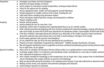
The quality of the laboratory diagnostic approach can be severely affected by a high number of pre-analytical factors of variation. It is essential for herd health examination, as well as for research projects to have proper knowledge about the importance and source of these factors to alter the entire diagnostic process. The impact varies highly from case to case and depends on the laboratory analyte of interest. Greatest efforts should be made to minimize controllable factors as far as possible. Analytical results have to be evaluated with care considering that pre-analytical factors are possible causes of misinterpretation. The use of a wide range of procedures worldwide emphasizes the caution needed when comparing study data with reference values or published data. It is desirable to establish and follow standardized guidelines in pre-analytical procedures in livestock. A proposal for a general checklist is given in Table 1. Close collaboration and prior consultation with specialized animal diagnostic laboratories help to coordinate good pre-analytical practice, to obtain high-quality analytical results, to avoid misinterpretation and to save money.
Blood sampling checklist

EDTA = ethylenediaminetetraacetic acid.