Introduction
Snoring and obstructive sleep apnoea (OSA) are common problems. However, there are few reports in the literature on the histomorphological findings and pathophysiology of these two medical and social problems, and even these published few lack consistency.Reference Metternich, Brusis, Koebke and Wenzel1 Formal trials have assessed histopathological changes in the uvula or palate of such patients, in order to clarify the pathophysiology of snoring and OSA. However, there is still much debate on the pathogenesis of OSA. Two theories have been proposed to explain the pathophysiology of OSA, namely, the obstructive theory and the neurogenic theory.
The obstructive theory states that there is hypertrophy of all the tissue components of the uvula and the palate, i.e. the muscles, glands, fat and blood vessels. This hypertrophy causes narrowing and subsequent collapse of the airway.
The neurogenic theory states that nerve degeneration from repeated vibratory trauma causes atrophic changes of the palatine muscles, with subsequent weakness and collapse of the muscles.
Histopathological study of the nerves in the palate or uvula of OSA patients may help indicate which theory is true. However, these nerve fibres cannot be clearly seen under the light microscope with haematoxylin and eosin (H&E) staining; the only way to study them is to use special stains with light microscopy or, preferably, to use transmission electron microscopy.
A literature review identified only a few articles addressing the ultrastructure of the soft palate and uvula in heavy snoring and OSA syndrome patients. These papers mainly focussed on the muscular and glandular structure rather than the neural tissues.
In the present study, we used transmission electron microscopy to examine the neural components of uvular specimens from OSA patients, and then compared these features to those of simple snorers. We aimed to clearly identify the exact pathophysiology of OSA; is it nerve atrophy with subsequent muscle atrophy or, on the contrary, muscle hypertrophy with no neural role?
Materials and methods
Selection of patients and controls
Twenty-five patients were included in this study; 10 had heavy snoring without OSA and 10 had severe OSA. Five non-snoring, non-apnoeic patients were used as controls. All of our patients were men. We defined apnoea as cessation of respiration for 10 seconds or more. Hypopnoea was defined as a fall of oxygen saturation by 4 per cent or more from baseline during a decrease in airflow of 50 per cent or more; this was consistent with the definitions of Goode and Stauffer et al. Reference Goode and Cummings2, Reference Stauffer, Buick, Bixler, Sharkey, Abt and Manders3
None of our patients had a history suggestive of general disease, such as hypothyroidism, general muscle disease or any type of peripheral neuritis. Smokers and diabetics were excluded (to remove cases of peripheral neuritis). Patients with nasal, retrolingual or hypopharyngeal causes of obstruction were also excluded, as were those with mixed or central apnoea.
Control samples were taken from the cadavers of patients who had died from causes unrelated to head and neck problems (mostly accidents). These patients had been non-snoring and non-apnoeic (confirmed by discussion with their sleeping partners) and had suffered no systemic illnesses (confirmed from their medical records). In all of these deceased patients, the same part of the uvula was removed immediately after death, in order to avoid autolysis.
Written, informed consent for participation in the research, and for the surgical procedure, was obtained from the patients, and the study design was approved by the Cairo university ethics committee.
The surgical procedures were carried out in Kasr el Aini Hospital between January 2003 and April 2004.
All patients underwent the following: general examination to locate general causes of snoring and sleep apnoea; full ENT examination; flexible endoscopy and Muller's manoeuvre to assess the site of airway obstruction and to decide if the patient would benefit from uvulopalatoplasty; and pre-operative polysomnography.
Methods
Sample collection
A section of the uvula was removed during laser assisted uvulopalatoplasty (LAUP) under general anaesthesia. A laser was used to remove the distal two-thirds of the uvula. A neo-uvula was fashioned immediately after creation of two vertical incisions and vaporisation of redundant parts of the posterior pillars. After removal, the uvular specimen was flattened on a cork-plate, with its nasal surface facing downwards and its oral surface facing upwards.
The specimen was then bisected midsagitally (i.e. longitudinally) in the midline from the tip to the base, creating two identical halves. One half was sent for electron microscopy (the middle parts of the specimen were used for scanning), and the other for light microscopy.
Electron microscopy
Transmission electron microscopy was performed according to the method described by Woodson et al. and Edström et al. Reference Woodson, Garancis and Toohill4, Reference Edström, Larsson and Larsson5 Preparation of samples included: preparation of the capsules; sectioning by ultramicrotome; picking the sections on copper grids (electron microscopy (EM) grade); staining the grid with a salt solution of heavy metals (uranyl acetate and lead citrate); and, lastly, examination using a Jeol EM 100S transmission electron microscope (Jeol Limited, Tokyo, Japan) at 60 kV (images were obtained at magnifications ranging from 3000 to 20 000).
Results
The mean age of the OSA group was 44.5 years, with a range of 32 to 55 years. The mean age of the heavy snoring group was 40.9 years, with a range of 31 to 50 years. The mean age of the control group was 32.4 years, with a range of 29 to 36 years. The mean respiratory disturbance index and mean body mass index (BMI) were 42.8 (range 38–53) and 33.9 for the 10 severe OSA patients and 1.2 (range 0–4) and 29.1 for the heavy snoring patients, respectively. Controls had a mean BMI of 23.9 (range 21.8–25.04).
Controls
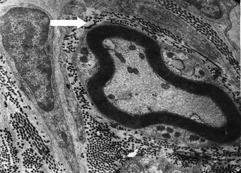
In all five control specimens, the nerve fibres were obvious and showed normal architecture and shape, without any apparent degenerative changes (Figure 1).
Transmission electron photomicrograph of the uvula of a control case, showing myelinated nerve fibres (arrow) which appear normal, with an intact myelin sheath and Schwann cells (lead citrate and urenyl acetate; ×8000).

Simple snorers
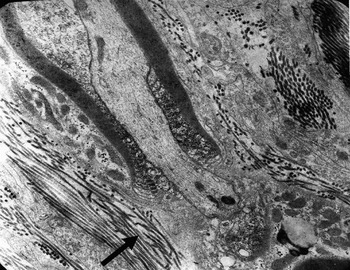
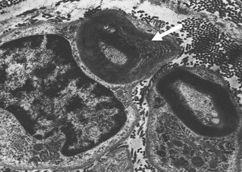
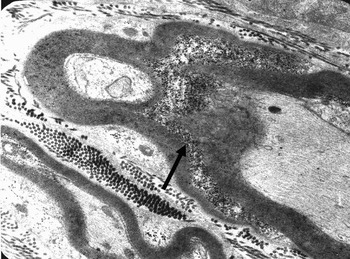
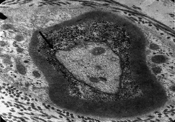
In these specimens, the nerve fibres showed mild degenerative changes. Six out of 10 specimens had partially degenerated and partially lamellated myelin sheaths. The remaining four specimens appeared almost normal (Figures 2 and 3).
Transmission electron photomicrograph of the uvula of a heavy snoring patient, showing longitudinal section (LS) of myelinated nerve with degeneration of the myelin sheath (arrow) (lead citrate and urenyl acetate; ×8000).

Transmission electron photomicrograph of the uvula of a heavy snoring patient, showing transverse section (TS) of myelinated nerve with degeneration and lamellation of the myelin sheath (arrow) (lead citrate and urenyl acetate; ×9000).

Obstructive sleep apnoea patients
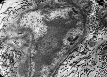
In these specimens, severe degenerative nerve fibre changes were noted in all cases (10/10). The myelin sheath of myelinated nerves had obvious, severe degeneration. Vacuolation, degenerated and irregular Schwann cells, lamellated bodies, neurofilament loss and fatty degeneration were all noted in all specimens (Figures 4, 5 and 6).
Transmission electron photomicrograph of an unmyelinated nerve in the uvula of an obstructive sleep apnoea patient, showing severe degeneration of the nerve, which is surrounded by excessive and haphazardly arranged collagenous fibrils (lead citrate and urenyl acetate; ×10 000).

Transmission electron photomicrograph of a uvula from a severe obstructive sleep apnoea patient, showing myelinated nerve fibres with severe degeneration of the myelin sheath (arrow) of the axon (lead citrate and urenyl acetate; ×8000).

Transmission electron photomicrograph of a uvula from another patient with severe obstructive sleep apnoea, showing severe degeneration of the myelin sheath (arrow) of the axon (lead citrate and urenyl acetate; ×15 000).

Although the statistical association was not formally assessed, we noted no direct correlation between the degree of OSA severity (as measured by the respiratory disturbance index) and the observed degree of neural degeneration; the severest degenerative changes were noticed in patients with milder degrees of OSA.
Discussion
Two theories have been proposed to explain the pathophysiology of snoring and OSA, namely, the obstructive theory and the neurogenic theory. Investigation of these two theories depends mainly on assessing the structure and ultrastructure of the palate or, preferably (as it is more accessible), the uvula, in order to study their nerves. According to the first theory, these nerves should appear normal; according to the second, they should show evidence of degeneration. As these nerve fibres cannot be clearly seen using H&E staining under light microscopy, it is possible to study them only by using special stains or electron microscopy. This requires difficult and time-consuming specimen preparation. Thus, all prior investigations which have attempted to differentiate between the two theories have been based on indirect study of the muscles rather than direct study of the nerves. For example, Stauffer et al. observed only muscle fibre hypertrophy in whole uvulae of OSA patients,Reference Stauffer, Buick, Bixler, Sharkey, Abt and Manders3 and Edström et al., Swift et al. and Yu et al. observed only muscle atrophy in the uvulae of OSA patients.Reference Edström, Larsson and Larsson5–Reference Yu, Liu and Zhang7
The most valuable and interesting finding in the present study was the observation of nerve fibre degeneration in all OSA patient specimens and in six out of 10 heavy snoring patient specimens, while the nerves in the control specimens appeared to have normal architecture, without any definitive evidence of degeneration.
In our 10 patients with severe OSA, we found degeneration of myelinated and unmyelinated nerve fibres in all 10. Four cases showed severe total degeneration of the whole nerve fibre; the degree of degeneration in the remaining patients varied. In our 10 heavy snoring patients, we found a lesser degree of degenerative changes in six out of 10.
• This study investigated histopathological changes of the uvula in patients with simple snoring and obstructive sleep apnoea, using electron microscopy
• Cases of obstructive sleep apnoea (OSA) were associated with definite degenerative changes in the myelinated and unmyelinated nerve endings
• Degenerative changes were present to a lesser degree, and in a smaller proportion, in cases of simple snoring
• A neurogenic basis to the pathophysiology of snoring and OSA seems possible
Using transmission electron microscopy, Woodson et al. found degenerative changes of the myelin sheath in only two out of four cases of OSA, in a study including only four OSA patients, four patients with severe snoring and four controls.Reference Woodson, Garancis and Toohill4 However, this is the only previously published study which used transmission electron microscopy to study the nerve fibres in heavy snoring and OSA patients, and which showed the image of the degenerated nerve fibre at ×9000 magnification.
Another paper, by Friberg et al., used immunohistochemical studies and found increased neuropeptidases (as protein-gene product 9.5, substance P and calcitonin gene peptide) within nerve terminals.Reference Friberg, Gazelius, Hökfelt and C-Nordlander8 The authors attributed these findings to regenerative changes following degeneration.
In the present study, the presence of nerve degeneration in uvular specimens from heavy snoring and OSA patients supports the neurogenic theory; i.e. that the severe vibratory trauma of snoring causes damage to the nerve fibres. It is well known that snoring produces a low frequency vibration and results in mechanical stretch trauma to the pharyngeal tissues.Reference Schafer9 Peripheral nerve fibres can be injured when exposed to such low frequency vibrations.Reference Takeuchi, Futatsuka and Imanishi10
Nerve degeneration (due to the vibratory trauma of snoring) will lead to muscle atrophy, manifested as weakness of the dilator muscles of the upper airway, which then cannot maintain its patency. Collapse will result, and heavy snoring patients will progress to OSA.
The presence of the same pathology – but to a lesser degree and in fewer patients – in simple snoring compared with OSA suggests that both conditions have the same pathophysiology, and that both represent different stages of the same disease.
In order to better interpret these results, a future trial with a larger number of cases is needed, with elimination of any confounding factors which might affect results (e.g. OSA-related hypoxia, age, and tissue changes due to laser surgery).
Conclusion
Nerve degeneration in the palate of OSA patients, and to lesser extent in simple snoring patients, was documented in our study. This supports the neurogenic theory of OSA, i.e. that the severe vibratory trauma of snoring damages nerve fibres. This mechanism seems to play an important role in the pathophysiology of both simple snoring and OSA.