Introduction
The squirrel monkey Saimiri collinsi, a neotropical primate endemic from the Amazon region of Brazil, is used as an animal model for conservational and biotechnology studies for species from the genus Saimiri (Oliveira et al., Reference Oliveira, Leão, Almeida, Santos and Domingues2015, Reference Oliveira, Santos, Leão, Brito, Lima, Sampaio and Domingues2016a, Reference Oliveira, Santos, Leão, Queiroz, Paim, Vianez-Júnior and Domingues2016b). Among these, S. oerstedii and S. vanzolini are listed as vulnerable, while S. ustus is almost threatened to extinction (IUCN, 2019).
Despite the relevant studies conducted to investigate the male reproductive physiology in this non-human primate using classical semen analysis (Oliveira et al., Reference Oliveira, Leão, Almeida, Santos and Domingues2015, Reference Oliveira, Santos, Leão, Brito, Lima, Sampaio and Domingues2016a, Reference Oliveira, Santos, Leão, Queiroz, Paim, Vianez-Júnior and Domingues2016b; Sampaio et al., Reference Sampaio, Oliveira, Leão, Caldas-Bussiere, Queiroz, Paim, Santos and Domingues2017; Almeida et al., Reference Almeida, Lima, Leão, Oliveira, Santos, Domingues and Miranda2018), knowledge on the morphology of sperm from S. collinsi is still limited. Seminal analysis or spermiogram is an important tool to evaluate and determine sperm morphological aspects that are directly related to potential male fertility (Visco et al., Reference Visco, Raffa, Elia, Delfino, Imbrogno, Torrisi and Mazzilli2010) and is crucial for the processes of artificial insemination, in vitro fertilization, and embryo development (Ozkavukcu et al., Reference Ozkavukcu, Erdemli, Ayca Isik, Oztuna and Karahuseyinoglu2008).
At a routine basis, sperm morphology from neotropical primates has been examined in semen smears with the main criteria for normality relying on morphological parameters of the sperm head, middle piece and flagellum (Leão et al., Reference Leão, Miranda, Brito, Lima, Santos and Domingues2015; Oliveira et al., Reference Oliveira, Leão, Almeida, Santos and Domingues2015, Reference Oliveira, Santos, Leão, Brito, Lima, Sampaio and Domingues2016a, Reference Oliveira, Santos, Leão, Queiroz, Paim, Vianez-Júnior and Domingues2016b; Swanson et al., Reference Swanson, Valle, Carvalho, Arakaki, Rodas-Martínez, Muniz and García-Herreros2016; Arakaki et al., Reference Arakaki, Carvalho, Castro, Muniz and Valle2017, Reference Arakaki, Nichi, Monteiro, Muniz, Guimarães and Valle2019; Sampaio et al., Reference Sampaio, Oliveira, Leão, Caldas-Bussiere, Queiroz, Paim, Santos and Domingues2017). In S. collinsi, sperm morphology has only been described by optical microscopy using eosin–nigrosine staining (Oliveira et al., Reference Oliveira, Leão, Almeida, Santos and Domingues2015, Reference Oliveira, Santos, Leão, Brito, Lima, Sampaio and Domingues2016a, Reference Oliveira, Santos, Leão, Queiroz, Paim, Vianez-Júnior and Domingues2016b; Sampaio et al., Reference Sampaio, Oliveira, Leão, Caldas-Bussiere, Queiroz, Paim, Santos and Domingues2017). However, sperm analysis using optical microscopy has low resolution, and does not allow the identification and analysis in more detail than at the micro-anatomical level that can affect the sperm and decrease sperm capacity to fertilize the oocyte (Visco et al., Reference Visco, Raffa, Elia, Delfino, Imbrogno, Torrisi and Mazzilli2010).
In this context, the scanning electron microscopy (SEM) and transmission electron microscopy (TEM) are technologies that provide a high-range view, on the nanometre scale, that can aid morphological description and detection of possible abnormalities in the sperm (Nussdorfer et al., Reference Nussdorfer, Cilenšek, Zorn and Petrovič2018). These techniques have already been used in domestic animals such as horses (Pesch et al., Reference Pesch, Bostedt, Failing and Bergmannc2006), sheep (López-Armengol et al., Reference López-Armengol, Jurado, Pelufo and Aisen2012), and wild animals such as the six-banded armadillo (Sousa et al., Reference Sousa, Santos, Bezerra, Lima, Castelo, Fontenele-Neto and Silva2013), collared peccaries (Bezerra et al., Reference Bezerra, Souza, Silva, Vasconcelos, Moura, Pereira, Oliveira and Silva2018), jaguar (Silva et al., Reference Silva, Nunes, Ribeiro, Freitas, Oliveira, Assis Neto, Silva and Silva2019) and some non-human primates (Bedford and Nicander, Reference Bedford and Nicander1971; Martin et al., Reference Martin, Gould and Warner1975; Gould, Reference Gould1980; Steinberg et al., Reference Steinberg, Nieves, Ascunce, Palermo and Mudry2009; Nakazato et al., Reference Nakazato, Yoshizawa, Isobe, Kusakabe, Kuraishi, Hattori, Matsumoto, Fukui, Kuwahata, Ochi, Kiso and Kai2015). For these microscopy technologies in the genus Saimiri, only SEM has been used to characterize sperm morphology in squirrel monkeys (S. sciureus) (Martin et al., Reference Martin, Gould and Warner1975; Gould, Reference Gould1980), or obtain morphometric sperm data as a tool for more accurate taxonomic identification of the S. boliviensis (Steinberg et al., Reference Steinberg, Nieves, Ascunce, Palermo and Mudry2009). In S. collinsi nothing is known about sperm components in higher resolution. Therefore, the aim of the present study was to describe the micromorphological and ultrastructural characteristics of squirrel monkeys (S. collinsi) sperm using SEM and TEM.
Materials and methods
Study facilities
Semen collection was carried out at the National Primate Center (CENP, Ananindeua, Pará, Brazil), SEM was conducted at the Analytical Center of the Universidade Federal do Ceará (UFC, Fortaleza, Ceará, Brazil), and transmission electron microscopy was conducted at the Laboratory of Electron Microscopy of the Evandro Chagas Institute (IEC, Belém, Pará, Brazil).
Animals and semen collection
Saimiri collinsi males originated from the Marajó Archipelago (0°58′S and 49°34′W) and maintained in captivity at the CENP (1°22′58′′S and 48°22′51′′W) and were used for collecting semen. The experimental group (n = 5 males, c. 15 years old) was selected by their physical characteristics, and clinical parameters such as complete haemogram, hepatic and renal function.
Animals were collectively housed in mixed groups (males and females in a varied number of members) in cages of 4.74 m × 1.45 m × 2.26 m (length, width and height, respectively), under natural photoperiod (i.e. 12 h of light and 12 h of dark). The climate is humid and tropical, with an average annual temperature of 28°C. The diet consisted of fresh fruits, vegetables, commercial pellet chow specific for neotropical non-human primates (Megazoo® P18, 18% protein, 6.5% fibre, Brazil) and cricket larvae (Zophobas morio). Vitamins, minerals and eggs were supplied once a week, and water was available ad libitum.
Semen collection
Semen was collected at the same period of the day (in the morning before feeding). Physical restraint was performed by a trained animal caretaker wearing leather gloves. All animals were anaesthetized with ketamine hydrochloride (20 mg/kg intramuscularly (IM); Vetanarcol, König S.A., Avellaneda, Argentina) and xylazine hydrochloride 1 mg/kg IM; Kensol, König S.A.), and monitored by a veterinarian.
Achieved total anaesthetic effect, the animals were placed in lateral recumbency, genital region was then sanitized with a mild soap and distilled water (1:10) and gauze. The prepuce was retracted with the thumb and index fingers for a more efficient cleaning of the penis with saline solution. Animals were stimulated with rectal electroejaculation (EEJ) procedure described by Oliveira et al. (Reference Oliveira, Leão, Almeida, Santos and Domingues2015). Ejaculates (liquid and coagulated fractions) were collected into microtubes (1.5 ml), then were placed in a water bath at 37°C. SEM and TEM were only performed in the liquid fraction of the ejaculate.
Sperm preparation
The sample (liquid fraction) were centrifuged at 500 g for 5 min, and the supernatant was discarded. Centrifugation was performed due to the low sperm concentration in the liquid fraction, and to remove the seminal plasma. The pellet formed was washed three times in sodium cacodylate and placed in Karnovsky fixative (4% paraformaldehyde, 2.5% glutaraldehyde in 0.1 M sodium cacodylate buffer, pH 7.2).
Semen evaluation
Seminal volume (liquid fractions) was evaluated in a graduated tube, with the aid of a pipette. Appearance and consistency were assessed subjectively, that is colour (colourless, yellowish, or whitish) and opacity (opaque or transparent) (Oliveira et al., Reference Oliveira, Leão, Almeida, Santos and Domingues2015). Sperm motility, vigour, and morphology were evaluated according to Oliveira et al. (Reference Oliveira, Leão, Almeida, Santos and Domingues2015, Reference Oliveira, Santos, Leão, Brito, Lima, Sampaio and Domingues2016a, Reference Oliveira, Santos, Leão, Queiroz, Paim, Vianez-Júnior and Domingues2016b). Sperm morphology and plasma membrane integrity were evaluated using a prepared smear, adding 5 µl eosin 1% (Vetec) and 5 µl nigrosine 1% (Vetec) to 5 µl of semen on a pre-warmed (37°C) glass slide. Sperm concentration was determined in a Neubauer chamber after dilution of 1 µl semen in 99 µl formalin solution 10%. Morphologic defects detected in sperm were classified as primary or secondary according Bloom (Reference Bloom1973). Plasma membrane functionality was assessed by hypoosmotic swelling test (HOST) after dilution of 5 µl of semen in 45 µl of hypoosmotic solution (0.73 g sodium citrate, 1.35 g fructose and 100 ml ultrapure water; pH 7.2 and 108 mOsm/l). After 45 min incubation in a water bath (37°C), number of sperm tail coiled was assessed by placing 10 µl of this solution on a pre-warmed (37°C) glass slide with coverslip, and at least 200 spermatozoa were counted. Spermatozoa with functional plasma membrane were those presenting a coiled tail. All evaluations were performed under a light microscope (Nikon E400, Japan) at a magnification of ×100.
Scanning electron microscopy
Sperm samples were placed in a coated coverslip with poly-d-lysine, air-dried it onto a filter and dehydrated in a series of acetone solution (50, 70, 90 and 100% × 3 or 5 min each). Afterwards, the samples were dried using the critical point method (EMS 850, Quorum Technologies, Lewes, UK) and then attached to supports using aluminium stubs with carbon tape. The samples were covered with a thin (20 nm) layer of gold (Q150T ES, Quorum Technologies, Lewes, UK) and observed under SEM (Quanta 450-FEG, Thermo Fisher Scientific, Massachusetts, USA). The description of S. collinsi sperm by SEM was conducted according to that previously reported for non-humans primates by Martin et al. (Reference Martin, Gould and Warner1975).
Transmission electron microscopy
For TEM, the sperm sample were washed in 0.05 M cacodylate buffer (40 min, three times), post-fixed with osmium tetroxide (OsO4) for 60 min and washed in 0.05 M cacodylate buffer with 0.08% potassium ferrocyanide (40 min, five times). Subsequently, the contracting block was performed using 2.5% uranyl acetate in 50% acetone for 1 h. Then, the samples were dehydrated in a series of acetone solutions (70, 80, 90 and 100%) for 10 min each, the solution of 100% acetone was carried out three times. After dehydration, the samples were infiltrated with epoxy resin (EponPolibed), using an increasing series of resin in acetone (dilution 1:2; 1:1; 2:1), until 100% Epon + 2,4,6-trisdimethylaminomethylphenol (DMP-30). Polymerization of the resin was performed at 60°C for 48 h. Semithin cuts of c. 70 nm thickness were obtained using an ultramicrotome with glass cutters (Leica EM UC7, Leica Microsystems, Wetzlar, Germany) and contrasted with 5% uranyl acetate for 20 min in 60°C. Observations of the sperm ultrastructure were performed using TEM (EM 900, Carl Zeiss, Brasil Ltd, São Paulo, Brazil).
Results
Semen evaluation
Mean (± standard error of the mean) of seminal volume was 339 ± 61 µl (15–500 µl; min–max). Liquid fractions were whitish or yellowish, and opaque. Sperm concentration was 1.718 ± 610 × 106 sperm/ml. In addition, the samples presented a mean of 67 ± 11% motility, 3 ± 0.3 vigour, 38 ± 13% and 43 ± 8% functional and intact sperm membranes, respectively. Evaluation of sperm morphology through light microscopy was 56 ± 5% normal sperm, 13 ± 4 bent tail, 27 ± 4 coiled tail and 3.6 ± 1.2 strongly coiled tail.
Scanning electron microscopy
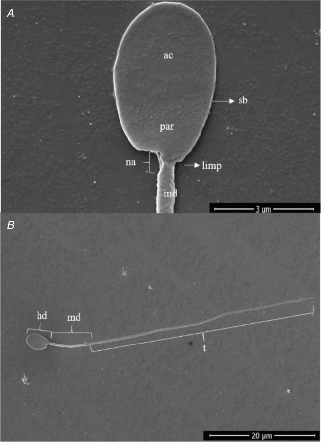
SEM revealed that the head of S. collinsi sperm was a flattened paddle-shaped with a uniform thickness throughout, measuring 5.14 ± 0.08 μm in length and 3.36 ± 0.03 μm in width, with an intact and uninterrupted surface. The acrosome occupied most of the head with length of 3.56 ± 0.06 μm (Fig. 1A). The insertion of the tail was lateral (abaxial; Fig. 1A), with length of middle piece at 9.49 ± 0.38 μm. The squirrel monkey sperm presented a total length of 71.7 ± 0.7 μm (Fig. 1B).
Scanning electron micrographs of Saimiri collinsi sperm. (A) Sperm head with plasma membrane integrity, acrosomal region (ac), serrated band (sb); post-acrosomal region (par), neck area (na), lateral insertion of the middle piece (limp) and middle piece (md). (B) Normal sperm: head (hd), middle piece (md) and tail (t).

Transmission electron microscopy
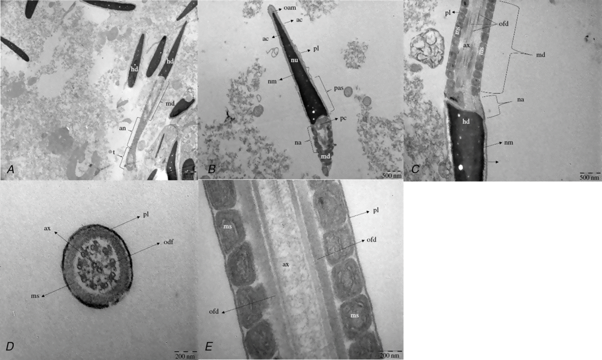
TEM images showed the S. collinsi sperm presents a flattened head in a longitudinal cut, containing a very electron-dense nucleus, which is larger at the base, narrowing as it goes to the tip of the head. There is a diffuse small electronic spot. The structures present in the sperm head are the nucleus, the acrosome, and membranous envelopes (Fig. 2A, B). The large nucleus is covered by the acrosome (ac), which occupies one-half to two-thirds of the anterior portion of the head. The acrosome is thickest around the anterior margin of the sperm head, and thinnest from the equator to the posterior third of the nucleus where it terminates (Fig. 2B). The neck area (na), which is the region located between the head and the middle piece, is characterized by the presence of a centriole, the proximal centriole (oriented transversally to the cell; pc), and segmented columns (Fig. 2B). The middle piece is characterized by a central 9 + 2 microtubule axoneme surrounded by nine dense fibres. This set is surrounded by a plasmalemma and a helical mitochondrial sheath (Fig. 2C, D), which identified the presence of an intact axoneme as well as outer dense fibres. The mitochondria were juxtaposed, therefore forming the mitochondrial sheath, which was surrounded by the plasmalemma with some undulations (Fig. 2E). It was possible to visualize about c. 72 mitochondria.
Transmission electron micrographs of the squirrel monkey (Saimiri collinsi) sperm. (A) hd: head; md: middle piece; an: annulus; t: tail. (B) oam: outer acrosome membrane; ac: acrosome; pl: plasmalemma; nu: nucleus; nm: nuclear membrane; pas: postacrosomal sheath; pc: principal centriole: na: neck area; md: middle piece. (C) pl: plasmalemma; nm: nuclear membrane: hd: head; na: neck area; md: middle piece; ax: axonema; ofd: outer dense fibres; ms: mitochondrial spiral. (D) pl: plasmalemma; ax: axonema; odf: outer dense fibres; ms: mitochondrial spiral; *: longitudinal cut. (E) ofd: outer dense fibres; ax: axonema; pl: plasmalemma, ms: mitochondrial spiral.

Discussion
Our study represents a valuable basis for understanding sperm physiology through the description of micromorphological and ultrastructural aspects in S. collinsi. Evaluation of sperm morphology has become much more precise, and rigorous analysis of these parameters is of pivotal importance in semen, as it permits detailed study of different sperm abnormalities that predict the fertile capacity of the male gamete (WHO, 2010).
In the genus Saimiri, SEM has been performed only in S. sciureus (Martin et al., Reference Martin, Gould and Warner1975) and S. boliviensis (Steinberg et al., Reference Steinberg, Nieves, Ascunce, Palermo and Mudry2009). In S. collinsi, the abaxial tail insertion was also observed by SEM, as previously described as a normal morphology characteristic in the morphometric analysis of sperm stained with eosin–nigrosine for S. collinsi and S. vanzolini (Sampaio et al., Reference Sampaio, Oliveira, Leão, Caldas-Bussiere, Queiroz, Paim, Santos and Domingues2017). The average total length of the S. collinsi sperm was similar to that described for other squirrel monkeys species in stained samples (Nakazato et al., Reference Nakazato, Yoshizawa, Isobe, Kusakabe, Kuraishi, Hattori, Matsumoto, Fukui, Kuwahata, Ochi, Kiso and Kai2015; Sampaio et al., Reference Sampaio, Oliveira, Leão, Caldas-Bussiere, Queiroz, Paim, Santos and Domingues2017). The morphology of the sperm head showed a thick, flattened, paddle-shape that resembled that in other primates, including humans (Martin et al., Reference Martin, Gould and Warner1975; Gould & Martin, Reference Gould and Martin1978). The measurements of head morphology for S. collinsi using SEM agreed with values obtained previously via optical microscopy (Sampaio et al., Reference Sampaio, Oliveira, Leão, Caldas-Bussiere, Queiroz, Paim, Santos and Domingues2017).
For the first time, an ultrastructure description of S. collinsi sperm by TEM was reported. Therefore, the results presented here will serve as a parameter for future morphological studies involving the preparation and evaluation of semen in non-human primates, as well as for studies using morphological data for the diagnosis of sperm changes, including acquired damage post-thawing, which is a relevant factor for development of reproduction (reproductive) biotechnology (Sousa et al., Reference Sousa, Santos, Bezerra, Lima, Castelo, Fontenele-Neto and Silva2013). TEM might be an additional diagnostic tool for the presence of asthenozoospermia or the absence of motility, which is important as the pattern of axoneme structure can be visualized in longitudinal sections and cross-sections using this technique. In humans, TEM has been used to diagnose sperm changes that have a possible genetic origin, known as systematic defects (Baccetti et al., Reference Baccetti, Capitani, Collodel, Di Cairano, Gambera, Moretti and Piomboni2001). The observation of sperm head ultrastructure revealed a large and obvious nucleus. The acrosome is the only cytoplasmic element in the sperm head. Similar to that described for human sperm, in S. collinsi, the acrosome was a relatively inconspicuous and covers the anterior two-thirds to one-half of the head, cap-like structure containing enzymes that are essential for fertilization (Bartoov et al., Reference Bartoov, Eltes, Weissenberg and Lunenfeld1980). In Rhesus monkeys sperm, the acrosome is thickest around the anterior margin of the sperm head and thinnest from the equator to the posterior third of the nucleus, where it terminates. The thin portion of the acrosome is usually referred to as the equatorial segment. As shown in human and Rhesus monkey (Zamboni et al.,Reference Zamboni, Zemjanis and Stefanini1971), in S. collinsi the plasma membrane surrounding the anterior portion of the head is continuous and often undulated.
The sperm head and tail are connected via the sperm neck, a region formed by the centriole and connecting piece (Bornens, Reference Bornens2012). In Rhesus macaque, the middle piece sheath consists of 84–86 mitochondria (Zamboni et al., Reference Zamboni, Zemjanis and Stefanini1971). In our study, the exact number of mitochondria in S. collinsi could not be measured due to the absence of longitudinal sections showing the middle piece to its full extent. These organelles function to provide energy for movement of the flagella, as the midpiece accommodates more mitochondria to produce more energy to fuel motility (Pesch et al., Reference Pesch, Bostedt, Failing and Bergmannc2006), and generates greater propulsion forces in the principal piece (Gomendio and Roldan, Reference Gomendio and Roldan1991). However, it has been demonstrated that swimming speed is not dependent on the tail size, but on head size, which in return may retard the propulsion generated by the sperm tail (Humphries et al., Reference Humphries, Evans and Simmons2008).
Regarding morphological defects found using light microscopy, according to Bloom (Reference Bloom1973) classification, some secondary defects were identified in the S. collinsi sperm, including bent tail, coiled tail and strongly coiled tail. Such defects have also been verified in the ejaculates of neotropical primates (Leão et al., Reference Leão, Miranda, Brito, Lima, Santos and Domingues2015; Oliveira et al., Reference Oliveira, Leão, Almeida, Santos and Domingues2015; Swanson et al., Reference Swanson, Valle, Carvalho, Arakaki, Rodas-Martínez, Muniz and García-Herreros2016; Arakaki et al., Reference Arakaki, Carvalho, Castro, Muniz and Valle2017, Reference Arakaki, Nichi, Monteiro, Muniz, Guimarães and Valle2019). Comparing our study with others already carried out for S. collinsi species (Oliveira et al., Reference Oliveira, Leão, Almeida, Santos and Domingues2015, Reference Oliveira, Santos, Leão, Brito, Lima, Sampaio and Domingues2016a, Reference Oliveira, Santos, Leão, Queiroz, Paim, Vianez-Júnior and Domingues2016b; Sampaio et al., Reference Sampaio, Oliveira, Leão, Caldas-Bussiere, Queiroz, Paim, Santos and Domingues2017), we observed a high percentage of sperm pathologies, which Sampaio et al. (Reference Sampaio, Oliveira, Leão, Caldas-Bussiere, Queiroz, Paim, Santos and Domingues2017) characterized as a group of animals with low seminal quality. Conversely, it is noteworthy that we only worked with the liquid fraction for sperm characterization, as the dilution process could generate changes in sperm morphology. It is known that the liquid fraction is the fraction of lower seminal volume and sperm concentration (Oliveira et al., Reference Oliveira, Leão, Almeida, Santos and Domingues2015), and may also contain sperm with higher percentages of sperm pathologies when compared with the coagulated fraction.
Saimiri collinsi ejaculates presented a normal range for sperm morphology similar to that accepted for the genus Saimiri. Additionally, they presented a general sperm structure, but with characteristics such as an abaxial middle piece insertion, seen using SEM.
Acknowledgements
We would also like to thank to the National Primate Center (CENP/Brazil), to Central Analytical of the Federal University of Ceará and Laboratory of Electronic Microscopy of Evandro Chagas Institute for the technical support during this research.
Financial support
The authors thank ‘Coordenação de Aperfeiçoamento de Pessoal de Nível Superior – Brasil (CAPES) – Finance Code 001, and ‘Conselho Nacional de Desenvolvimento Científico e Tecnológico (Projeto Universal 01-2016/ Processo no. 421649/2016-0)’ for financial support.
Conflicts of interest
The authors declare that there is no conflict of interest that can be perceived as prejudicing the impartiality of the research reported.
Ethical standards
This study was approved by the Ethical Committee in Animal Research (no. 02/2015/CEPAN/IEC/SVS/MS) and by the System of Authorization and Information in Biodiversity (SISBIO/ICMBio/MMA no. 47051-2). All procedures were performed under the supervision of a veterinarian.