Introduction
Antarctic notothenioid fishes thrive in the freezing waters of the Southern Ocean, one of the most inhospitable marine environments on Earth. Their success in this environment is largely attributable to the synthesis of antifreeze glycoproteins (AFGPs), which protect against the life threatening potential of internalized ice. Although AFGPs were first identified and characterized over forty years ago (DeVries & Wohlschlag Reference DeVries and Wohlschlag1969, DeVries Reference DeVries1971) we still remain ignorant of many of the details of how they function in conferring freeze-avoidance and exactly how fishes cope with internal ice. The prevailing paradigm envisages blood-borne AFGPs binding to ice crystals that enter or form in the circulatory system, conferring protection by preventing further crystal growth (Raymond & DeVries Reference Raymond and DeVries1977). An essential condition of this adsorption-inhibition hypothesis is that ice must be present in the body fluids of a fish for AFGPs to function. Here we show how both Arctic (cf. northern) gadids (typified by the polar cod, Boreogadus saida (Lepechin, 1774)) and Antarctic notothenioids have evolved almost identical solutions to the problem of how to deal with ice in the circulation.
Antarctic notothenioid AFGPs are represented by a family of glycoproteins, members of which characteristically express a repetitive element based on a tripeptide (alanine-alanine-threonine or AAT) in which each threonine is O-linked to the disaccharide β-D-galactosyl-(1,3)-α-D-N-acetylgalactosamine (reviewed in Fletcher et al. Reference Fletcher, Hew and Davies2001, DeVries & Cheng Reference DeVries and Cheng2005, Peltier et al. Reference Peltier, Brimble, Wojnar, Williams, Evans and DeVries2010). Eight size classes of AFGPs were originally recognized based on their electrophoretic characteristics, with the classes ranging in size from c. 2600 Da with 4 AAT repeats (AFGP 8) to at least c. 34 000 Da with 55 AAT repeats (AFGP 1). Although more recent evidence indicates a large number of intermediate-sized forms, reference to the prominent electrophoretic size classes remains in common practice. The AFGPs are encoded by multiple genes, which direct the synthesis of large polyprotein precursors. These polyproteins consist predominantly of stretches of AAT repeats of varying length, with the stretches separated by three amino acid linking motifs. The linkers are cleaved post-translationally to yield the active AFGP sequences (Hsiao et al. Reference Hsiao, Cheng, Fernandes, Detrich and DeVries1990), which circulate in the body fluids at concentrations between 10 and 35 mg ml-1 (DeVries Reference DeVries1983).
In a remarkable example of convergent evolution, phylogenetically distant Arctic gadids not only display AFGPs with similar amino acid sequences to the Antarctic notothenioids (based on the repetitive AAT tripeptide), but they are also synthesized as polyprotein precursors (although with different cleavable linkers), exist in comparable size classes, and contain the same disaccharide moiety (Chen et al. Reference Chen, DeVries and Cheng1997b). Indeed, apart from the presence of small amounts of arginine in the polar cod forms, the primary structures of the AFGPs of Antarctic notothenioids and B. saida are indistinguishable. These similarities do not extend to the genetic level, however, since AFGPs from the two different groups of fishes do not share significant coding sequence identity and nor do they have similar intron-exon structure. Further analysis has shown that the Antarctic notothenioid AFGPs have evolved from a trypsinogen-like precursor, whereas the evolutionary origin of the Arctic cod AFGPs is different, but remains unknown (Chen et al. Reference Chen, DeVries and Cheng1997a).
Although notothenioid AFGPs were originally thought to be made in the liver (Hudson et al. Reference Hudson, DeVries and Haschemeyer1979, O'Grady et al. Reference O'Grady, Schrag, Raymond and DeVries1982) it is now known that they are synthesized predominantly in the exocrine pancreas, which is consistent with their evolutionary origin from a trypsinogen-like molecule. From here they are discharged into the gastrointestinal tract (Cheng et al. Reference Cheng, Cziko and Evans2006). The only other region where AFGPs are believed to be synthesized in notothenioid fishes based on molecular data is from sites in the anterior stomach wall near the oesophageal junction, which also presumably discharge into the gastrointestinal tract. These converging secretory pathways emphasize the primary importance of protection against ice internalized during drinking and feeding, but they raise a paradox in that there is no known direct anatomical pathway by which high levels of AFGPs can reach the blood.
Northern blot analysis of B. saida liver RNA provided evidence that AFGPs are synthesized in this organ (Chen et al. Reference Chen, DeVries and Cheng1997b), consistent with their different evolutionary origin, but could this too be a case of misinterpretation? Here we use immunohistology to explore the major sites of AFGP synthesis in the Arctic cod and follow the pathways of AFGP secretion in both Arctic cods and Antarctic notothenioids. We then construct a model illustrating parallels in the pathways of AFGP synthesis, secretion and recycling in the two phylogenetically disparate groups of fishes, which also resolves the problem of how AFGPs reach the blood in notothenioids.
Irrespective of their sites of AFGP synthesis, both Arctic cods and Antarctic notothenioids are faced with the same problem of how to deal with internal ice. In previous work we modelled the fate of internal ice by injecting AFGP-nanoparticle complexes into host fish as a proxy for AFGPs adsorbed onto ice (Evans et al. Reference Evans, Gubala, Nooney, Williams, Brimble and DeVries2011). These experiments suggested the spleen was probably involved in trapping internal AFGP-adsorbed ice crystals. We now employ immunohistological methods to gain further insight into this poorly understood phenomenon by looking for concentrations of AFGPs in tissues where they are not synthesized but probably accumulated as a consequence of the endocytosis of AFGPs adsorbed to ice.
Methods
Specimen collection
The Arctic cod B. saida was collected from coastal regions west of Spitzbergen in January 2011 when the seawater temperature was between -1.5 and -1.8°C. Trawls were made in c. 100 m of seawater covered by ice c. 30 cm thick. Fish were dissected immediately after catch and isolated tissues and organs were then fixed in chilled 4% paraformaldehyde until required for histological examination.
Specimens of the Antarctic toothfish Dissostichus mawsoni Norman, 1937 and the bald notothen Pagothenia borchgrevinki (Boulenger, 1902) were caught in the vicinity of McMurdo Sound, Antarctica using baited hooks set at c. 550 m depth for D. mawsoni or just below the sea ice surface for P. borchgrevinki. Specimens of the striped notothen Trematomus hansoni Boulenger, 1902 and the spotted notothen Trematomus nicolai (Boulenger, 1902) were collected from c. 10–25 m of water in Winter Quarters Bay, Ross Island, Antarctica. Fish were returned to aquaria at Scott Base (Ross Island) containing running seawater at about -1.0°C and maintained without feeding until required.
AFGP isolation and fluorescent labelling
Antifreeze glycoproteins were prepared from Antarctic toothfish serum and separated into large (AFGP 1–5) and small (AFGP 7, 8) size classes on a Sephacryl HR-100 column (2.5 × 150 cm) essentially as described previously (Evans et al. Reference Evans, Gubala, Nooney, Williams, Brimble and DeVries2011). The size classes within each column fraction were verified using non-denaturing polyacrylamide gel electrophoresis after labelling with fluorescamine (O'Grady et al. Reference O'Grady, Schrag, Raymond and DeVries1982, Ahlgren et al. Reference Ahlgren, Cheng, Schrag and DeVries1988) and lyophilized for storage. Ten mg of the lyophilized AFGP 7, 8 size class fraction were labelled with fluorescein isothiocyanate (FITC) as described (Evans et al. Reference Evans, Gubala, Nooney, Williams, Brimble and DeVries2011) and separated from unbound or hydrolysed FITC using a Sephadex G-25 column (2.5 × 30 cm) equilibrated with water. After elution the fluorescent conjugate (FL-AFGP) was lyophilized and stored at 4°C. Prior to intubation the FL-AFGP was dissolved in local seawater at 16 mg ml-1 and c. 0.2 ml was delivered directly into the stomachs of two adult P. borchgrevinki using a needleless 1 ml syringe. The fish were scanned every day using a hand-held UV source to check for external fluorescence (Evans et al. Reference Evans, Gubala, Nooney, Williams, Brimble and DeVries2011). The first fish was killed 72 h after a single intubation with FL-AFGP to check on the passage of FL-AFGP through the gut, and organ samples were taken for histology. The second fish was re-injected 72 h later and monitored over a total of 13 days to allow for transport through the gastrointestinal tract and absorption into the circulatory system. This fish was then killed, and serum and organ samples taken for subsequent analysis by mass spectroscopy and fluorescence microscopy.
Immunohistology
Tissue samples dissected from T. hansoni, T. nicolai and B. saida fixed in 4% paraformaldehyde were soaked overnight in 70% ethanol, transferred to paraffin using a Tissue Tek VIP (MilesCo Scientific, MN, USA) automatic tissue processor and mounted in paraffin blocks using a Leica E6 1150 H tissue embedder (Bio-Strategy, Auckland, NZ). Sections (5 μm) cut using a Microm HM330 microtome (Heidelberg, Germany) were transferred to glass slides, de-waxed and incubated for 30 min at room temperature (RT) with a blocking solution (Dulbecco's phosphate buffered saline (PBS) containing 1% dimethyl sulphoxide and 1% Ig-free bovine serum albumin). The sections were then treated (2 h RT) with a custom-prepared primary anti-AFGP antibody (1:1000 dilution in blocking solution) prepared in rabbits, washed in blocking solution (30 min RT) and then further reacted (2 h RT) with a secondary Alexa Fluor 488 (Life Technologies, USA) goat anti-rabbit antibody (1:1000 dilution in blocking solution). After immunostaining, the sections were washed once (5 min RT) with blocking solution, incubated (5 min RT) with 4,6′-diamidino-2-phenylindole, dihydrochloride (DAPI; 300 nM in PBS) to counterstain nuclei blue, rewashed (4 × 5 min) and mounted in Fluoroguard (Bio-Rad, CA, USA). Control preparations carried out in parallel omitted the primary antibody. Alternate sections were stained with haematoxylin and eosin for routine examination (Presnell et al. Reference Presnell, Schreibman and Humason1997). Sections were examined in a Leica DMR microscope using bright field, phase contrast and differential interference contrast (DIC) optics, or with appropriate filter sets for fluorescence, and photographed using a Leica DC500 camera (Bio-Strategy, Auckland, NZ). Resulting images were edited and merged using Adobe Photoshop software (Adobe Systems Inc, CA, USA).
Solid phase extraction and mass spectroscopy (LC MS/MS)
Aliquots (50 μl) of serum samples collected from P. borchgrevinki before and after intubation with FL-AFGP were diluted to 500 μl with 0.1% formic acid and extracted on a 10 mg Oasis HLB Solid Phase Extraction cartridge (Waters, Milford, MA, USA) and eluted with 40% acetonitrile in 0.1% formic acid. The eluates were then concentrated in a SPD121P vacuum centrifuge (Thermo Savant, Holbrook, NY, USA) before being made up to 25 μl with 0.1% formic acid. A 10 μl injection was then made of a sevenfold dilution in Buffer A of each sample onto a 0.3 × 5 mm PepMap C18 cartridge (LC Packings, Amsterdam, Netherlands) followed by separation on a Zorbax 300SB-C18 0.3 × 100 mm column (Agilent, Santa Clara, CA, USA) using the following gradient at 6 μl min-1: 0–3 min 98A:2B; 53 min 65A:35B; 56 min 5A:95B; 59 min 5A:95B; 60.5 min 98A:2B; and 65 min 98A:2B, where Buffer A was 0.1% formic acid in water and Buffer B was 0.1% formic acid in acetonitrile. The column eluent was directed into the Ionspray source of a QSTAR XL hybrid mass spectrometer (Applied Biosystems, Foster City, CA, USA) scanning from 300–1600 m/z, followed by two Product Ion Scans for m/z 1012.3 using collision energies of 25 and 35 eV. The resulting MS/MS datasets for the intubated fish were manually inspected to confirm the identity of the major chromatographic peak for m/z 1012.3 as being FL-AFGP 8. Extracted ion chromatograms were then created for the triple-charged precursor ions of both AFGP 8 (m/z 882.6–884.6) and FL-AFGP 8 (m/z 1012.2–1014.2).
Results
AFGP is synthesized in the liver and pancreas of B. saida
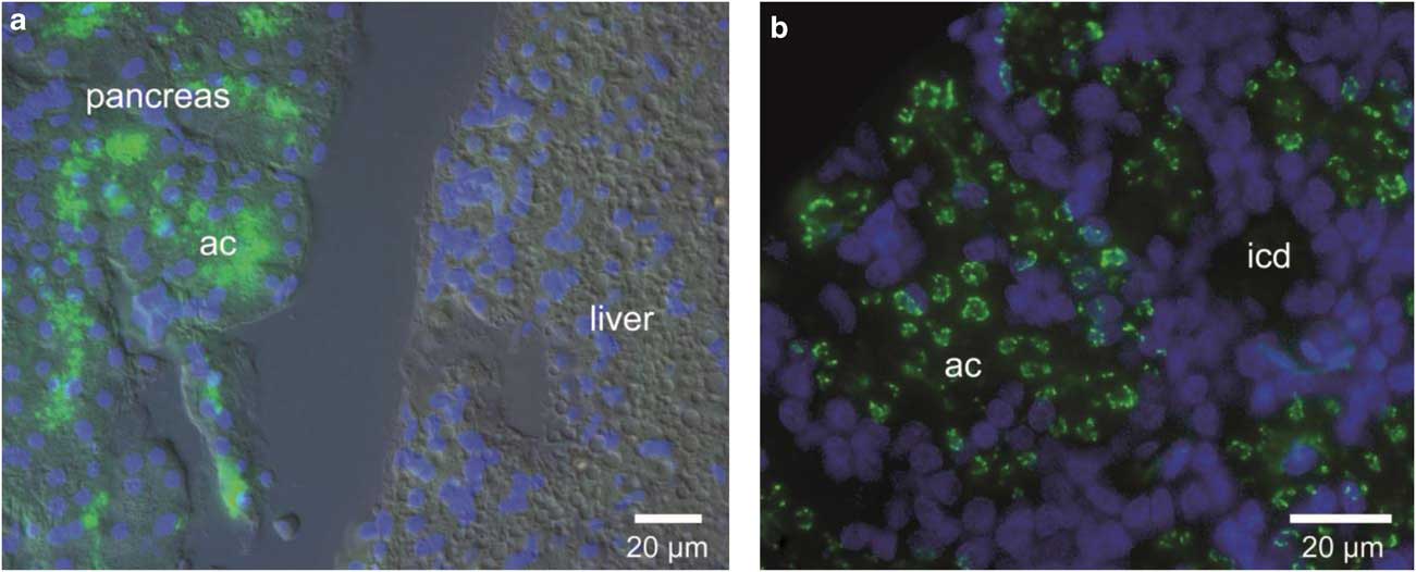
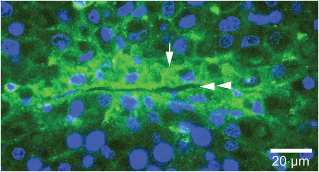
Immunostaining of paraffin sections of pancreatic tissue from representatives of the Antarctic notothenioids (T. nicolai) and the northern gadids (B. saida) using a polyclonal anti-AFGP antibody showed a positive reaction confined to zymogen granules of the exocrine acinar cells (Fig. 1a & b). Samples of liver from both groups of fishes showed a positive response only for B. saida, QJ;where immunostaining was most prominent in the apicolateral regions of hepatocytes and the sinusoid lumens (Fig. 2).
The exocrine pancreas is a major source of antifreeze glycoproteins (AFGPs). Pancreatic acini stain positively for AFGPs in both a. the Antarctic notothenioid Trematomus nicolai, and b. the polar cod Boreogadus saida. Note the liver of T. nicolai is negative for AFGP immunostaining. a. T. nicolai, merged green immunofluorescence, DAPI and DIC images. b. B. saida, merged green immunofluorescence and DAPI images only. Antifreeze glycoproteins are shown green (fluorescent secondary antibody) and nuclei blue (DAPI). ac = acinus, icd = intralobular collecting duct.

The liver of B. saida secretes AFGPs. Hepatocytes (arrow) lining the sinusoids (double arrowhead) display heightened AFGP immunoreactivity. Merged green immunofluorescent and DAPI images.

Fluorescent AFGP introduced into the gastrointestinal tract is adsorbed in the rectum
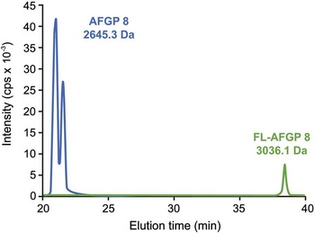
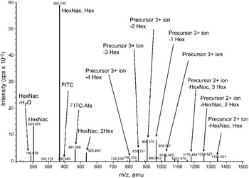
Neither fish showed externally visible fluorescence 72 h after first intubation with FL-AFGP. When checked using a hand-held UV source, the fish killed at this time showed fluorescence in the all parts of the gastrointestinal tract except the stomach. All other major tissues, organs and body fluids were negative, including the brain, muscle, urine, bile and serum suggesting a longer post-intubation period was required for significant FL-AFGP uptake. The second fish was intubated again at 72 h and monitored daily until external fluorescence was observable eight days after the first intubation. This signal had begun to decline after a total of 13 days at which time the fish was killed and sampled. FL-AFGP was subsequently detected in the serum by LC-MS/MS. Two peaks (shoulders) of non-fluorescent endogenous AFGP 8 of equivalent mass (2645.3 Da) were detectable in the extracted MS trace (Fig. 3) eluting at c. 21 min, clearly separate from the later eluting FL-AFGP 8 (3036.1 Da). Further analysis using MS/MS confirmed that the species eluting at 38 min was indeed FL-AFGP 8 (Fig. 4) as evidenced by the presence of cleaved FITC, FITC-labelled alanine (the N-terminal amino acid of AFGP 8) and the expected sugars, an N-acetyl hexosamine (HexNac) and a hexose (Hex), predominantly as the disaccharide but also as cleavage products or in various experimentally introduced combinations. No attempt was made to quantify the yield of FL-AFGP 8.
Fluorescent conjugates (FL-AFGPs) traverse the gastrointestinal tract to reach the blood. Extracted ion chromatograms for endogenous AFGP 8 (blue trace) and experimentally introduced FL-AFGP 8 (green) in control and intubated Pagothenia borchgrevinki serum following LC-MS/MS. AFGP 8 (2645.3 Da) typically eluted earlier (c. 21 min) than the heavier (3026.1 Da) and more hydrophobic FL-AFGP 8 (c. 38 min). The presence of two peaks in the AFGP 8 trace probably represents two amino acid sequence isoforms that can be resolved chromatographically. Other AFGPs that were detected with this experimental protocol are not shown.

Fluorescent conjugates (FL-AFGPs) are resorbed from the gastrointestinal tract and released into the circulation. Fragment ion spectrum for FL-AFGP 8 detected in the serum obtained from P. borchgrevinki intubated with FL-AFGPs. Two series of fragment ions representing FL-AFGP 8 in different charge states with the loss of various numbers of HexNac and/or Hex sugars from the glycosylated threonine residues are prominent. Other fragment ions correspond to free FITC and FITC-labelled alanine (the N-terminal amino acid of AFGP 8) both released on dissociation of the parent molecule, and Hex and HexNac sugars (the natural components of the threonine linked disaccharide in AFGP) in the native state and as an artefactual trisaccharide (m/z 528.2) as a consequence of experimentally induced recombination after dissociation from FL-AFGP 8.

AFGPs are transported across rectal epithelial cells
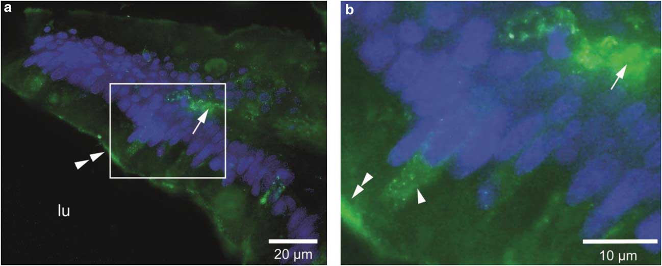
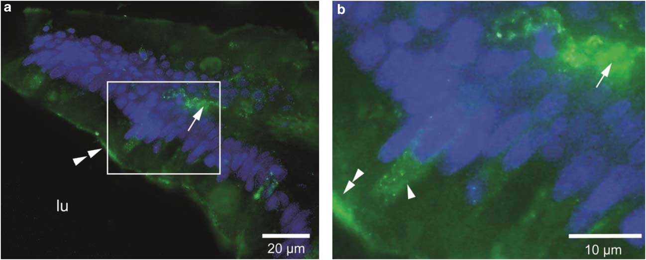
Fluorescent immunostaining using anti-AFGP as the primary antibody revealed that AFGPs accumulate in adsorptive cells in the rectum of both T. hansoni (Figs 5a & b, 6a & b) and B. saida (Fig. 7a & b). Positive staining was more extensive in the apical regions of the absorptive cells of T. hansoni, but both species showed immunoreactivity in the lamina propria suggestive of intraepithelial transport from the rectal lumen.
Antifreeze glycoprotein immunoreactivity in the notothenioid rectum. a. The Antarctic notothenioid Trematomus hansoni shows AFGP accumulation in the apical regions of absorptive cells in the rectal epithelium (arrow). Merged green immunofluorescent, DAPI and DIC images. b. Detail of the boxed area in the left panel showing an absorptive cell at the villus tip with an extensive accumulation of cytoplasmic AFGPs. Note the apical displacement of the nucleus (arrowhead). Another smaller, possibly intraepithelial cell also contains AFGPs. Merged green immunofluorescent and DAPI images. gc = mucus secreting goblet cell.

Antifreeze glycoprotein immunoreactivity in the rectum of the polar cod B. saida. a. The tip of a villus showing AFGP immunoreactivity in the lamina propria (arrow) and in association with the absorptive cell surface (double arrowhead). Merged green immunofluorescent, DAPI and DIC images. b. Detail of the boxed area in the left panel showing cytoplasmic granules immunoreactive for AFGPs (arrowhead) and the accumulation of AFGPs near the cell surface (double arrowhead) and in the lamina propria (arrow). Merged green immunofluorescent and DAPI images.
Spleen ellipsoids contain AFGPs
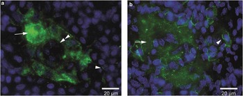
Positive immunostaining of AFGP in the spleens of T. nicolai and B. saida was detected in macrophages contributing to the periarteriolar sheaths of the ellipsoids in both species (Fig. 8a & b). Some cells of uncertain derivation in the spleen of B. saida showed strong peripheral reactivity possibly representing pericytoplasmic AFGPs.
Antifreeze glycoprotein accumulates in spleen ellipsoids. a. An ellipsoid in the spleen of the notothenioid T. nicolai displays a cuff of AFGP immunoreactivity coincident with the location of periarteriolar macrophages (arrow). Endothelial cells do not accumulate significant amounts of AFGPs (arrowhead) and most parenchymal splenocytes are unreactive. A few cells more distant from the ellipsoid appear to have small accumulations of AFGPs (arrowhead), but these were not as extensive as those in B. saida. Merged green immunofluorescent and DAPI images. b. An ellipsoid in the spleen of the polar cod B. saida displaying a peri-arteriolar cuff of AFGP immunoreactivity (arrow) as seen in T. nicolai. Some cells of uncertain origin but typically located on the periphery of the ellipsoid were immunoreactive (arrowhead). Immunoreactivity in these cells was most prominent in the pericytoplasmic region, in close association with the plasma membrane. Merged green immunofluorescent and DAPI images.

Discussion
Identification of the site of AFGP synthesis using northern blot analysis or reverse transcription-polymerase chain reaction (RT-PCR) amplification is potentially compromised when organs are distributed diffusely through the body, as is the case with the pancreas in some teleosts, which may extend into the liver, around the portal vein, and over the surface of the gall bladder (Oliveira Ribeiro & Fanta Reference Oliveira Ribeiro and Fanta2000). Resolution of this potential problem requires certainty that there is no contamination from other tissues. Using immunohistology to identify tissue source, we have been able to confirm that AFGPs in B. saida are not only synthesized in the liver (Fig. 1a) but they are synthesized in the pancreas as well (Fig. 1b), in agreement with molecular assays (Chen et al. Reference Chen, DeVries and Cheng1997b, Cheng et al. Reference Cheng, Cziko and Evans2006). Thus secretion in the liver of this species is not a case of misinterpretation as it was in the Antarctic notothenioids (Cheng et al. Reference Cheng, Cziko and Evans2006).
Our results leave no doubt that both Antarctic notothenioids and the Arctic cod synthesize AFGPs in the pancreas in addition to other sites (such as the liver in B. saida). The pancreatic AFGPs in both cases are confined to zymogen granules of acinar cells in the exocrine portion of the pancreas, which is responsible for the secretion of various digestive enzymes (including the protease precursors trypsinogen and chymotrypsinogen) into the intestine. Antifreeze glycoproteins probably follow the same secretory pathway, being discharged from the zymogen granules at the apical surface of the acinar cells into lumen of the acinus, which drains into the anterior intestine (duodenum) via a system of ducts.
The liver secretory pathway of B. saida enables AFGPs to reach the circulation directly, while secretion from the pancreas protects the gastrointestinal lumen against ingested ice. The situation in Antarctic notothenioids presents a significant paradox, however, since there is no identifiable anatomical pathway by which high levels of AFGPs can reach the circulation directly from their known sites of synthesis, all of which discharge into the gastrointestinal tract. In order to resolve this paradox we used FITC to label the N-terminal alanine of the small AFGP fraction prepared from serum of the Antarctic toothfish D. mawsoni and introduced this by intubation into the stomach of another notothenioid, P. borchgrevinki. Blood sampled 13 days after first intubation and analysed by LC-MS/MS showed the presence of FL-AFGP 8, indicating that at least some of the AFGP fraction can reach the circulatory system from the digestive tract (Fig. 3). External fluorescence was first detectable after eight days, suggesting uptake from the gut commences before this time.
Endogenous AFGP 8 from a control P. borchgrevinki appeared as two peaks (shoulders) on the extracted ion chromatogram (Fig. 3). Since these two peaks have identical molecular weights (2645.5 Da), amino acid and sugar content, they presumably represent protein isomers varying in the linear arrangement of the constituent amino acids. For example, two isomeric sequences for AFGP 8, AATAATPATAATPA and AATAATAATPATPA, have been identified as polyprotein cleavage peptides in a number of Antarctic notothenioids including the black rockcod Notothenia coriiceps Richardson, 1884 (Cheng Reference Cheng1996) and the naked dragonfish Gymnodraco acuticeps Boulenger, 1902 (Evans unpublished observations). Analysis of the MS/MS data for the m/z 1012.3 species showed the presence of fragment ions representing the expected disaccharide (HexNac-Hex) and FITC-labelled alanine (the N-terminal amino acid in AFGP), in addition to a small amount of free FITC (Fig. 4). There is some experimentally induced gas-phase rearrangement of the sugars giving, for example, the peak at m/z 528.197 for the trisaccharide HexNac-Hex-Hex.
Although our LC-MS methodology is targeted at identifying AFGP 8, and thus we followed the transport of FL-AFGP 8 from the gastrointestinal tract to the blood, we argue that the entire range of AFGPs could follow a similar absorptive pathway and there are several lines of evidence in support of this possibility. The size classes within the AFGP family differ significantly only in their molecular weights, for example, with very little amino acid sequence variation and identical sugar components. Thus they probably behave similarly. All members are believed to have the same tertiary structure based on a polyproline type II helix (Bush & Feeney Reference Bush and Feeney1986) and all display similar resistance to enzymatic digestion once the linkers in the polyprotein precursor have been cleaved (Hsiao et al. Reference Hsiao, Cheng, Fernandes, Detrich and DeVries1990), a property which probably contributes to them passing intact through the gastrointestinal tract. Additionally, there are a number of reports that the fish rectum can absorb large molecular weight compounds, at least up to 40 kDa (McLean & Ash Reference McLean and Ash1987, McLean et al. Reference McLean, Rønsholdt and Sten1999, Berge et al. Reference Berge, Goodman, Espe and Lied2003) and probably much greater. Hernandez-Blazquez & Cunha da Silva (Reference Hernandez-Blazquez and Cunha da Silva1998), for example, reported that the Antarctic notothenioid Notothenia neglecta Nybelin, 1951 could absorb horse spleen ferritin (the globular apoprotein is c. 450 kDa) through the rectum, where it was identified in cytoplasmic vesicles of the absorptive cells.
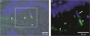
In order to resolve the pathway involved in absorption we sectioned portions of the gastrointestinal tract from T. hansoni and found AFGPs in absorptive cells of the rectum using immunofluorescence microscopy (Figs 5 & 6). Very few epithelial cells in the rest of the gastrointestinal tract displayed clear signs of cytoplasmic AFGP (results not shown) indicating that absorption of AFGP is largely the province of the rectum. The distribution of the AFGPs within the rectum suggested a pathway of absorption from the lumen through the apical to the basal surface of the lining epithelial cells, with subsequent discharge into vessels within the lamina propria. From this region of the rectum, distribution via the blood and interstitial fluid is probably rapid and extensive (Evans et al. Reference Evans, Gubala, Nooney, Williams, Brimble and DeVries2011). Sections of the rectum from B. saida immunostained with AFGP antibodies showed AFGPs on the apical surface of rectal absorptive cells and within intracellular vesicles as well as in the lamina propria (Fig. 7). Taken together these results suggest that the Arctic cod utilizes a similar rectal absorptive pathway for AFGPs as do the Antarctic notothenioids, providing yet another example of functional convergence in these two groups of polar fishes.
Antifreeze glycoprotein absorptive cells in the notothenioid rectal epithelium. a. Antifreeze glycoprotein accumulation is apparent in the apical regions of the absorptive cells of T. hansoni (arrowhead). Merged green immunofluorescent, DAPI and DIC images. b. Detail of the boxed area in the left panel highlighting apical accumulation of AFGPs (arrowhead) and the displaced nucleus (double arrowhead). Small, rounder nuclei are most probably from intraepithelial lymphocytes. Merged green immunofluorescent and DAPI images. gc = mucus secreting goblet cell, lu = lumen of the rectum.

We next addressed the problem of how the two groups of polar fishes cope with internal ice, a situation potentially lethal for both of them since they both inhabit freezing waters. The presence of high levels of AFGPs in gastrointestinal fluids will limit the growth of ingested ice crystals, minimizing potential tissue damage until the ice and adsorbed AFGPs is discharged along with the faeces. We assume that only free (soluble) AFGPs are resorbed in the rectum since it would be counterproductive to resorb AFGPs bound to potentially lethal ice crystals - these would be discharged with the faeces. Our interpretation is consistent with the resistance of AFGPs to enzymatic degradation and their extreme solubility in aqueous solutions, allowing for pinocytotic uptake in the rectum.
Ice that reaches the circulation presents a more significant risk factor than ice in the gastrointestinal tract, with the potential to become entrapped and damage narrow diameter blood vessels. From a pathophysiological perspective, there would appear to be distinct advantages in rapidly removing ice crystals from the circulation so they do not accumulate and damage sensitive tissues of the body, such as the gills and brain. Initial experiments using silica nanoparticles coated with AFGPs as a proxy for AFGP-adsorbed ice crystals suggested spongy macrophages in ellipsoids of the spleen as a probable site for the uptake of ice in the circulation (Evans et al. Reference Evans, Gubala, Nooney, Williams, Brimble and DeVries2011). Because ice crystals melt during tissue preparation we used immunohistology to identify concentrations of AFGPs likely to reflect sites where AFGP-adsorbed ice crystals accumulate. The only significant intracellular concentrations of AFGPs in Antarctic notothenioids (other than at the sites of synthesis) were found within the spleen (Fig. 8a), including within ellipsoidal macrophages. We reason that AFGPs accumulate in spleen macrophages as a consequence of being endocytosed along with ice crystals to which they are absorbed. This interpretation is in agreement with our initial studies, which showed AFGP-adsorbed nanoparticles (used as a proxy for AFGPs adsorbed to ice) accumulate within ellipsoidal macrophages (Evans et al. Reference Evans, Gubala, Nooney, Williams, Brimble and DeVries2011) and other studies which showed the notothenioid spleen to contain ice crystals (Praebel et al. Reference Praebel, Hunt, Hunt and DeVries2009). Ellipsoids in fishes, as in other vertebrates, have a role in the selective filtration and retention of blood-borne particles (Espenes et al. Reference Espenes, Press, Dannevig and Landsverk1994), rendering them ideal for the removal of circulating AFGP-adsorbed ice crystals in the blood of polar fishes.
In a further example of functional convergence, AFGPs were also found within the splenic ellipsoids of the Arctic cod (Fig. 8b), suggesting that they have evolved a similar mechanism to deal with internal ice. Some cells towards the periphery of the ellipsoids in B. saida displayed relatively strong AFGP immunoreactivity (Fig. 8b), which appeared to be concentrated in their outer cytoplasmic regions adjacent to the plasma membrane. The nature and function of these cells is uncertain, but their disposition suggests they may be migratory within the spleen, possibly transporting AFGPs to other locations. Possibly similar cells in the notothenioid T. nicolai had more patchy immunoreactivity (Fig. 8a).
Following entrapment of AFGP-adsorbed ice crystals in ellipsoidal macrophages, the next major problem for both groups of polar fishes is how to melt the engulfed ice so that it does not accumulate to lethal proportions. Although a number of theoretical mechanisms could be proposed, we think the most likely option is that the ice remains entrapped in the spleen until such time as the fish is exposed to an appropriate warming event. As far as the Arctic cod is concerned, although it is often found in freezing water it is also found in large schools during the summer in warmer water (2.8–4.4°C) where internal ice would be expected to melt (Schurmann & Christiansen Reference Schurmann and Christiansen1994). With respect to the Antarctic notothenioids, seasonal warming events sufficient to melt internal ice can occur at high latitudes when local marine environments become ice-free during the summer months (Hunt et al. Reference Hunt, Hoefling and Cheng2003). One testable corollary of our proposal is that a fish never exposed to a warming event probably has more ice crystals in its spleen than one of similar age occupying a similar habitat, but regularly exposed to warming on a seasonal basis. Such situations exist in McMurdo Sound, Antarctica where benthic notothenioids with restricted home ranges, as illustrated by various Trematomus species (Evans et al. Reference Evans, Hills and Dickson2000), can be exposed to either perennially freezing water (e.g. in relation to supercooled water emerging from under an ice shelf) or to warming events as reported in the vicinity of Ross Island (Hunt et al. Reference Hunt, Hoefling and Cheng2003). Another testable corollary is that fish never exposed to ice should not contain AFGPs in their spleens. Such fish might include the deepwater notothen Trematomus loennbergii Regan, 1913 although uncertainties surround the habit of its larval and juvenile forms.
Taken together with our previous results that AFGPs circulating in the blood can reach the bile by traversing the liver (Evans et al. Reference Evans, Gubala, Nooney, Williams, Brimble and DeVries2011), we can now elaborate on how AFGPs circulate within polar fishes. Using notothenioids as an example, AFGPs synthesized in the exocrine pancreas and anterior stomach are discharged into the gut where they are reabsorbed in the rectum to pass into the blood and interstitial fluid. Blood-borne AFGPs traversing the liver are transferred into the bile and subsequently released again into the gastrointestinal tract, thus establishing a recycling loop that conserves energy expenditure and enables relatively high AFGP levels to be maintained in the blood (Fig. 9). Ice crystals ingested with food and water are coated with AFGPs (secreted into the gastrointestinal tract) to inhibit further growth and expelled from the body with the faeces. Ice crystals that enter the circulation also adsorb AFGPs, but instead of being expelled they are stored in ellipsoidal macrophages until such time as they are able to be melted. The subsequent fate of AFGPs (freed from melted ice crystals) within ellipsoidal macrophages is uncertain, although given their resistance to degradation it is conceivable that they are ultimately released intact back into the circulation. What is remarkable about this proposed recycling loop is the extent to which it is shared by Antarctic notothenioids and Arctic cods, illustrating that these two divergent groups of polar fishes display elements of convergent evolution not only in terms of AFGP structure, but also in terms of its physiological deployment.
A proposed model for AFGP synthesis and recycling in polar fishes. Antifreeze glycoproteins synthesized in the exocrine pancreas of both Antarctic notothenioids and Arctic gadids (as well as the anterior stomach of notothenioids) are discharged into the gastrointestinal tract to protect against ingested ice. Antifreeze glycoproteins not lost along with the faeces can be resorbed by epithelial cells predominantly in the rectum and subsequently transferred into the blood and interstitial fluid. Antifreeze glycoproteins reaching the liver in the blood are transferred to the bile, stored in the gall bladder and released back into the gastrointestinal tract along with other bile components. The liver of Arctic gadids (boxed) also secretes AFGPs, releasing them directly into the blood from where they enter into the recycling pathway.

Acknowledgements
This research was supported by the Human Frontier Science Program (grant RGP003/2009-C). We thank Antarctica New Zealand for logistic support in Antarctica, colleagues at Scott Base for assistance in the field, Dr Adrian Turner for advice with microscopy, and Vivian Ward for graphics. We are particularly grateful to Prof Jorgen Christiansen of the University of Tromso, Norway and the crew of the RV Jan Mayen for assistance in obtaining the polar cod. The constructive comments of the reviewers are also gratefully acknowledged.