Introduction
The head mirror is the most recognisable symbol of an otorhinolaryngologist. In certain cultures, the head mirror is as common as the stethoscope, if not more so, in artists' renderings and popular depictions of doctors. Introduced in 1743 by the French accoucheur Levert for examining the larynx, today's head mirror has withstood the test of time.1 However, an increasing number of doctors are starting to use battery-powered headlights instead of head mirrors. Some otolaryngologists question whether the head mirror can continue to sustain its relevance and trump its younger technological sibling, the battery-powered or fibre-optic headlight.
During our training years, many of us were told of the multiple advantages of the head mirror, the most important of which was the ability to align one's eye sight and the light source axis, which theoretically facilitated the identification of details within narrow and often tubular spaces during the otorhinolaryngological examination.Reference Hannan2 However, there is no clear evidence to support this statement.
In this study, we aimed to evaluate and compare the head mirror and various headlights, through the measurement of illumination, visual identification and visual acuity, while using these instruments under conditions simulating a clinical otorhinolaryngological examination.
Materials and methods
Head mirror and headlights
The head mirror used in the study was manufactured by Nagashima (Tokyo, Japan) and had the following specifications: 89 mm diameter and 13 mm round aperture, with a fibre forehead band.
Three types of headlight were tested: the Kimscope 1 W (model: SLL 01 Warm) (Seahanul Biotech, Anyang, South Korea); the Kimscope 3 W (model: SLL 02 Warm; Seahanul Biotech); and the LumiView Portable Binocular Microscope (Welch Allyn, Skaneatelese Falls, New York, USA). All these headlights emitted warm light, similar to the light source when using a head mirror.
Illuminance
Illuminance was measured using a portable lux meter (Sanpometer LX 1010BS; Sanpo Instrument, Shenzhen, China). The mean spectral sensitivity ± standard deviation (SD) of the meter at 600–620 nm (warm light) was approximately 55 ± 5 per cent, as claimed by the manufacturer.
The lux meter was placed and secured on an examination chair headrest. Illuminance was measured and recorded while the head mirror or headlight directly illuminated the meter sensor from a distance of 50 cm, simulating a common examination condition in daily otorhinolaryngological practice.Reference Mester3
Illumination field
For the rest of the study, the Kimscope 3 W headlight was selected as the representative headlight due to its superior illuminance.
To test illumination field, the examined head was replicated using a full-scale skull model (First Class Human Skull; Anatomical Chart Company, Lippincott, Williams & Wilkins, Philadelphia, Pennsylvania, USA) with highly accurate replication of the nasal cavity, nasal turbinates and nasopharynx. A visual target was attached to the posterior nasopharyngeal wall such that it could be visualised through the nasal cavity and the choana. This visual target was a 4 × 4 cm plain sheet imprinted with a reference grid scale. The distance between the visual target and the light source was 35 cm. The illuminated area was recorded and compared for the head mirror and the Kimscope 3 W headlight.
Visual identification and acuity
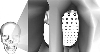
Under similar settings as mentioned above, the visual target was replaced with a down-scaled Landolt C table.Reference Danilova and Bondarko4 This table was reduced to 1/12 of its original size in order to match the requirements for a standard visual acuity test at a distance of 50 cm. Sixty-four ‘c' letters ranging in diameter from 2.5 to 16.67 mm were included in the chart, arranged in various orientations (see Figure 1).
Diagram showing positioning of the target for visual identification (a down-scaled Landolt C table) at the nasopharynx within a full-scale skull model.

Thirteen medical students and 10 otolaryngology specialists were enrolled as volunteer subjects. The participants were asked to identify the visual target at the nasopharynx, as effectively as possible, using the head mirror and headlight, within a period of 3 minutes. In each group, half started the test with the head mirror and the other half started with the headlight. Participants were asked to read out the target Landolt C table as far as possible, without assistance. In the student group, only the number of correctly identified letters was recorded and compared. In the consultant group, visual acuity (i.e. the correct interpretation of the direction of the c letter) was also recorded and compared.
Statistical methods
Data from each group were compared using Student's t-test (paired, two-tailed). A p value of less than 0.05 was taken to indicate a statistically significant difference.
Results and analysis
Illuminance
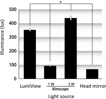
The mean illuminance ± SD for the LumiView, Kimscope 1 W and Kimscope 3 W headlights and the head mirror was 352.3 ± 9, 92.3 ± 4.5, 438 ± 15.7 and 68.3 ± 1.2 lux, respectively. The Kimscope 3 W headlight gave the highest illuminance of all the devices tested (Figure 2).
Comparison of illuminance for the three headlights and the head mirror. P < 0.05, comparing all sources.

Illumination field
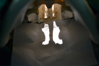
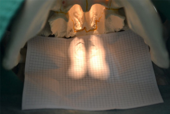
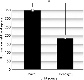
Figures 3 and 4 show the typical illumination fields generated using the Kimscope 3 W headlight and the head mirror, respectively. The mean ± SD illumination field of the head mirror was 348 ± 29.8 grid squares, significantly greater than that of the Kimscope 3 W headlight (183 ± 9.2 grid squares) (p = 0.0017, two-tailed t-test) (Figure 5). Each grid square was equal to 3.579 mm2.
Photograph showing typical illumination field projected through the nasospharynx by the Kimscope 3 W headlight.

Photograph showing typical illumination field projected through the nasospharynx by the head mirror.

Comparison of the illumination field of the head mirror and Kimscope 3 W headlight. P < 0.05, comparing both sources.

Visual identification and acuity
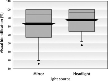
In the student group, the mean ± SD number of letters identified with the headlight and head mirror were respectively 56.2 ± 9 and 53.3 ± 14.1; this difference was not statistically significant (p = 0.3) (Figure 6).
Comparison of visual identification percentage for the head mirror and the Kimscope 3 W headlight in the student group.

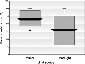
In the consultant group, surprisingly, the mean ± SD number of letters identified with the head mirror was significantly higher than with the headlight (59.7 ± 3.3 vs 55.2 ± 5.8, respectively; p = 0.0035) (Figure 7). Despite this difference, the consultants' visual acuity was the same regardless of the device used (i.e. whether using a head mirror or headlight, the line number of the smallest correctly identified letters remained identical).
Comparison of visual identification percentage for the head mirror and the Kimscope 3 W headlight in the consultant group.

Discussion
In 1585, Aranzi was the first to use a light source for an endoscopic procedure, by focusing sunlight through a flask of water and projecting the light into the nasal cavity.
In 1806, Philip Bozzini built an instrument that could be introduced into the human body to visualise the internal organs, and is thus regarded as the father of endoscopy. He used an aluminium tube to visualise the interior body in a novel way: the tube, illuminated by a wax candle, had a fitted mirror to reflect images. In 1901, Dimitri Ott wore a headband mirror to reflect light and augment his visualisation. To this end, he used an access technique whereby a speculum was introduced through an incision in the posterior vaginal fornix.
Thus, head mirrors have a time-honoured history, and are typically used today for the examination of the ear, nose and throat. In recent times, the head mirror has gradually become a symbolic tool used by otorhinolaryngologists in daily practice.
Commonly, the head mirror comprises a circular, concave mirror with a small hole in the middle, which is attached to a head band. The key feature of the head mirror is the combination of the concave mirror and the centre aperture, allowing the axis of the light source to align with the visual axis. This feature theoretically allows the examiner to look deeply into a narrow cavity.
Typically, the patient sits and faces the physician. A bright lamp is positioned adjacent to the patient's head, pointing toward the physician's face and thus toward the head mirror. The light from the lamp reflects off the mirror, along the line of sight of the user. Theoretically, the head mirror provides excellent, shadow-free illumination when used properly.
The main drawback of head mirrors is generally that they require some skill to use well.
Head mirrors are rarely used outside the otorhinolaryngology setting, having been largely replaced by pen lights among general practitioners.
In recent decades, the development and the usage of battery-powered headlights have increased remarkably. This rise in popularity may be related to progress in the field of electronics, especially the introduction of light-weight, rechargeable batteries and light-emitting diode light sources. Headlights no longer require a connection to an external light source with optical fibre cables. Nevertheless, no matter how the headlight is designed, it is difficult to render the axis of the light source and the visual axis coaxial, which can lead to a decrease in visible field while examining a narrow, deep space. However, these opinions remain to be tested scientifically.
Few related studies have been published to date. One study that attempted to justify and compare the use of headlights and head mirrors was performed by Rowlands et al. Reference Rowlands, Mal and Thornton5 These authors recruited 48 medical students and asked them to complete a simple task using a headlight and using a head mirror. The students required more time to complete the task when using the mirror, and overwhelmingly preferred the headlight to the mirror. The authors concluded that headlights should replace head mirrors in routine clinical practice, particularly for newcomers to the specialty.
We observed similar trends in our study. Although the student group's visual identification was slightly better using the headlight rather than the head mirror, this difference was not statistically significant. However, surprisingly, the consultant group had significantly better visual identification when using the head mirror as opposed to the headlight. We presume that, in skilful hands, the light reflected from the mirror is coaxial with the visual axis, and the large area of the concave mirror surface converges a broader area of light into narrow cavities, significantly reducing shadows and ‘blind spots’.
• Head mirrors and headlights are frequently used in otolaryngology
• There has been little scientific evaluation of their performance
• The ideal device would provide maximal illuminance and illumination field, aiding detailed visualisation in narrow spaces
• In this study, headlights provided best illumination but head mirrors threw fewer shadows
• Head mirrors performed better in experienced hands
There is evidence supporting a large difference in vision quality under conditions of strong versus weak light intensity.Reference Sjostrand6 Adequate illumination is crucial to achieving good diagnostic accuracy.Reference Hausner7, Reference Perrig, Kazennikov and Wiesendanger8 However, the present study found that, despite the headlight's greater illuminance, visual acuity was similar with both the head mirror and the headlight.
Conclusion
Despite the headlight's greater illuminance, head mirrors provide illumination with fewer shadows. While there seemed to be little difference between headlights and head mirrors amongst inexperienced students, surprisingly, head mirrors performed better in experienced hands.
Acknowledgment
The authors greatly appreciate the assistance of Professor Roger W Chan in the statistical analysis and revision of this manuscript.